BIOSTATIC ENDOSCOPIC ETHMOID SURGERY A New Approach ...
BIOSTATIC ENDOSCOPIC ETHMOID SURGERY A New Approach ...
BIOSTATIC ENDOSCOPIC ETHMOID SURGERY A New Approach ...

You also want an ePaper? Increase the reach of your titles
YUMPU automatically turns print PDFs into web optimized ePapers that Google loves.
6<br />
Biostatic Endoscopic Ethmoid Surgery – B.E.E.S. – A <strong>New</strong> <strong>Approach</strong> to<br />
Functional Endoscopic Sinus Surgery for Acute Recurrent Rhinosinusitis<br />
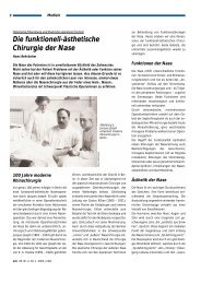
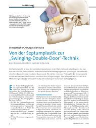
Emil Zuckerkandl (1848—1910)<br />
(Photo: Prof. Daniel Simmen, Zurich,<br />
Switzerland)<br />
Introduction<br />
Historical Considerations<br />
After initially pursuing a career as a virtuoso violinist, Emil Zuckerkandl<br />
(1849–1910) became an extraordinary professor of anatomy at 31 years of<br />
age, without having to present the customary postdoctoral credentials. In<br />
1882 Zuckerkandel became a full professor in Graz, Austria, and six years<br />
later he assumed the first anatomy chair in Vienna. The poet Arthur Schnitzler<br />
studied medicine in Vienna and shared a vivid memory of Zuckerkandl in his<br />
autobiography, Youth in Vienna, describing him as “… a pale young man<br />
with a dark goatee and black eyes. In his academic robes he closely re -<br />
sembled one of those anatomists familiar to us from the famous Rembrandt<br />
portraits, while the legendary stories of his rakish youth, filled with drinking<br />
and fencing, seemed to hover about him.” He also had a reputation for “…<br />
going straight to work from some tavern or perhaps even from the arms of a<br />
beautiful woman and launching directly into his daily routine, teaching and<br />
studying with prodigious energy far into the night.”<br />
This intense drive for scientific knowledge surely formed the basis for his<br />
pamphlet on Normal and Pathologic Anatomy of the Nasal Cavity and its<br />
Pneumatic Appendages, published in 1882. In this work, Zuckerkandl was<br />
the first author to give a detailed anatomical description of the ethmoid<br />
bone and all the paranasal sinuses, thus creating a scientific basis for<br />
understanding their anatomy. He also drew attention to specific structures<br />
and narrow passageways that contribute to the pathogenesis of rhino -<br />
sinusitis and are still relevant today – such as the ethmoid infundibulum and<br />
variants in the pneumatization and curvature of the middle nasal turbinate.<br />
He described in detail the cellular anatomy of the ethmoid labyrinth, noting<br />
that “…the ethmoid bulla is highly variable in its development, and its<br />
importance rests not only on its relationship to the middle turbinate. An<br />
ethmoid cell belonging to the lower portion of the labyrinth, the ethmoid<br />
bulla presents a convex medial surface to the nasal cavity and is bounded<br />
laterally by the lamina papyracea of the ethmoid bone or may be separated<br />
from it by another, intervening ethmoid cell…”<br />
Zuckerkandl’s writings on the variability of the middle turbinate included the<br />
following: “The variations relate both to the shape and size of the [middle]<br />
turbinate. The turbinate may be so markedly curved that it occludes the<br />
olfactory groove and engages against the nasal septum. The transformation<br />
of the anterior end of the turbinate into a large bony bulla is a common<br />
occurrence and was even described in the past century by Giovanni<br />
Santorini in his ‘Observationes anatomicae’. In cases of this kind, the<br />
turbinate contains a cavity, at times even subdivided by a septum, that<br />
communicates openly with the middle meatus.”<br />
ZUCKERKANDL E: Normal and Pathologic<br />
Anatomy of the Nasal Cavity and its<br />
Pneumatic Appendages. Vol. 2, Vienna.<br />
Wilhelm Braumüller (1892, 1893).