BIOSTATIC ENDOSCOPIC ETHMOID SURGERY A New Approach ...
BIOSTATIC ENDOSCOPIC ETHMOID SURGERY A New Approach ...
BIOSTATIC ENDOSCOPIC ETHMOID SURGERY A New Approach ...

You also want an ePaper? Increase the reach of your titles
YUMPU automatically turns print PDFs into web optimized ePapers that Google loves.
8<br />
Biostatic Endoscopic Ethmoid Surgery – B.E.E.S. – A <strong>New</strong> <strong>Approach</strong> to<br />
Functional Endoscopic Sinus Surgery for Acute Recurrent Rhinosinusitis<br />
The results: Occasional postoperative CT scans obtained for<br />
various indications (they are not routinely necessary) have shown<br />
the following typical problems:<br />
Significant scarring and contraction may occur after a complete<br />
ethmoidectomy.<br />
These effects can obstruct surgically created drainage routes from<br />
the frontal and maxillary sinuses.<br />
The middle turbinate is very susceptible to postoperative laterali<br />
zation.<br />
Mucosal lesions are associated with a high risk of synechia formation<br />
and ethmoid atelectasis.<br />
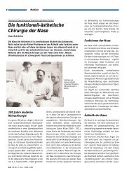
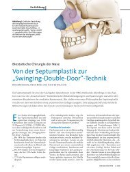
The ethmoid “matchbox.” The principal<br />
supporting structures of the anterior ethmoid<br />
are the ethmoid bulla (red) and the<br />
middle turbinate with its basal lamina,<br />
which separates the anterior and posterior<br />
ethmoid cells and stabilizes the width of<br />
the ethmoid bone.<br />
Other structures:<br />
brown = Agger nasi cell<br />
green<br />
= Uncinate process<br />
blue<br />
= Retrobullar cell<br />
gray and yellow = Posterior ethmoid cells<br />
shading = Ethmoid infundibulum<br />
and frontal recess.<br />
The most important postulate of F.E.S.S. in the treatment of patients with<br />
recurrent rhinosinusitis is the improvement of ventilation and drainage,<br />
which is essential for mucosal regeneration. This can be accomplished only<br />
by taking into account biostatic principles, which must be applied in order<br />
to achieve the main goal: adequate and permanent postoperative patency<br />
of the ethmoid sinuses.<br />
Establishing drainage from the frontal sinuses will be ineffective if post -<br />
operative contraction of the ethmoid causes lateralization of the middle<br />
turbinate and possible synechia formation, leading to stenosis of the<br />
drainage pathway.<br />
Biostatic principles<br />
In simplified terms, the ethmoid bone has the approximate size and<br />
shape of a matchbox stood on edge. The width of the “matchbox”<br />
is variable and depends on the degree of ethmoid pneumatization<br />
and on the variable symmetry of the ethmoid cells.<br />
The supporting structure of the ethmoid bone, which forms cavities<br />
to maintain ventilation and drainage of the maxillary and frontal<br />
sinuses, shows varying patterns of pneumatization.