BIOSTATIC ENDOSCOPIC ETHMOID SURGERY A New Approach ...
BIOSTATIC ENDOSCOPIC ETHMOID SURGERY A New Approach ...
BIOSTATIC ENDOSCOPIC ETHMOID SURGERY A New Approach ...

Create successful ePaper yourself
Turn your PDF publications into a flip-book with our unique Google optimized e-Paper software.
12<br />
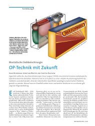
Biostatic Endoscopic Ethmoid Surgery – B.E.E.S. – A <strong>New</strong> <strong>Approach</strong> to<br />
Functional Endoscopic Sinus Surgery for Acute Recurrent Rhinosinusitis<br />
Hypothesis<br />
Today, endoscopic surgery of the paranasal sinuses has assumed a<br />
broad range of indications. Based on our own experience, we feel that<br />
strict distinctions should be drawn among the subsets of indications<br />
described below in order to fully utilize the potential of differentiated<br />
microsurgery and prevent postoperative stenosis.<br />
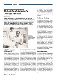
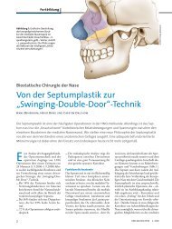
Coronal CT scan of a woman who underwent<br />
a previous complete right ethmoidectomy and<br />
B.E.E.S. in which the upper cap of the<br />
ethmoid bulla was preserved.<br />
Medialized middle turbinates. The bulla<br />
lamellae have been preserved on both<br />
sides.<br />
Acute recurrent rhinosinusitis is marked by inflammatory exacerbations,<br />
typically involving the frontal and maxillary sinuses. Complete<br />
anterior ethmoidectomy should never be considered a routine<br />
procedure. If exenteration of the anterior ethmoid is required, the<br />
surgeon should make every effort to preserve the lamella or the<br />
upper cap of the ethmoid bulla in order to prevent narrowing of the<br />
anterior ethmoid and frontal recess. The parietal mucosa of the<br />
lamina papyracea, anterior skull base, and middle turbinate should<br />
be preserved. Enlarging the frontal recess in a type I to IIb drainage<br />
procedure is easily accomplished under vision with a 45° endo -<br />
scope while preserving the bulla lamella. Infundibular and agger<br />
nasi cells that obstruct the frontal recess can be removed by using<br />
the “uncapping the egg” technique of Stammberger, which involves<br />
a posterior-to-anterior dissection. A maximum amount of mucosa<br />
should be preserved in the frontal recess. The basal lamina of the<br />
middle turbinate may be trephined over a circumscribed area, but it<br />
should not be fractured.<br />
Chronic rhinosinusitis with nasal polyps or sinonasal polyposis<br />
currently represents a large subset of indications for endoscopic<br />
sinus surgery. Both biomechanical and immunologic factors contribute<br />
significantly to the pathogenesis of chronic rhinosinusitis.<br />
Based on regional variations in the texture of the ethmoid sinus<br />
mucosa, such as an abundance of glands on the anterior surface of<br />
the bulla, sites of predilection exist for the development of polyps.<br />
These polyps destroy the ethmoid cells and their infrastructure. Due<br />
to pressure effects from the polyps and the absence of scar<br />
contractures with an intact parietal mucosa, sinus contraction does<br />
not occur. Typically, moreover, these patients rarely complain of<br />
headaches. Cell remnants that have already been destroyed should<br />
be removed at operation, and the parietal mucosa on the lamina<br />
papyracea, skull base and middle turbinate, for example, should be<br />
preserved.<br />
Coronal CT one year after bilateral medialization<br />
of the middle turbinates.<br />
Principle of enlarging the frontal recess,<br />
illustrated here in a patient with infundibular<br />
or agger nasi cells.<br />
The basal lamina (green) of the ethmoid<br />
bulla is preserved. The anterior ethmoidal<br />
artery usually runs approximately 2 mm<br />
behind the bulla lamella.