BIOSTATIC ENDOSCOPIC ETHMOID SURGERY A New Approach ...
BIOSTATIC ENDOSCOPIC ETHMOID SURGERY A New Approach ...
BIOSTATIC ENDOSCOPIC ETHMOID SURGERY A New Approach ...

Create successful ePaper yourself
Turn your PDF publications into a flip-book with our unique Google optimized e-Paper software.
18<br />
Biostatic Endoscopic Ethmoid Surgery – B.E.E.S. – A <strong>New</strong> <strong>Approach</strong> to<br />
Functional Endoscopic Sinus Surgery for Acute Recurrent Rhinosinusitis<br />
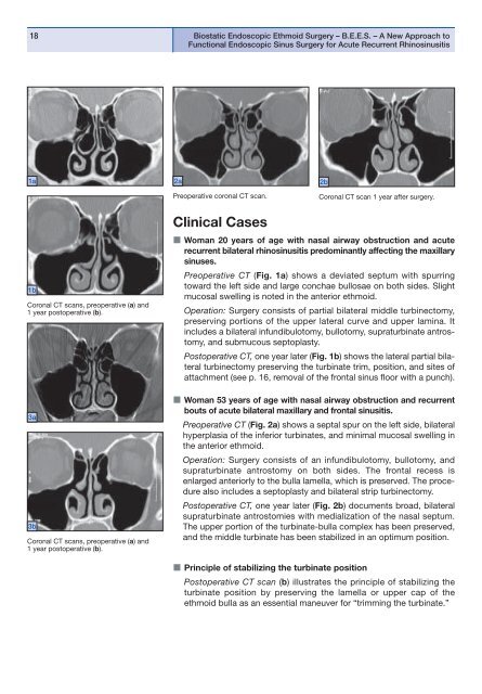
1a<br />
2a<br />
2b<br />
Preoperative coronal CT scan.<br />
Coronal CT scan 1 year after surgery.<br />
1b<br />
Coronal CT scans, preoperative (a) and<br />
1 year postoperative (b).<br />
3a<br />
3b<br />
Coronal CT scans, preoperative (a) and<br />
1 year postoperative (b).<br />
Clinical Cases<br />
Woman 20 years of age with nasal airway obstruction and acute<br />
recurrent bilateral rhinosinusitis predominantly affecting the maxillary<br />
sinuses.<br />
Preoperative CT (Fig. 1a) shows a deviated septum with spurring<br />
toward the left side and large conchae bullosae on both sides. Slight<br />
mucosal swelling is noted in the anterior ethmoid.<br />
Operation: Surgery consists of partial bilateral middle turbinectomy,<br />
preserving portions of the upper lateral curve and upper lamina. It<br />
includes a bilateral infundibulotomy, bullotomy, supraturbinate antros -<br />
tomy, and submucous septoplasty.<br />
Postoperative CT, one year later (Fig. 1b) shows the lateral partial bilateral<br />
turbinectomy preserving the turbinate trim, position, and sites of<br />
attachment (see p. 16, removal of the frontal sinus floor with a punch).<br />
Woman 53 years of age with nasal airway obstruction and recurrent<br />
bouts of acute bilateral maxillary and frontal sinusitis.<br />
Preoperative CT (Fig. 2a) shows a septal spur on the left side, bilateral<br />
hyperplasia of the inferior turbinates, and minimal mucosal swelling in<br />
the anterior ethmoid.<br />
Operation: Surgery consists of an infundibulotomy, bullotomy, and<br />
supraturbinate antrostomy on both sides. The frontal recess is<br />
enlarged anteriorly to the bulla lamella, which is preserved. The procedure<br />
also includes a septoplasty and bilateral strip turbinectomy.<br />
Postoperative CT, one year later (Fig. 2b) documents broad, bila teral<br />
supraturbinate antrostomies with medialization of the nasal septum.<br />
The upper portion of the turbinate-bulla complex has been preserved,<br />
and the middle turbinate has been stabilized in an optimum position.<br />
Principle of stabilizing the turbinate position<br />
Postoperative CT scan (b) illustrates the principle of stabilizing the<br />
turbinate position by preserving the lamella or upper cap of the<br />
ethmoid bulla as an essential maneuver for “trimming the turbinate.”