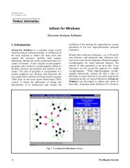
MICRO AREA X-RAY DIFFRACTION TECHNIQUES - Rigaku
MICRO AREA X-RAY DIFFRACTION TECHNIQUES - Rigaku
MICRO AREA X-RAY DIFFRACTION TECHNIQUES - Rigaku

You also want an ePaper? Increase the reach of your titles
YUMPU automatically turns print PDFs into web optimized ePapers that Google loves.
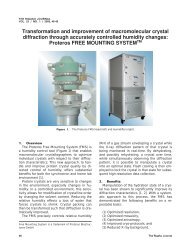
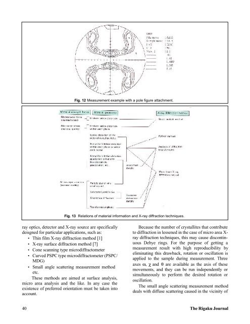
Fig. 12 Measurement example with a pole figure attachment.<br />
Fig. 13 Relations of material information and X-ray diffraction techniques.<br />
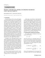
ray optics, detector and X-ray source are specifically<br />
deisgned for particular applications, such as:<br />
• Thin film X-ray diffraction method [1]<br />
• X-ray surface diffraction method [7]<br />
• Cone scanning type microdiffractometer<br />
• Curved PSPC type microdiffractometer (PSPC/<br />
MDG)<br />
• Small angle scattering measurement method<br />
etc.<br />
These methods are aimed at surface analysis,<br />
micro area analysis and the like. In any case the<br />
existence of preferred orientation must be taken into<br />
account.<br />
Because the number of crystallites that contribute<br />
to diffraction in lessened in the case of micro area X-<br />
ray diffraction techniques, this may cause discontinuous<br />
Debye rings. For the purpose of getting a<br />
measurement result with high reproducibility by<br />
eliminating this drawback, rotation or oscillation is<br />
applied to the sample during measurement. Three<br />
axes ω, χ and θ are available as the axis of these<br />
movements, and they can be run independently or<br />
simultaneously to perform the desired rotation or<br />
oscillation.<br />
The small angle scattering measurement method<br />
deals with diffuse scattering caused in the vicinity of<br />
40 The <strong>Rigaku</strong> Journal