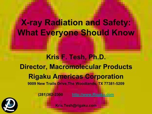
X-ray Radiation and Safety: What Everyone Should Know - Rigaku
X-ray Radiation and Safety: What Everyone Should Know - Rigaku
X-ray Radiation and Safety: What Everyone Should Know - Rigaku

You also want an ePaper? Increase the reach of your titles
YUMPU automatically turns print PDFs into web optimized ePapers that Google loves.
X-<strong>ray</strong> <strong>Radiation</strong> <strong>and</strong> <strong>Safety</strong>:<br />
<strong>What</strong> <strong>Everyone</strong> <strong>Should</strong> <strong>Know</strong><br />
Kris F. Tesh, Ph.D.<br />
Director, Macromolecular Products<br />
<strong>Rigaku</strong> Americas Corporation<br />
9009 New Trails Drive,The Woodl<strong>and</strong>s, TX 77381-5209<br />
(281)362-2300 http://www.<strong>Rigaku</strong>.com<br />
Kris.Tesh@rigaku.com
Outline<br />
1. Basics of X-<strong>ray</strong> Diffraction<br />
2. Where are the X-<strong>ray</strong>s<br />
3. General X-<strong>ray</strong> <strong>Safety</strong><br />
-definitions<br />
-procedures<br />
-videos<br />
-h<strong>and</strong>outs<br />
4. At the Instrument<br />
5. Some Software Instruction
Personnel Training<br />
All personnel involved in the installation, maintenance, repair or use<br />
of analytical X-<strong>ray</strong> units must be registered with the <strong>Radiation</strong> <strong>Safety</strong><br />
Office. Prior to beginning work with an analytical unit, the user<br />
shall attend a radiation safety training session provided by the<br />
<strong>Radiation</strong> <strong>Safety</strong> Office. This session is intended to provide basic<br />
safety information <strong>and</strong> to introduce the administrative procedures of<br />
the <strong>Safety</strong> Office at <strong>Rigaku</strong> Americas Corporation.<br />
Detailed instructions on the operations, hazards <strong>and</strong> radiation<br />
warning devices of a specific analytical unit , must be provided by<br />
the owner of the equipment. Before starting to work on an analytical<br />
unit, make sure you receive specific instruction on the unit’s<br />
operation from the person responsible for the unit.
General <strong>Radiation</strong><br />
• <strong>Radiation</strong> is energy in transit in the form of high<br />
speed particles <strong>and</strong> electromagnetic waves. We<br />
encounter electromagnetic waves every day. They<br />
make up our visible light, radio <strong>and</strong> television<br />
waves, ultra violet (UV), <strong>and</strong> microwaves with a<br />
spectrum of energies. These examples of<br />
electromagnetic waves do not cause ionizations<br />
of atoms because they do not carry enough<br />
energy to separate molecules or remove electrons<br />
from atoms.
General <strong>Radiation</strong><br />
• Ionizing radiation is radiation with enough<br />
energy so that during an interaction with an atom,<br />
it can remove tightly bound electrons from their<br />
orbits, causing the atom to become charged or<br />
ionized. Ionizing radiation deposits energy at the<br />
molecular level, causing chemical changes which<br />
lead to biological changes. These include cell<br />
death, cell transformation, <strong>and</strong> damage which<br />
cells cannot repair. Effects are not due to heating.
General <strong>Radiation</strong><br />
• X-<strong>ray</strong>s are a form of ionizing radiation. They<br />
are electromagnetic waves emitted by<br />
energy changes in electrons. These energy<br />
changes are either in electron orbital shells<br />
that surround an atom (<strong>Rigaku</strong> FRE+ or<br />
Micromax 007HF generators) or in the<br />
process of slowing down (synchrotron).
General X-<strong>ray</strong><br />
• X-<strong>ray</strong>s are produced from the excitation of<br />
electrons followed by the cascading of these<br />
electrons back down to the ground state<br />
• The typical X-<strong>ray</strong>s used in crystallography<br />
range from 0.6 to 2.5Å<br />
• Your instrument ideally emits X-<strong>ray</strong>s of only<br />
one wavelength (1.54Å or 0.7107Å) out of<br />
the end of the collimator:<br />
But other wavelengths are produced while<br />
the primary wavelength is being produced<br />
X-<strong>ray</strong>s
Where are the X-<strong>ray</strong>s?<br />
Rotating Anode/Confocal Optic<br />
Systems
Where are the X-<strong>ray</strong>s?<br />
Rotating Anode/Confocal Optic<br />
Systems
Three Regions of<br />
High Exposure Concern<br />
1. Primary Beam<br />
The critical radiation exposure problem with analytical X-<strong>ray</strong> equipment is the primary<br />
beam. Exposure to the primary beam can cause localized acute exposure. Consequently,<br />
the analytical operator must never intentionally place any part of their body in the<br />
primary beam. Typically, these beams are relatively “soft” X-<strong>ray</strong>s resulting in maximal<br />
energy deposition in epithelial tissues. Erythema or reddening of the skin can occur<br />
when skin is acutely exposed to 300 R (much less than a second). <strong>Radiation</strong> burns may<br />
occur from longer exposures.<br />
2. Scattered <strong>Radiation</strong><br />
When the primary beam intersects a material such as a sample or elements of the X-<strong>ray</strong><br />
unit including the beam stop, some of the radiation is scattered out of the primary beam.<br />
While these radiation fields are considerably less intense than the primary beam, they<br />
still represent a potential hazard. Scattered radiation fields can be measured by the<br />
analytical operators with a survey meter.<br />
3. Leakage<br />
Some radiation may leak around the tube housing structure. State law requires that<br />
source housing construction shall be that when all the shutters are closed the leakage<br />
radiation must not exceed that of radiation limits for the general public.
Rotating Anode Systems:<br />
<strong>What</strong> are the danger areas?<br />
3. Leakage<br />
1. Primary Beam<br />
2. Scattered <strong>Radiation</strong>
Rotating Anode Systems:<br />
<strong>What</strong> are the danger areas?
Rotating Anode Systems:<br />
<strong>What</strong> are the danger areas?<br />
3. Leakage<br />
1. Primary Beam 2. Scattered <strong>Radiation</strong>
Emergency Procedures<br />
If an exposure is suspected, do the following:<br />
1. Report all potential exposures of this kind<br />
immediately to your supervisor <strong>and</strong>/or person<br />
responsible for the analytical unit.<br />
2. The supervisor in turn needs to immediately<br />
notify the <strong>Radiation</strong> <strong>Safety</strong> Office so that<br />
evaluation, corrective action <strong>and</strong> if necessary,<br />
medical evaluation can be initiated.
Definitions<br />
• Chronic vs. Acute dose<br />
• Somatic vs. Genetic vs. Teratogenic<br />
effects<br />
• Stochastic vs. Non-Stochastic effects<br />
• Units of <strong>Radiation</strong>
Types of Exposure<br />
• A Chronic dose means a person received a<br />
radiation dose over a long period of time.<br />
• An Acute dose means a person received a<br />
radiation dose over a short period of time.
Effects of Exposure<br />
• Somatic effects are effects from some agent, like<br />
radiation that are seen in the individual who receives the<br />
agent.<br />
• Genetic effects are effects from some agent, that are<br />
seen in the offspring of the individual who received the<br />
agent. The agent must be encountered pre-conception.<br />
• Teratogenic effects are effects from some agent, that<br />
are seen in the offspring of the individual who received the<br />
agent. The agent must be encountered during the<br />
gestation period.
Effects of Exposure<br />
• Stochastic effects are effects that occur on a r<strong>and</strong>om<br />
basis with its effect being independent of the size of dose.<br />
The effect typically has no threshold <strong>and</strong> is based on<br />
probabilities, with the chances of seeing the effect<br />
increasing with dose. Cancer is a stochastic effect.<br />
• Non-stochastic effects are effects that can be<br />
related directly to the dose received. The effect is more<br />
severe with a higher dose, i.e., the burn gets worse as<br />
dose increases. It typically has a threshold, below which<br />
the effect will not occur. A skin burn from radiation is a<br />
non-stochastic effect.
Common Units of <strong>Radiation</strong><br />
• The Roentgen (R) is a unit used to measure<br />
a quantity called exposure. This can only be<br />
used to describe an amount of gamma <strong>and</strong><br />
X-<strong>ray</strong>s, <strong>and</strong> only in air.<br />
• One roentgen is equal to depositing in dry air<br />
enough energy to cause 2.58x 10 -4 coulombs per kg.<br />
It is a measure of the ionizations of the molecules in<br />
a mass of air.<br />
- The main advantage of this unit is that it<br />
is easy to measure directly, but it is<br />
limited because it is only for deposition<br />
in air, <strong>and</strong> only for gamma <strong>and</strong> X-<strong>ray</strong>s.
Common Units of <strong>Radiation</strong><br />
• The rad (radiation absorbed dose) is a unit used<br />
to measure a quantity called absorbed dose. This<br />
relates to the amount of energy actually absorbed<br />
in some material, <strong>and</strong> is used for any type of<br />
radiation <strong>and</strong> any material.<br />
• One rad is defined as the absorption of 100 ergs per gram<br />
of material. The unit rad can be used for any type of<br />
radiation, but it does not describe the biological effects of<br />
the different forms of radiation.
Common Units of <strong>Radiation</strong><br />
• The rem (Roentgen equivalent man) is a unit<br />
used to derive a quantity called equivalent dose.<br />
This relates the absorbed dose in human tissue to<br />
the effective biological damage of the radiation.<br />
• Not all radiation has the same biological effect, even for<br />
the same amount of absorbed dose. Equivalent dose is<br />
often expressed in terms of thous<strong>and</strong>ths of a rem, or<br />
mrem.<br />
• (rem) = (rad) X (Q)<br />
– Where Q is the quality factor that is unique to<br />
the type of incident radiation
Common Units of <strong>Radiation</strong><br />
• The sievert (Sv) is a unit used to derive a<br />
quantity called equivalent dose. This relates the<br />
absorbed dose in human tissue to the effective<br />
biological damage of the radiation.<br />
– Not all radiation has the same biological effect, even for<br />
the same amount of absorbed dose. Equivalent dose is<br />
often expressed in terms of millionths of a sievert, or<br />
micro-sievert.<br />
– To determine equivalent dose<br />
• (Sv) = (Gy) x (Q)<br />
• One sievert is equivalent to 100 rem.
Other Units of <strong>Radiation</strong><br />
• The curie(Ci) is a unit used to measure a radioactivity. One curie is that quantity of a<br />
radioactive material that will have 37,000,000,000 transformations in one second. Often<br />
radioactivity is expressed in smaller units like: thous<strong>and</strong>ths (mCi), millionths (uCi) or<br />
even billionths (nCi) of a curie. The relationship between becquerels <strong>and</strong> curies is: 3.7 x<br />
10 10 Bq in one curie.<br />
• The g<strong>ray</strong> (Gy) is a unit used to measure a quantity called absorbed dose. This relates<br />
to the amount of energy actually absorbed in some material, <strong>and</strong> is used for any type of<br />
radiation <strong>and</strong> any material. One g<strong>ray</strong> is equal to one joule of energy deposited in one kg<br />
of a material. The unit g<strong>ray</strong> can be used for any type of radiation, but it does not<br />
describe the biological effects of the different radiations. Absorbed dose is often<br />
expressed in terms of hundredths of a g<strong>ray</strong>, or centi-g<strong>ray</strong>s. One g<strong>ray</strong> is equivalent to<br />
100 rads.<br />
• The Becquerel (Bq) is a unit used to measure a radioactivity. One Becquerel is that<br />
quantity of a radioactive material that will have 1 transformations in one second. Often<br />
radioactivity is expressed in larger units like: thous<strong>and</strong>s (kBq), one millions (MBq) or<br />
even billions (GBq) of a becquerels. As a result of having one Becquerel being equal to<br />
one transformation per second, there are 3.7 x 10 10 Bq in one curie.
Federal Maximum Exposure<br />
Limits<br />
Limits for Exposures<br />
Occupational Dose limit (US - NRC)<br />
Occupational Exposure Limits for Minors (10%)<br />
Occupational Exposure Limits for Fetus<br />
Public dose limits (ouside radiation area)<br />
Occupational Limits (eye)<br />
Occupational Limits (skin)<br />
Occupational Limits (extremities)<br />
Exposure<br />
50 mSv/year (5 rem)<br />
0.5 rem/year<br />
0.5 rem/9 months<br />
1 mSv/year (0.1 rem)<br />
15 rem/year<br />
50 rem/year<br />
50 rem/year<br />
ALARA: The above limits are the Maximum Permissible<br />
Doses allowed by regulation. However, all doses should be<br />
maintained As Low As Reasonably Achievable (ALARA).<br />
ANSI/HPS N43.2-2001 <strong>and</strong> Federal CFR
Federal<br />
Maximum<br />
Exposure<br />
Limits<br />
(areas)
Personnel Monitoring<br />
Ring/Badge Dosimeters<br />
Operators of analytical X-<strong>ray</strong> equipment will be provided<br />
with a finger (ring) <strong>and</strong> body (badge) monitoring device.<br />
The dosimeter is designed to record information about the<br />
amount of radiation which you receive during the course of<br />
your work. However, it is important to note that the crosssectional<br />
area of the primary radiation beam is usually small<br />
<strong>and</strong> that the monitoring device may not indicate the<br />
maximum exposure to the operator.
Dosimeter Use Practices<br />
1. Ring/Badge dosimeters are issued for a specific period of time. The beginning<br />
<strong>and</strong> ending date is printed on the face of the dosimeter. At the end of each<br />
wear period, a replacement set will be issued through the ring/badge<br />
coordinator.<br />
2. It is important to exchange the ring/badge dosimeter promptly so that<br />
exposures may be evaluated in a timely fashion. Prompt reading on the<br />
dosimeters will insure accurate information.<br />
3. Chronic late ring/badge dosimeter returns may jeopardize your right to work<br />
with the instrumentation.<br />
4. The ring dosimeter should be worn on the h<strong>and</strong> that will be nearest the<br />
primary beam. For example, if the operator sets up an experiment working<br />
mainly with the right h<strong>and</strong>, the ring dosimeter should be worn on the at h<strong>and</strong>.<br />
5. When not wearing the dosimeters, do not store it in an area where it may<br />
receive a radiation exposure.
Dosimeter Use Practices (cont.)<br />
6. H<strong>and</strong> carry your badge through Airport Security…Do not allow it to be X-<br />
<strong>ray</strong>ed!<br />
7. If you lose your ring or badge dosimeter, promptly inform your <strong>Radiation</strong><br />
<strong>Safety</strong> Officer for a replacement. If the lost dosimeter is subsequently<br />
recovered, return it to the <strong>Radiation</strong> <strong>Safety</strong> Office for processing <strong>and</strong> continue<br />
to wear the replacement dosimeter.<br />
8. If your dosimeter is damaged, return it to the <strong>Radiation</strong> <strong>Safety</strong> Office for<br />
replacement.<br />
9. Do not lend your ring or badge dosimeter to another person; <strong>and</strong> do not wear<br />
another person’s dosimeter.<br />
10.Do not wear your dosimeter during personal medical procedures involving<br />
nuclear medicine or X-<strong>ray</strong> radiation. The exposure recorded by the dosimeter<br />
must be restricted to your occupational exposure. If you inadvertently wear<br />
the dosimeter while being exposed to radiation for medical reasons, promptly<br />
report this to the <strong>Radiation</strong> <strong>Safety</strong> Office <strong>and</strong> obtain a replacement dosimeter.
Annual estimated average effective dose equivalent received by a<br />
member of the population of the United States.<br />
Average annual effective dose<br />
Source<br />
equivalent<br />
Natural<br />
Exposure<br />
table <strong>and</strong><br />
graph<br />
(mrem) (µSv) (percent of total)<br />
Inhaled (Radon <strong>and</strong> Decay Products) 200 2 55%<br />
Cosmic <strong>Radiation</strong> 27 0.27 8%<br />
Terrestrial <strong>Radiation</strong> 28 0.28 8%<br />
Other Internally Deposited Radionuclides 39 0.39 11%<br />
Cosmogenic Radioactivity 1 10 0%<br />
Total Natural 300 3 82%<br />
Artificial<br />
Other<br />
Medical X <strong>ray</strong> 39 0.39 11%<br />
Nuclear medicine 14 0.14 4%<br />
Consumer products 10 0.1 3%<br />
Occupational 0.9 1
Typical Exposure <strong>and</strong> Dose<br />
S ource of Exposure<br />
Exposure (Range)<br />
Average Dose to US public from All sources<br />
360 mrem/year<br />
Average Dose to US Public From Natural Sources<br />
300 mrem/year<br />
Average Dose to US Public From Medical Sources<br />
53 mrem/year<br />
Average Dose to US Public from Weapons Fallout<br />
< 1 mrem/year<br />
Average Dose to US Public From Nuclear Power<br />
< 0.1 mrem/year<br />
Coal Burning Power Plant<br />
0.165 mrem/year<br />
X-<strong>ray</strong>s from TV set (1 inch)<br />
0.500 mrem/hour<br />
Airplane ride (39,000 ft.)<br />
0.500 mrem/hour<br />
Nuclear Power Plant (normal operation at plant boundary) 0.600 mrem/year<br />
Natural gas in home<br />
9 mrem/year<br />
Average Natural Background 0.008 mR/hour (0.006-0.015)<br />
Average US Cosmic <strong>Radiation</strong><br />
27 mrem/year<br />
Average US Terrestrial <strong>Radiation</strong><br />
28 mrem/year<br />
Terrestrial background (Atlantic coast)<br />
16 mrem/year<br />
Terrestrial background (Rocky Mountains)<br />
40 mrem/year<br />
Cosmic <strong>Radiation</strong> (Sea level)<br />
26 mrem/year<br />
Cosmic <strong>Radiation</strong> (Denver)<br />
50 mrem/year<br />
Background <strong>Radiation</strong> Total (East, West, Central US) 46 mrem/year (35-75)<br />
Background <strong>Radiation</strong> Total (Colorado Plateau) 90 mrem/year (75-140)<br />
Background <strong>Radiation</strong> Total (Atlantic <strong>and</strong> Gulf in US) 23 mrem/year (15-35)<br />
Radionuclides in the body (i.e., potassium)<br />
39 mrem/year<br />
Building materials (concrete)<br />
3 mrem/year<br />
Drinking Water<br />
5 mrem/year<br />
Pocket watch (radium dial)<br />
6 mrem/year
Typical Exposure <strong>and</strong> Dose<br />
S ource of Exposure<br />
Exposure (Range)<br />
Chest X-<strong>ray</strong> 8 mrem (5-20)<br />
Extemities X-<strong>ray</strong><br />
1 mrem<br />
Dental X-<strong>ray</strong><br />
10 mrem<br />
Head/neck X-<strong>ray</strong><br />
20 mrem<br />
Cervical Spine X-<strong>ray</strong><br />
22 mrem<br />
Lumbar spinal X-<strong>ray</strong>s<br />
130 mrem<br />
Pelvis X-<strong>ray</strong><br />
44 mrem<br />
Hip X-<strong>ray</strong><br />
83 mrem<br />
Shoe Fitting Fluroscope (not in use now)<br />
170 mrem<br />
Upper GI series<br />
245 mrem<br />
Lower GI series<br />
405 mrem<br />
CT (head <strong>and</strong> body)<br />
1,100 mrem<br />
Therapeutic thyroid treatment (dose to the thyroid) 10,000,000 mrad<br />
Therapeutic thyroid treatment (dose to the whole body) 7,000 mrem (5,000-15,000)<br />
Earliest Onset of <strong>Radiation</strong> Sickness<br />
75,000 mrad<br />
Onset of hematopoietic syndrome 300,000 mrad (100,000 - 800,000)<br />
Onset of gastrointestinal syndrome 1,000,000 mrad (500,000 - 1,200,000)<br />
Onset of cerebrovacular syndrome 10,000,000 mrad (>500,000)<br />
Thershold for cataracts (dose to the eye)<br />
200,000 mrad<br />
Expected 50% death without medical attention 400,000 mrad (300,000 - 500,000)<br />
Doubling dose for genetic effects<br />
100,000 mrad<br />
Doubling dose for cancer<br />
500,000 mrad<br />
Dose for increase cancer risk of 1 in a 1,000<br />
1,250 mrem<br />
Consideration of theraputic abortion threshold (dose in utero) 10,000 mrem
Commonly Used Radioactive Elements<br />
Americium -241: Used in many smoke detectors for homes <strong>and</strong> business...to measure levels of<br />
toxic lead in dried paint samples...to ensure uniform thickness in rolling processes like steel <strong>and</strong><br />
paper production...<strong>and</strong> to help determine where oil wells should be drilled.<br />
Cadmium -109: Used to analyze metal alloys for checking stock, sorting scrap.<br />
Calcium - 47: Important aid to biomedical researchers studying the cell function <strong>and</strong> bone<br />
formation of mammals.<br />
Californium - 252: Used to inspect airline luggage for hidden explosives...to gauge the<br />
moisture content of soil in the road construction <strong>and</strong> building industries...<strong>and</strong> to measure the<br />
moisture of materials stored in silos.<br />
Carbon - 14: Helps in research to ensure that potential new drugs are metabolized without<br />
forming harmful by-products.<br />
Cesium - 137: Used to treat cancers...to measure correct patient dosages of radioactive<br />
pharmaceuticals...to measure <strong>and</strong> control the liquid flow in oil pipelines...to tell researchers<br />
whether oil wells are plugged by s<strong>and</strong>...<strong>and</strong> to ensure the right fill level for packages of food,<br />
drugs <strong>and</strong> other products. (The products in these packages do not become radioactive.)<br />
Chromium - 51: Used in research in red blood cell survival studies.<br />
Cobalt - 57: Used in nuclear medicine to help physicians interpret diagnosis scans of patients'<br />
organs, <strong>and</strong> to diagnose pernicious anemia.<br />
Cobalt - 60 : Used to sterilize surgical instruments...to improve the safety <strong>and</strong> reliability of<br />
industrial fuel oil burners...<strong>and</strong> to preserve poultry fruits <strong>and</strong> spices.<br />
Copper - 67: When injected with monoclonal antibodies into a cancer patient, helps the<br />
antibodies bind to <strong>and</strong> destroy the tumor.<br />
Curium - 244: Used in mining to analyze material excavated from pits slurries from drilling<br />
operations.<br />
Iodine - 123: Widely used to diagnose thyroid disorders.<br />
Iodine - 129: Used to check some radioactivity counters in vitro diagnostic testing laboratories.<br />
Iodine - 131: Used to diagnose <strong>and</strong> treat thyroid disorders. (Former President George Bush <strong>and</strong><br />
Mrs. Bush were both successfully treated for Grave's disease, a thyroid disease, with<br />
radioactive iodine.)<br />
Iridium - 192: Used to test the integrity of pipeline welds, boilers <strong>and</strong> aircraft parts.<br />
Iron - 55: Used to analyze electroplating solutions.<br />
Krypton - 85: Used in indicator lights in appliances like clothes washer <strong>and</strong> dryers, stereos <strong>and</strong><br />
coffee makers...to gauge the thickness of thin plastics <strong>and</strong> sheet metal, rubber, textiles <strong>and</strong><br />
paper...<strong>and</strong> to measure dust <strong>and</strong> pollutant levels.<br />
Nickel - 63: Used to detect explosives...<strong>and</strong> as voltage regulators <strong>and</strong> current surge protectors<br />
in electronic devices.<br />
Phosphorus - 32: Used in molecular biology <strong>and</strong> genetics research.<br />
Plutonium - 238: Has safely powered at least 20 NASA spacecraft since 1972.<br />
Polonium - 210: Reduces the static charge in production of photographic film <strong>and</strong> phonograph<br />
records.<br />
Promethium - 147: Used in electric blanket thermostats...<strong>and</strong> to gauge the thickness of thin<br />
plastics, thin sheet metal, rubber, textiles, <strong>and</strong> paper.<br />
Radium - 226: Makes lightning rods more effective.<br />
Selenium - 75: Used in protein studies in life science research.<br />
Sodium - 24: Used to locate leaks in industrial pipelines...<strong>and</strong> in oil well studies.<br />
Strontium - 85: Used to study bone formation <strong>and</strong> metabolism.<br />
Technetium - 99m: The most widely used radioactive isotope for diagnostic studies in nuclear<br />
medicine. Different chemical forms are used for brain, bone, liver, spleen <strong>and</strong> kidney imaging<br />
<strong>and</strong> also for blood flow studies.<br />
Thallium - 204: Measures the dust <strong>and</strong> pollutant levels on filter paper...<strong>and</strong> gauges the<br />
thickness of plastics, sheet metal, rubber, textiles <strong>and</strong> paper.<br />
Thoriated tungsten: Used in electric arc welding rods in the construction, aircraft,<br />
petrochemical <strong>and</strong> food processing equipment industries. It produces easier starting, greater arc<br />
stability <strong>and</strong> less metal contamination.<br />
Thorium - 229: Helps fluorescent lights to last longer.<br />
Thorium - 230: Provides coloring <strong>and</strong> fluorescence in colored glazes <strong>and</strong> glassware.<br />
Tritium: Used for life science <strong>and</strong> drug metabolism studies to ensure the safety of potential<br />
new drugs... for self-luminous aircraft <strong>and</strong> commercial exit signs... for luminous dials, gauges<br />
<strong>and</strong> wrist watches...<strong>and</strong> to produce luminous paint.<br />
Uranium - 234: Used in dental fixtures like crowns <strong>and</strong> dentures to provide a natural color <strong>and</strong><br />
brightness.<br />
Uranium - 235: Fuel for nuclear power plants <strong>and</strong> naval nuclear propulsion systems...also used<br />
to produce fluorescent glassware, a variety of colored glazes <strong>and</strong> wall tiles.<br />
Xenon - 133: Used in nuclear medicine for lung ventilation <strong>and</strong> blood flow studies.<br />
Adapted from Nuclear Energy Institute, 17706 I Street, N.W., Suite 400Washington, DC 20006-3708
Risks:<br />
Reduced Life Expectancy<br />
Health Risk<br />
Smoking 20 cigs a day<br />
Overweight (15%)<br />
Alcohol (US Ave)<br />
All Accidents<br />
All Natural Hazards<br />
All Industries<br />
Agriculture<br />
Construction<br />
Mining <strong>and</strong> quarrying<br />
Manufacturing<br />
Occupational dose (1 rem/yr)<br />
Occupational dose (300 mrem/yr)<br />
Est. life expectancy loss<br />
6 years<br />
2 years<br />
1 year<br />
207 days<br />
7 days<br />
60 days<br />
320 days<br />
227 days<br />
167 days<br />
40 days<br />
51 days<br />
15 days<br />
NRC Draft guide DG-8012, adapted from B.L Cohen <strong>and</strong> I.S. Lee, "Catalogue of<br />
Risks Extended <strong>and</strong> Updates", Health Physics, Vol. 61, September 1991.
Risks: 1 in a Million<br />
Another way of looking at risk, is to look at the<br />
Relative Risk of 1 in a million chances of dying of<br />
activities common to our society.<br />
•Smoking 1.4 cigarettes (lung cancer)<br />
•Eating 40 tablespoons of peanut butter<br />
•Spending 2 days in New York City (air pollution)<br />
•Driving 40 miles in a car (accident)<br />
•Flying 2500 miles in a jet (accident)<br />
•Canoeing for 6 minutes<br />
•Receiving 10 mrem of radiation (cancer)<br />
Adapted from DOE <strong>Radiation</strong> Worker Training, based on work by B.L Cohen, Sc.D.
Ways to Reduce Risk<br />
There are 3 general ways to reduce exposure risk<br />
• Time: Reduce the amount of time you are<br />
near the source of radiation<br />
• Distance: Get as far away from the source as<br />
possible<br />
• Shielding: Place something between you<br />
<strong>and</strong> the source to absorb approaching X-<strong>ray</strong>s
Administrative Controls<br />
Equipment Registration<br />
All analytical X-<strong>ray</strong> equipment shall be registered with the<br />
<strong>Radiation</strong> <strong>Safety</strong> Office. The <strong>Radiation</strong> <strong>Safety</strong> Office must<br />
be notified prior to initial use, if the unit is moved, modified<br />
or serviced.
Administrative Controls<br />
Operating Procedures<br />
Detailed written operating procedures shall be available to<br />
each registered unit. These procedures shall include all<br />
routine operating conditions for which the instrument will be<br />
used. At a minimum this shall include: sample insertion <strong>and</strong><br />
manipulation, equipment alignment, routine maintenance, as<br />
well as emergency procedures.<br />
-User Manual is a good start<br />
for your procedures.
Administrative Controls<br />
<strong>Safety</strong> Overrides<br />
Under some circumstances it may be necessary to override<br />
the analytical unit’s safety devices. All overrides must be<br />
approved in writing by the <strong>Radiation</strong> <strong>Safety</strong> Officer.
<strong>Safety</strong> Devices<br />
<br />
<br />
Analytical units shall have the following safety devices as required<br />
by State Regulations.<br />
Unused ports shall be secure in a manner which will prevent accidental<br />
opening. Open beam units shall have a shutter over the port which<br />
cannot be opened unless a collimator or coupling has been connected.<br />
<strong>Safety</strong> interlocks shall not be used to de-activate the X-<strong>ray</strong> beam except<br />
in an emergency or during testing of the interlock system.<br />
Warning Devices<br />
<br />
<br />
All units with an open beam configuration shall have an easily identified<br />
device located near the radiation source housing <strong>and</strong> labeled what gives a<br />
clear, visible indication of the X-<strong>ray</strong> generation status (on-off)<br />
<strong>Safety</strong> interlocks shall not be used to de-activate the X-<strong>ray</strong> beam except<br />
in an emergency or during testing of the interlock system.
Warning Labels<br />
<br />
<br />
A label which bears the following or similar words shall be placed on the<br />
X-<strong>ray</strong> source housing:<br />
CAUTION - HIGH INTENSITY X-RAY BEAM<br />
A label which bears the following or similar wording shall be placed on<br />
the control console of each unit near any switch which energizes the<br />
source:<br />
CAUTION - RADIATION<br />
THIS EQUIPMENT PRODUCES<br />
RADIATION WHEN ENERGIZED
Warning Labels
Warning Labels
Warning Lights<br />
<br />
An easily visible warning light labeled with these or similar words “X-<br />
RAY ON” shall be placed near any switch that energizes an X-<strong>ray</strong><br />
source, <strong>and</strong> shall be illuminated only when the generator is energized,<br />
<strong>and</strong> have fail-safe characteristics.<br />
Shutters<br />
<br />
Each port shall be equipped with a shutter that cannot be opened unless a<br />
collimator or a coupling device has been connected to the port.
Emergency Stop Buttons<br />
“Panic Buttons”<br />
All instruments are designed with a panic button which<br />
powers off the generator immediately upon activating.<br />
In an emergency, the X-<strong>ray</strong> On lamp can be jarred <strong>and</strong> the<br />
filament broken (if for example there is water on the floor).
<strong>Radiation</strong> Surveys<br />
The <strong>Radiation</strong> <strong>Safety</strong> Office will perform a survey annually <strong>and</strong><br />
following major repairs <strong>and</strong>/or system modifications. This survey<br />
will include inspection of all safety systems <strong>and</strong> a radiation<br />
exposure survey. The results of the survey will be kept on file in<br />
the <strong>Radiation</strong> <strong>Safety</strong> Office.<br />
Users of analytical equipment should also routinely perform<br />
radiation surveys. The surveys should include monitoring for st<strong>ray</strong><br />
radiation in the immediate vicinity of the X-<strong>ray</strong> apparatus.<br />
All labs should have a radiation survey meter readily available!!!
Survey Meter<br />
Instrumentation<br />
Survey should be performed with a portable Geiger-Mueller<br />
survey instrument although the results are not necessarily<br />
quantitative. If accurate measurements are desired, the<br />
instrument should be calibrated with the source of low energy<br />
X-<strong>ray</strong>s. Consideration should also be given to possible<br />
monitoring errors due to the cross-sectional area of the<br />
monitored radiation beam being smaller than the sensitive area<br />
of the survey meter.
When the Operator <strong>Should</strong><br />
Perform a <strong>Radiation</strong> Survey<br />
1. Upon installation of your instrument.<br />
2. After any major changes in equipment configuration or minor system<br />
maintenance to insure that no unanticipated exposure hazards exist.<br />
3. Following any maintenance requiring the disassembly or removal of<br />
local components.<br />
4. During the performance of maintenance <strong>and</strong> alignment procedures.<br />
5. When visual inspection of the local components in the system reveals an<br />
abnormal condition.
General Precautions<br />
<br />
<br />
<br />
<br />
<br />
<br />
<br />
<br />
Only Trained personnel shall be permitted to operate an analytical unit.<br />
Be familiar with the procedure to be carried out.<br />
Never expose any part of your body to the primary beam.<br />
Turn the X-<strong>ray</strong> beam OFF before attempting to make any changes to the<br />
experimental set-up (except for beam alignment)<br />
While the beam is on DO NOT attempt to h<strong>and</strong>le, manipulate or adjust<br />
any object (sample, sample holder, collimator, etc.) which is in the direct<br />
beam path (except for beam alignment procedures).<br />
Examine the system carefully for any system modifications or<br />
irregularities.<br />
Follow the operating procedures carefully. DO NOT take short cuts!<br />
Never leave the energized system unattended in an area where access in<br />
not controlled.
General Precautions<br />
<br />
<br />
<br />
<br />
<br />
Survey the area frequently to evaluate scatter <strong>and</strong> leakage radiation<br />
fields.<br />
Never remove auxiliary shielding without authorization from the owner<br />
of the analytical equipment or <strong>Radiation</strong> <strong>Safety</strong> Officer.<br />
Never bypass safety circuits, such as interlocks.<br />
Report all unusual occurrences to the owner of the analytical unit for<br />
possible corrective actions.<br />
Only authorized, trained individuals as specified by the unit’s owner <strong>and</strong><br />
the <strong>Radiation</strong> <strong>Safety</strong> Office may repair, align or make modifications to<br />
the X-<strong>ray</strong> apparatus.
Notice to Employees
Theoretical Intensity Calculations for Cu Kα radiation at 1.54 Angstrom<br />
-ln ( I/Io)= μt<br />
μ=ρΣgi(μ/ρ)i<br />
ex: N2(air)<br />
Intensity at front of material 10000 1000 100 100 100 100 atom μ/ρ cm2/g<br />
ntensity out back of material 1 1 1 50 90 99 H 0.40<br />
V ratio of I/Io 0.000100 0.001000 0.010000 0.500000 0.900000 0.990000 N 7.50<br />
V μ/ρ cm2/g 7.5 7.5 7.5 7.5 7.5 7.5 O 11.50<br />
V ρ g/cm3 0.001210 0.001210 0.001210 0.001210 0.001210 0.001210 Pb 232.00<br />
Absorption<br />
V μ cm−1 0.009075 0.009075<br />
Copper<br />
0.009075 0.009075 0.009075<br />
Kα<br />
0.009075<br />
V<br />
V ln I/Io -9.210340 -6.907755 -4.605170 -0.693147 -0.105361 -0.010050<br />
V -ln I/Io 9.210340 6.907755 4.605170 0.693147 0.105361 0.010050<br />
Thickness of material in mm 10149 7612 5075 764 116 11<br />
ex: water (body fluids)<br />
Intensity at front of material 10000 1000 100 100 100 100 atom gi<br />
ntensity out back of material 1 1 1 50 90 99 H 2/18<br />
V ratio of I/Io 0.000100 0.001000 0.010000 0.500000 0.900000 0.990000 O 16/18<br />
V Σgi(μ/ρ)i cm2/g 10.23 10.23 10.23 10.23 10.23 10.23<br />
V ρ g/cm3 1.00 1.00 1.00 1.00 1.00 1.00<br />
V μ cm−1 10.23 10.23 10.23 10.23 10.23 10.23<br />
V<br />
V ln I/Io -9.210340 -6.907755 -4.605170 -0.693147 -0.105361 -0.010050<br />
V -ln I/Io 9.210340 6.907755 4.605170 0.693147 0.105361 0.010050<br />
Thickness of material in mm 9.00 6.75 4.50 0.68 0.10 0.01<br />
ex: lead (beam stop)<br />
Intensity at front of material 10000 1000 100 100 100 100 1.00E+300<br />
ntensity out back of material 1 1 1 50 90 99 1<br />
V ratio of I/Io 0.000100 0.001000 0.010000 0.500000 0.900000 0.990000 1.00E-300<br />
V μ/ρ cm2/g 232.00 232.00 232.00 232.00 232.00 232.00 232.00<br />
V ρ g/cm3 11.30 11.30 11.30 11.30 11.30 11.30 11.30<br />
V μ cm−1 2621.60 2621.60 2621.60 2621.60 2621.60 2621.60 2621.60<br />
V<br />
V ln I/Io -9.210340 -6.907755 -4.605170 -0.693147 -0.105361 -0.010050 -690.78<br />
V -ln I/Io 9.210340 6.907755 4.605170 0.693147 0.105361 0.010050 690.78<br />
Thickness of material in mm 3.51E-02 2.63E-02 1.76E-02 2.64E-03 4.02E-04 3.83E-05 2.63E+00
Walk In <strong>Radiation</strong> Enclosure
L<strong>and</strong>auer Service Guide 1
L<strong>and</strong>auer Service Guide 2
L<strong>and</strong>auer Service Guide 3
L<strong>and</strong>auer Service Guide 4
L<strong>and</strong>auer Service Guide 5
Sources of Information<br />
University of Pittsburgh<br />
V<strong>and</strong>erbilt University<br />
International Energy Agency, Division of Public Information<br />
UCLA <strong>Radiation</strong> <strong>Safety</strong> H<strong>and</strong>out (8/92)<br />
http://www.tdh.state.tx.us/ech/rad/pages/brc.htm<br />
-Texas Department of Health, Bureau of <strong>Radiation</strong> Control<br />
http://www.physics.isu.edu/radinf/index.html<br />
http://www.physics.isu.edu/radinf/law.htm<br />
-Idaho State University<br />
http://liley.physics.swin.oz.au/~dtl/sp407/projrad/<br />
-University of Swinburne Technology<br />
http://www.umich.edu/~radinfo/<br />
-University of Michigan<br />
http://www.access.gpo.gov/nara/<br />
-National Archives <strong>and</strong> Records Administration, Office of the Federal Register<br />
http://www.dhs.ca.gov/rhb/<br />
-California Department of Health Services, Radiologic Health Branch<br />
http://www.hhmi.org/home/publication/3.html<br />
http://www.ntis.gov/nac/index.html
THANK YOU FOR YOUR INTEREST<br />
<strong>Rigaku</strong> Americas<br />
Corporation<br />
9009 New Trails Drive,The Woodl<strong>and</strong>s, TX<br />
77381-5209<br />
(281)362-2300 http://www.<strong>Rigaku</strong>.com<br />
Info@rigaku.com