twrama 1990_final oc.. - AMA WA
twrama 1990_final oc.. - AMA WA
twrama 1990_final oc.. - AMA WA

Create successful ePaper yourself
Turn your PDF publications into a flip-book with our unique Google optimized e-Paper software.
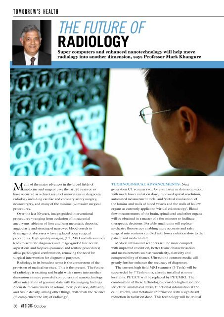
TOMORROW’S HEALTH<br />
The future of<br />
RADIOLOGY<br />
Super computers and enhanced nanotechnology will help move<br />
radiology into another dimension, says Professor Mark Khangure<br />
Many of the major advances in the broad fields of<br />
medicine and surgery over the last 80 years or so<br />
have <strong>oc</strong>curred as a direct result of innovations in diagnostic<br />
radiology including cardiac and coronary artery surgery,<br />
neurosurgery, and many of the minimally-invasive surgical<br />
pr<strong>oc</strong>edures.<br />
Over the last 30 years, image-guided interventional<br />
pr<strong>oc</strong>edures – ranging from <strong>oc</strong>clusion of intracranial<br />
aneurysms, ablation of liver and lung metastatic deposits,<br />
angioplasty and stenting of narrowed blood vessels to<br />
drainages of abscesses – have replaced open surgical<br />
pr<strong>oc</strong>edures. High quality imaging (CT, MRI and ultrasound)<br />
leads to accurate diagnoses and image-guided fine needle<br />
aspirations and biopsies (common and routine pr<strong>oc</strong>edures)<br />
allow pathological confirmation, removing the need for<br />
surgical intervention for diagnostic purposes.<br />
Radiology in its broadest terms is the cornerstone of the<br />
provision of medical services. This is the present. The future<br />
of radiology is exciting and bright with a move into another<br />
dimension as more powerful computers and nanotechnology<br />
allow integration of genomic data with the imaging findings.<br />
Accurate measurements of volume, flow, perfusion, diffusion,<br />
and tissue density, among other things, will create the ‘science<br />
(to complement the art) of radiology’.<br />
Technological advancements: Next<br />
generation CT scanners will be even faster in data acquisition<br />
with much lower radiation dose, improved spatial resolution,<br />
automated measurement tools, and ‘virtual visulisation’ of<br />
the lumina and walls of blood vessels and the walls of hollow<br />
organs as currently applied to ‘virtual colonoscopy’. Blood<br />
flow measurements of the brain, spinal cord and other organs<br />
will be obtained in a matter of a few minutes to facilitate<br />
therapeutic decisions. Portable small units will replace<br />
in-theatre fluoroscopy enabling more accurate and safer<br />
surgical interventions coupled with lower radiation dose to the<br />
patient and medical staff.<br />
Medical ultrasound scanners will be more compact<br />
with improved resolution, better tissue characterisation<br />
and measurements such as vascularity, elasticity and<br />
compressibility of tissues. Ultrasound contrast media will<br />
greatly further enhance the accuracy of diagnoses.<br />
The current high field MRI scanners (3 Tesla) will be<br />
superseded by 7 Tesla units, already installed at some<br />
l<strong>oc</strong>ations. PET/CT will be replaced by PET/MRI. The<br />
combination of these technologies provides high-resolution<br />
structural anatomical detail, functional information at the<br />
cellular level, and metabolic information with a significant<br />
reduction in radiation dose. This technology will be crucial<br />
38 MEDICUS October