The design and imaging characteristics of dynamic, solid-state, flat ...
The design and imaging characteristics of dynamic, solid-state, flat ...
The design and imaging characteristics of dynamic, solid-state, flat ...

You also want an ePaper? Increase the reach of your titles
YUMPU automatically turns print PDFs into web optimized ePapers that Google loves.
1082 A.R. Cowen et al.<br />
becomes more pronounced as the x-ray quantum<br />
noise diminishes with increasing dose. In <strong>solid</strong><strong>state</strong><br />
detectors fixed-pattern noise is eliminated<br />
during system calibration, <strong>and</strong> this holds across<br />
the <strong>dynamic</strong> range. Overall the graphs presented<br />
in Fig. 7 confirm that modern, indirect-conversion,<br />
<strong>dynamic</strong>, <strong>solid</strong>-<strong>state</strong> detectors are dose-efficient<br />
<strong>imaging</strong> devices, which can support the full spectrum<br />
<strong>of</strong> clinical applications previously underwritten<br />
by digital x-ray IITV systems.<br />
New directions in digital fluoroscopy<br />
3D-enhanced fluoroscopy<br />
<strong>The</strong> 1990s saw a growth in the use <strong>of</strong> digital x-ray<br />
IITV systems in 3D reconstruction <strong>imaging</strong>, based<br />
upon a rotating C-arm <strong>imaging</strong> geometry. 44 Before<br />
clinically acceptable reconstructions can be computed,<br />
extensive data processing is required to<br />
correct for defects such as the changing geometrical<br />
distortion (which occurs as the image intensifier<br />
rotates around the patient). 45 For reasons<br />
explained above <strong>dynamic</strong>, <strong>solid</strong>-<strong>state</strong> detectors essentially<br />
produce distortion-free image data. Consequently,<br />
these new detectors yield 3D image<br />
reconstructions with greater detail resolution 46,47<br />
<strong>and</strong> fewer artefacts. 48,49 3D reconstruction <strong>imaging</strong><br />
is typically used to improve the visualization<br />
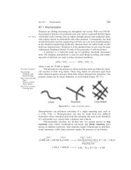
Figure 8 3D roadmap image <strong>of</strong> the iliac arteries with<br />
the catheter in situ acquired using a <strong>dynamic</strong>, <strong>solid</strong><strong>state</strong><br />
detector. Reproduced with the permission <strong>of</strong><br />
Philips Healthcare.<br />
<strong>of</strong> complex bone structures during orthopaedic<br />
surgery 50 or a tortuous network <strong>of</strong> blood vessels<br />
in endovascular procedures. 47 Reportedly the latter<br />
can aid the clinician in navigating <strong>and</strong> deploying<br />
interventional devices, thereby reducing<br />
procedure times <strong>and</strong> patient/staff radiation<br />
dose. 47,51 <strong>The</strong> availability <strong>of</strong> <strong>dynamic</strong> <strong>solid</strong>-<strong>state</strong><br />
detectors now makes it possible to reconstruct<br />
3D (<strong>and</strong> 2D sectional) images <strong>of</strong> not only high contrast<br />
details, but also s<strong>of</strong>t-tissue structures <strong>of</strong><br />
comparatively low subject contrast 52 ; (digital<br />
x-ray IITV systems lack the contrast resolution<br />
<strong>and</strong> <strong>dynamic</strong> range required to reliably achieve<br />
the latter). To illustrate the quality <strong>of</strong> 3D reconstructive<br />
<strong>imaging</strong> achievable with a <strong>solid</strong>-<strong>state</strong> detector<br />
let us focus on the technique known as<br />
‘‘<strong>dynamic</strong> 3D road-mapping’’. 53,54 This visualization<br />
tool makes it possible to project (<strong>and</strong> automatically<br />
register) the live 2D fluoroscopy image upon<br />
a 3D reconstruction <strong>of</strong> relevant vasculature, (<strong>and</strong><br />
when useful, a CT-like sectional slice through the<br />
surrounding s<strong>of</strong>t-tissue). A 3D roadmap composition<br />
<strong>of</strong> the iliac arteries (with a catheter in situ) acquired<br />
using a <strong>dynamic</strong> <strong>solid</strong>-<strong>state</strong> detector is presented<br />
in Fig. 8. 3D-enhanced digital fluoroscopy<br />
is set to proliferate <strong>and</strong> increase in clinical utility,<br />
for example, incorporating real-time interventional<br />
procedure evaluation <strong>and</strong> device tracking. 55<br />
Such advances will facilitate the increasingly<br />
sophisticated <strong>and</strong> precise interventions that will<br />
be realized in the future.<br />
Increasing detector sensitivity<br />
Further innovations in basic <strong>dynamic</strong> <strong>solid</strong>-<strong>state</strong><br />
detector <strong>design</strong> are anticipated. <strong>The</strong>se possibly<br />
include increases in detector sensitivity, by boosting<br />
signal gain <strong>and</strong>/or reducing electronic noise.<br />
Solutions considered include modifying the architecture<br />
<strong>of</strong> the readout array to maximize the pixel<br />
fill-factor 56 <strong>and</strong> implementing signal amplification<br />
at a pixel-level. 57 In the future signal digitization<br />
is likely to be increased to 16 bit grey-scale resolution<br />
(<strong>and</strong> in time possibly higher) to improve detector<br />
performance in 3D reconstructive <strong>imaging</strong><br />
applications. Automatic switching <strong>of</strong> the amplifier<br />
gain setting can also be used to extend detector<br />
<strong>dynamic</strong> range in these applications. 58 It is conceivable<br />
that direct-conversion <strong>dynamic</strong> detectors<br />
might mature to the point where they can challenge,<br />
or even out-perform indirect-conversion detectors.<br />
This could follow the adoption <strong>of</strong> more<br />
efficient x-ray photoconductive converter materials<br />
than a-Se, such as poly-crystalline HgI 2, PbI 2<br />
or PbO. 59,60 Reportedly, however, significant