The design and imaging characteristics of dynamic, solid-state, flat ...
The design and imaging characteristics of dynamic, solid-state, flat ...
The design and imaging characteristics of dynamic, solid-state, flat ...

You also want an ePaper? Increase the reach of your titles
YUMPU automatically turns print PDFs into web optimized ePapers that Google loves.
1078 A.R. Cowen et al.<br />
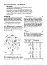
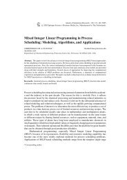
Figure 5 Comparison <strong>of</strong> the geometrical distortion exhibited by a large-field x-ray image intensifier (a), compared<br />
with the distortion-free image <strong>of</strong> a large-field, <strong>dynamic</strong>, <strong>solid</strong>-<strong>state</strong> detector (b). Figure courtesy <strong>of</strong> Pat Turner.<br />
between five <strong>and</strong> 10. 21 Consequently, <strong>dynamic</strong>,<br />
<strong>solid</strong>-<strong>state</strong> detectors produce images with superior<br />
contrast <strong>and</strong> wider <strong>dynamic</strong> range than x-ray IITV<br />
systems; this is reflected in the excellent reproduction<br />
<strong>of</strong> the high-contrast structures depicted<br />
in Fig. 4.<br />
Spatial resolution<br />
<strong>The</strong> spatial resolution <strong>of</strong> a <strong>solid</strong>-<strong>state</strong> detector is<br />
affected by a number <strong>of</strong> physical <strong>and</strong> technical<br />
factors. <strong>The</strong>se include light scatter in the x-ray<br />
absorption layer (in the case <strong>of</strong> indirect-conversion<br />
devices), the detector pixel sampling interval <strong>and</strong><br />
aperture size, <strong>and</strong> the b<strong>and</strong>width <strong>of</strong> the readout<br />
electronics. Dynamic, <strong>solid</strong>-<strong>state</strong>, <strong>flat</strong>-panel detectors<br />
come in a variety <strong>of</strong> sizes, form factors <strong>and</strong><br />
pixel resolutions, matched to their target clinical<br />
application(s). Typical values <strong>of</strong> spatial <strong>imaging</strong><br />
<strong>characteristics</strong> for indirect-conversion <strong>dynamic</strong><br />
detectors <strong>design</strong>ed for cardiac, vascular, <strong>and</strong><br />
radiography <strong>and</strong> fluoroscopy applications are listed<br />
in Table 1 (Readers should note that relevant <strong>characteristics</strong>,<br />
including pixel sampling interval <strong>and</strong><br />
Nyquist frequency, were defined in a previous review.<br />
6 ). Equivalent values for a large-field digital<br />
x-ray IITV system are included for reference. <strong>The</strong><br />
maximum spatial resolution <strong>of</strong> a large-field, <strong>dynamic</strong>,<br />
<strong>solid</strong>-<strong>state</strong> detector exceeds that <strong>of</strong> the<br />
digital x-ray IITV system by a factor <strong>of</strong> over two.<br />
It should be noted that the spatial resolution <strong>of</strong><br />
digital x-ray IITV systems deteriorates toward the<br />
periphery <strong>of</strong> the image field. <strong>The</strong> spatial resolution<br />
<strong>of</strong> <strong>solid</strong>-<strong>state</strong> detectors is maintained throughout<br />
the whole field <strong>of</strong> view. Dynamic, <strong>solid</strong>-<strong>state</strong> detectors<br />
<strong>of</strong>fer multiple (sometimes up to five) ancillary<br />
zoom-field selections. <strong>The</strong>se zoom-fields are<br />
used to magnify the presented image <strong>and</strong>, therefore,<br />
improve the resolution <strong>of</strong> fine-detail structures<br />
(albeit for a reduced field <strong>of</strong> view). <strong>The</strong><br />
spatial resolution <strong>of</strong> <strong>solid</strong>-<strong>state</strong> detectors remains<br />
essentially constant, independent <strong>of</strong> the field<br />
size selected. <strong>The</strong> frame rate <strong>of</strong> a <strong>dynamic</strong> <strong>solid</strong><strong>state</strong><br />
detector can be increased, say from 15 to<br />
Table 1 Typical spatial <strong>imaging</strong> <strong>characteristics</strong> <strong>of</strong> <strong>dynamic</strong>, <strong>solid</strong>-<strong>state</strong> detectors <strong>design</strong>ed for three clinical application areas,<br />
compared with a digital IITV system (results are quoted for the largest field selection in each case)<br />
Cardiac detector Vascular detector Radiography <strong>and</strong><br />
fluoroscopy detector<br />
Digital IITV<br />
Maximum field <strong>of</strong> Square field 24.8 24.8 Rectangular field Square field<br />
Circular field<br />
view (cm)<br />
38.2 29.4<br />
42.6 42.6<br />
35 cm diameter<br />
Pixel sampling<br />
interval (mm)<br />
184 154 148 341<br />
Maximum pixel array 956 954 2480 1910 2880 2881 1024 1024<br />
Nyquist frequency<br />
(lp/mm)<br />
2.72 3.25 3.38 1.46