The design and imaging characteristics of dynamic, solid-state, flat ...
The design and imaging characteristics of dynamic, solid-state, flat ...
The design and imaging characteristics of dynamic, solid-state, flat ...

You also want an ePaper? Increase the reach of your titles
YUMPU automatically turns print PDFs into web optimized ePapers that Google loves.
1080 A.R. Cowen et al.<br />
artefacts. 32 Memory effect reflects a non-uniform<br />
variation in detector response depending upon<br />
the exposure history. With regard to indirect-conversion<br />
detectors, memory effect represents an<br />
increase in CsI:Tl light emission <strong>and</strong> a-Si:H photodiode<br />
gain, following the x-ray exposure. <strong>The</strong>se<br />
effects manifest themselves as a spurious increase<br />
in detector conversion efficiency, producing a<br />
so-called bright-burn artefact in images acquired<br />
subsequently. Conversely, in direct-conversion<br />
detectors memory effect reflects a reduction<br />
(fatigue) in photoconductor response. Memory effect<br />
can be a particular problem in mixed-mode<br />
<strong>imaging</strong> applications, specifically where lowdose<br />
fluoroscopy might follow immediately after<br />
an image is acquired at a high fluorographic dose<br />
level, e.g., as occurs in DSA. 27 <strong>The</strong> x-ray dose per<br />
frame during fluoroscopy can be as low as one<br />
thous<strong>and</strong>th <strong>of</strong> that used during serial image acquisition;<br />
therefore, even a modest degree <strong>of</strong> memory<br />
effect may intrude upon the fluoroscopic images<br />
that follow.<br />
Lag is the property that quantifies the ability <strong>of</strong><br />
an image detector to accurately record timevarying<br />
changes in image content; the larger the<br />
lag, the poorer the temporal response <strong>and</strong> vice<br />
versa. Lag results from the carry-over <strong>of</strong> a proportion<br />
<strong>of</strong> recorded signal content into succeeding<br />
frames in the sequence. In the case <strong>of</strong> indirectconversion<br />
detectors a small contribution <strong>of</strong> lag<br />
arises from afterglow in the CsI:Tl layer, (but this is<br />
rarely significant in routine fluoroscopy). In practice,<br />
lag largely results from the relatively slow<br />
temporal response <strong>of</strong> a-Si:H. More specifically lag<br />
arises from the trapping <strong>and</strong> subsequent slow<br />
release (de-trapping) <strong>of</strong> charge carriers in the<br />
photodiode array. 34 In direct-conversion detectors<br />
lag is compounded by charge trapping/de-trapping<br />
mechanisms in the a-Se photoconductor. 32 Without<br />
correction lag causes unacceptable unsharpness<br />
(smearing) <strong>of</strong> rapidly moving <strong>and</strong> time-varying<br />
image structures.<br />
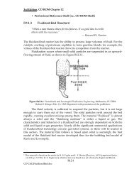
Dynamic <strong>solid</strong>-<strong>state</strong> detectors incorporate measures<br />
to minimize lag <strong>and</strong> memory effect. Many<br />
modern <strong>dynamic</strong> detectors achieve this using a socalled<br />
refresh (or reset) light, which reconditions<br />
the detector prior to each new image acquisition<br />
cycle. 9,20,28,34 <strong>The</strong> refresh light usually takes the<br />
form <strong>of</strong> an array <strong>of</strong> light-emitting diodes (see<br />
Fig. 1), which floods the detector with light photons,<br />
saturating charge-trapping sites in the<br />
a-Si:H prior to each x-ray exposure. As a result,<br />
lag (<strong>and</strong> memory effect) is reduced to an acceptably<br />
low level ensuring a suitably fast detector response.<br />
Signal retention due to lag in a modern<br />
indirect conversion detector is reportedly as low<br />
as 0.3% at a time 1 s after termination <strong>of</strong> the<br />
x-ray exposure; after 10 s the lag reduces by a further<br />
order <strong>of</strong> magnitude. 27 This ensures that the<br />
temporal resolution is adequate for high-speed<br />
<strong>imaging</strong> applications, such as paediatric cardiac<br />
fluoroscopy. Equivalent lag figures for direct conversion<br />
detectors are reportedly higher. 27 In some<br />
clinical applications a moderate degree <strong>of</strong> lag can<br />
be tolerated, <strong>and</strong> is used to improve fluoroscopic<br />
image quality by time-averaging (smoothing) noise<br />
fluctuations. Depending upon the type <strong>of</strong> clinical<br />
application, a suitable degree <strong>of</strong> lag is normally<br />
synthesized using digital recursive filtering.<br />
DQE<br />
DQE is the most effectual physical parameter used<br />
to quantify <strong>and</strong> compare the performance <strong>of</strong> different<br />
x-ray image detectors objectively. 35 To simplify<br />
the discussion, here it is assumed that the fluoroscopic<br />
image detector exhibits zero lag, (or any lag<br />
that does exist is fully corrected). <strong>The</strong> DQE <strong>of</strong> the<br />
detector can then be defined by the ratio, 6,35<br />
DQE detector ¼ SNR 2<br />
recorder =SNR2<br />
input<br />
2<br />
Where SNRinput<br />
is the square <strong>of</strong> the signal-to-<br />
noise ratio at the input <strong>of</strong> the image detector.<br />
This is defined by the fluence <strong>of</strong> x-ray photons<br />
(number per unit area) contributing to an individ-<br />
ual frame in the fluoroscopic image sequence.<br />
2<br />
SNRrecorded is the square <strong>of</strong> the signal-to-noise ratio<br />
recorded by the image detector. <strong>The</strong> value <strong>of</strong><br />
2<br />
SNRrecorded can be computed from the output<br />
2<br />
data. In terms <strong>of</strong> counting statistics SNRrecorded is<br />
an estimate <strong>of</strong> the fluence <strong>of</strong> information carriers<br />
that the recorded image frame is actually worth.<br />
<strong>The</strong> information content <strong>of</strong> a recorded image<br />
frame can never exceed that delivered to the<br />
detector in the incident x-ray beam, therefore,<br />
0 DQEdetector 1<br />
A DQE <strong>of</strong> unity implies that the recording <strong>of</strong> x-ray<br />
image information by the detector is perfect. At the<br />
other extreme a DQE <strong>of</strong> zero implies that no information<br />
at all is recorded. Real-world x-ray image<br />
detectors obviously <strong>of</strong>fer a DQE value falling somewhere<br />
between these two extremes. <strong>The</strong> deterioration<br />
in recorded information is for two principal<br />
reasons. First, no detector can absorb all the incident<br />
x-ray photons with 100% efficiency. Inevitably<br />
some x-ray photons pass straight through the<br />
x-ray absorber, while others that are absorbed may<br />
then be re-emitted <strong>and</strong> escape the detector. This<br />
loss in primary information is compounded by any<br />
noise sources arising in the detector itself (e.g.,