The design and imaging characteristics of dynamic, solid-state, flat ...
The design and imaging characteristics of dynamic, solid-state, flat ...
The design and imaging characteristics of dynamic, solid-state, flat ...

You also want an ePaper? Increase the reach of your titles
YUMPU automatically turns print PDFs into web optimized ePapers that Google loves.
1076 A.R. Cowen et al.<br />
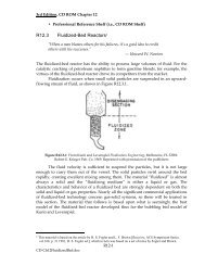
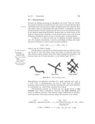
Figure 1 Cross-section through a <strong>dynamic</strong>, <strong>solid</strong>-<strong>state</strong>, <strong>flat</strong>-panel detector, identifying the surface reflector, CsI:Tl<br />
layer, a-SiH active matrix array, <strong>and</strong> refresh light (Trixell SA Pixium 4800 detector). Reproduced with the permission <strong>of</strong><br />
Medicamundi.<br />
Physical <strong>imaging</strong> <strong>characteristics</strong><br />
<strong>The</strong> physical image quality <strong>of</strong> <strong>dynamic</strong> digital x-ray<br />
image detectors can be evaluated using a toolkit <strong>of</strong><br />
parameters such as: <strong>dynamic</strong> range; geometrical<br />
distortion, vignetting, <strong>and</strong> veiling glare; spatial<br />
resolution; temporal resolution (lag <strong>and</strong> memory<br />
effect); <strong>and</strong> detective quantum efficiency (DQE).<br />
<strong>The</strong>se parameters are transportable across different<br />
<strong>design</strong>s <strong>of</strong> x-ray image detector, <strong>and</strong> can be<br />
used to compare <strong>imaging</strong> system performance on<br />
an objective basis.<br />
Figure 2 A modern cardiac catheterization laboratory<br />
incorporating an Allura Xper FD10 <strong>solid</strong>-<strong>state</strong>, cardiac,<br />
<strong>flat</strong>-panel detector in the Yorkshire Heart Centre, Leeds.<br />
Reproduced with the permission <strong>of</strong> Medicamundi.<br />
Dynamic range<br />
<strong>The</strong> <strong>dynamic</strong> range <strong>of</strong> a digital x-ray image detector<br />
describes the maximum range <strong>of</strong> entrance<br />
doses over which substantive image information is<br />
recorded. In basic terms <strong>dynamic</strong> range is described<br />
by the ratio <strong>of</strong> the maximum to the<br />
minimum detector usable operating dose levels;<br />
(specifically the former is defined by the maximum<br />
signal capability <strong>and</strong> the latter by noise). Dynamic<br />
x-ray image detectors require a wider <strong>dynamic</strong><br />
range than DR detectors, as they are multi-<br />
Figure 3 A modern neurovascular intervention laboratory<br />
incorporating a Philips Allura Xper FD20 large-field,<br />
<strong>dynamic</strong>, <strong>solid</strong>-<strong>state</strong> detector. Reproduced with the permission<br />
<strong>of</strong> Philips Healthcare.