

PDF Download - Glidewell Dental Labs
PDF Download - Glidewell Dental Labs
PDF Download - Glidewell Dental Labs

You also want an ePaper? Increase the reach of your titles
YUMPU automatically turns print PDFs into web optimized ePapers that Google loves.
educing treatment time with digital dentistry<br />
all leveraged digital dentistry technologies.<br />
The convergence of these<br />
technologies is enabling simpler, more<br />
convenient and more affordable restorative<br />
protocols.<br />
Diagnosis<br />
The patient had no medical contraindications<br />
for implant surgery. She<br />
was on blood thinners, which were<br />
discontinued for a period of four days<br />
before and two days after the surgery<br />
to mitigate the amount of bleeding<br />
during and after implant placement.<br />
Her periodontal tissues were generally<br />
healthy and free from irritation<br />
because of a recent reline and adjustment<br />
of the lower denture to relieve<br />
sore spots. Her occlusion with the relined<br />
dentures was good, and she had<br />
been functioning in these dentures<br />
for years without any TMJ issues. Her<br />
lower denture’s lack of retention was<br />
her primary motivation for considering<br />
implant therapy.<br />
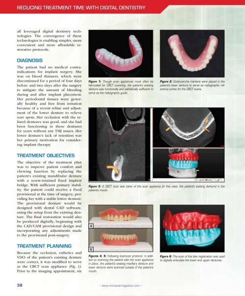
Figure 1: Though scan appliances must often be<br />
fabricated for CBCT scanning, the patient’s existing<br />
denture was functionally and esthetically sufficient to<br />
serve as the radiographic guide.<br />
Figure 2: Gutta-percha markers were placed in the<br />
patient’s lower denture to serve as radiographic reference<br />
points for the CBCT scans.<br />
Treatment Objectives<br />
The objective of the treatment plan<br />
was to improve patient comfort and<br />
chewing function by replacing the<br />
patient’s existing mandibular denture<br />
with a screw-retained fixed implant<br />
bridge. With sufficient primary stability,<br />
the patient could receive a fixed<br />
provisional at the time of surgery, providing<br />
her with a stable lower denture.<br />
The provisional denture would be<br />
designed with dental CAD software,<br />
using the setup from the existing denture.<br />
The final restoration would also<br />
be produced digitally, beginning with<br />
the CAD/CAM provisional design and<br />
incorporating any adjustments made<br />
to the provisional post-surgery.<br />
Figure 3: A CBCT scan was taken of the scan appliance (in this case, the patient’s existing denture) in the<br />
patient’s mouth.<br />
4<br />
Treatment Planning<br />
Because the occlusion, esthetics and<br />
VDO of the patient’s existing denture<br />
were correct, it was modified to serve<br />
as the CBCT scan appliance (Fig. 1).<br />
Prior to the imaging appointment, six<br />
5<br />
Figures 4, 5: Following dual-scan protocol, in addition<br />
to scanning the patient with the scan appliance<br />
in place, the patient’s existing maxillary denture and<br />
lower denture were scanned outside of the patient’s<br />
mouth.<br />
Figure 6: The scan of the bite registration was used<br />
to digitally articulate the lower and upper dentures.<br />
38<br />
– www.inclusivemagazine.com –


