

Chairside - Glidewell Dental Labs
Chairside - Glidewell Dental Labs
Chairside - Glidewell Dental Labs

You also want an ePaper? Increase the reach of your titles
YUMPU automatically turns print PDFs into web optimized ePapers that Google loves.
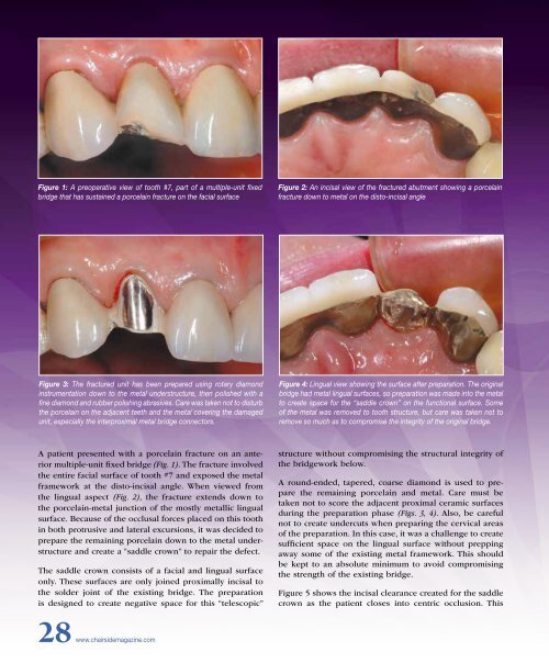
Figure 1: A preoperative view of tooth #7, part of a multiple-unit fixed<br />
bridge that has sustained a porcelain fracture on the facial surface<br />
Figure 2: An incisal view of the fractured abutment showing a porcelain<br />
fracture down to metal on the disto-incisal angle<br />
Figure 3: The fractured unit has been prepared using rotary diamond<br />
instrumentation down to the metal understructure, then polished with a<br />
fine diamond and rubber polishing abrasives. Care was taken not to disturb<br />
the porcelain on the adjacent teeth and the metal covering the damaged<br />
unit, especially the interproximal metal bridge connectors.<br />
Figure 4: Lingual view showing the surface after preparation. The original<br />
bridge had metal lingual surfaces, so preparation was made into the metal<br />
to create space for the “saddle crown” on the functional surface. Some<br />
of the metal was removed to tooth structure, but care was taken not to<br />
remove so much as to compromise the integrity of the original bridge.<br />
A patient presented with a porcelain fracture on an anterior<br />
multiple-unit fixed bridge (Fig. 1). The fracture involved<br />
the entire facial surface of tooth #7 and exposed the metal<br />
framework at the disto-incisal angle. When viewed from<br />
the lingual aspect (Fig. 2), the fracture extends down to<br />
the porcelain-metal junction of the mostly metallic lingual<br />
surface. Because of the occlusal forces placed on this tooth<br />
in both protrusive and lateral excursions, it was decided to<br />
prepare the remaining porcelain down to the metal understructure<br />
and create a “saddle crown” to repair the defect.<br />
The saddle crown consists of a facial and lingual surface<br />
only. These surfaces are only joined proximally incisal to<br />
the solder joint of the existing bridge. The preparation<br />
is designed to create negative space for this “telescopic”<br />
structure without compromising the structural integrity of<br />
the bridgework below.<br />
A round-ended, tapered, coarse diamond is used to prepare<br />
the remaining porcelain and metal. Care must be<br />
taken not to score the adjacent proximal ceramic surfaces<br />
during the preparation phase (Figs. 3, 4). Also, be careful<br />
not to create undercuts when preparing the cervical areas<br />
of the preparation. In this case, it was a challenge to create<br />
sufficient space on the lingual surface without prepping<br />
away some of the existing metal framework. This should<br />
be kept to an absolute minimum to avoid compromising<br />
the strength of the existing bridge.<br />
Figure 5 shows the incisal clearance created for the saddle<br />
crown as the patient closes into centric occlusion. This<br />
28 www.chairsidemagazine.com


