Slicer4Minute tutorial - 3D Slicer
Slicer4Minute tutorial - 3D Slicer
Slicer4Minute tutorial - 3D Slicer

You also want an ePaper? Increase the reach of your titles
YUMPU automatically turns print PDFs into web optimized ePapers that Google loves.
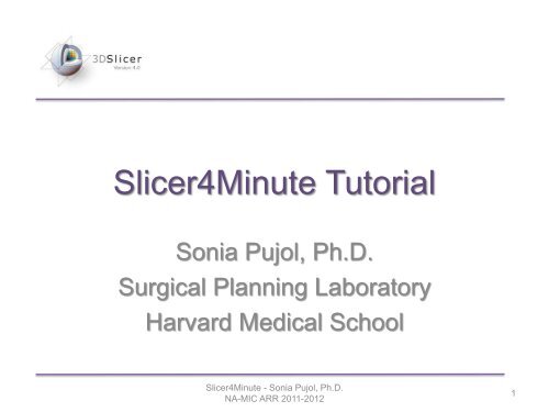
<strong><strong>Slicer</strong>4Minute</strong> Tutorial<br />
Sonia Pujol, Ph.D.<br />
Surgical Planning Laboratory<br />
Harvard Medical School<br />
<strong><strong>Slicer</strong>4Minute</strong> - Sonia Pujol, Ph.D.<br />
NA-MIC ARR 2011-2012<br />
1
<strong>Slicer</strong>4 minute <strong>tutorial</strong><br />
This <strong>tutorial</strong> is a 4-minute introduction to the <strong>3D</strong> visualization<br />
capabilities of the <strong>Slicer</strong>4 software for medical image analysis.<br />
<strong><strong>Slicer</strong>4Minute</strong> - Sonia Pujol, Ph.D.<br />
NA-MIC ARR 2011-2012<br />
2
<strong>Slicer</strong>4 software<br />
The <strong>Slicer</strong>4 software is available for download:<br />
http://download.slicer.org<br />
<strong><strong>Slicer</strong>4Minute</strong> - Sonia Pujol, Ph.D.<br />
NA-MIC ARR 2011-2012<br />
3
<strong>Slicer</strong>4 minute dataset<br />
Download the slicer4minute-data.zip training dataset<br />
http://wiki.slicer.org/slicerWiki/index.php/Training/4.0#<strong><strong>Slicer</strong>4Minute</strong>_Tutorial<br />
<strong><strong>Slicer</strong>4Minute</strong> - Sonia Pujol, Ph.D.<br />
NA-MIC ARR 2011-2012<br />
4
<strong>3D</strong><strong>Slicer</strong> version 4.0<br />
Start <strong>Slicer</strong> on your<br />
computer: the Welcome<br />
module is the default<br />
start-up module.<br />
<strong><strong>Slicer</strong>4Minute</strong> - Sonia Pujol, Ph.D.<br />
NA-MIC ARR 2011-2012<br />
5
<strong>3D</strong> <strong>Slicer</strong> Scene<br />
A <strong>Slicer</strong> scene is a MRML file which, contains<br />
the list of elements loaded into <strong>Slicer</strong><br />
(volumes, models, fiducials…)<br />
The following example uses a <strong>3D</strong> Scene,<br />
which contains images and <strong>3D</strong> surface<br />
models of the head.<br />
<strong><strong>Slicer</strong>4Minute</strong> - Sonia Pujol, Ph.D.<br />
NA-MIC ARR 2011-2012<br />
6
<strong>3D</strong><strong>Slicer</strong> version 4.0<br />
Select File Load Scene<br />
from the main menu, and load<br />
slicer4minute.mrml<br />
<strong><strong>Slicer</strong>4Minute</strong> - Sonia Pujol, Ph.D.<br />
NA-MIC ARR 2011-2012<br />
7
<strong>Slicer</strong>4 minute Scene<br />
<strong>Slicer</strong> displays<br />
the elements<br />
of the<br />
slicer4minute<br />
scene, which<br />
contains the<br />
MR volume of<br />
the brain and<br />
a series of <strong>3D</strong><br />
surface<br />
models.<br />
<strong><strong>Slicer</strong>4Minute</strong> - Sonia Pujol, Ph.D.<br />
NA-MIC ARR 2011-2012<br />
8
Slice4 minute scene<br />
Select Conventional<br />
Widescreen from the<br />
viewing mode menu<br />
<strong><strong>Slicer</strong>4Minute</strong> - Sonia Pujol, Ph.D.<br />
NA-MIC ARR 2011-2012<br />
9
<strong>Slicer</strong>4minute Scene<br />
Select Models from the Modules<br />
menu<br />
<strong><strong>Slicer</strong>4Minute</strong> - Sonia Pujol, Ph.D.<br />
NA-MIC ARR 2011-2012<br />
10
<strong>3D</strong> Visualization<br />
The Models module GUI displays the list of models<br />
loaded in the slicer4minute scene, their color and the<br />
value of their opacity (between 0.0 an 1.0)<br />
<strong><strong>Slicer</strong>4Minute</strong> - Sonia Pujol, Ph.D.<br />
NA-MIC ARR 2011-2012<br />
11
<strong>3D</strong> Visualization<br />
Click on the pin icon on the top left corner of the red<br />
slice to display the slice viewer menu then click on the<br />
eye icon to display the axial slice in the <strong>3D</strong> Viewer<br />
<strong><strong>Slicer</strong>4Minute</strong> - Sonia Pujol, Ph.D.<br />
NA-MIC ARR 2011-2012<br />
12
<strong>3D</strong> Visualization<br />
Use the slider of the red viewer<br />
to browse through the axial MR<br />
slices.<br />
<strong>Slicer</strong> simultaneously displays<br />
the slices in the <strong>3D</strong> viewer.<br />
<strong><strong>Slicer</strong>4Minute</strong> - Sonia Pujol, Ph.D.<br />
NA-MIC ARR 2011-2012<br />
13
<strong>3D</strong> Visualization<br />
Lower the opacity of the Skin.vtk<br />
model in the Display tab<br />
The skull_bone.vtk model appears through the skin.<br />
<strong><strong>Slicer</strong>4Minute</strong> - Sonia Pujol, Ph.D.<br />
NA-MIC ARR 2011-2012<br />
14
<strong>3D</strong> Visualization<br />
Position the mouse in the <strong>3D</strong> viewer.<br />
• Click the left-mouse button to drag and rotate<br />
the model.<br />
• Click the right-mouse button to zoom in and out.<br />
<strong><strong>Slicer</strong>4Minute</strong> - Sonia Pujol, Ph.D.<br />
NA-MIC ARR 2011-2012<br />
15
Anatomical Views<br />
Click on the pin icons in<br />
the top left corners of the<br />
red and green viewers to<br />
display the viewers’<br />
menu, and click on the<br />
eye icon to display the<br />
axial and coronal slice in<br />
the <strong>3D</strong> viewer.<br />
<strong><strong>Slicer</strong>4Minute</strong> - Sonia Pujol, Ph.D.<br />
NA-MIC ARR 2011-2012<br />
16
<strong>3D</strong> Visualization<br />
Turn off the visibility of<br />
the skull bone to display<br />
the hemispheric white<br />
matter model<br />
<strong><strong>Slicer</strong>4Minute</strong> - Sonia Pujol, Ph.D.<br />
NA-MIC ARR 2011-2012<br />
17
<strong>3D</strong> Visualization<br />
The white matter surface, as well as the left<br />
and right optic nerves appear in the viewer<br />
<strong><strong>Slicer</strong>4Minute</strong> - Sonia Pujol, Ph.D.<br />
NA-MIC ARR 2011-2012<br />
18
<strong>3D</strong> Visualization<br />
Select the hemispheric_white_ matter.vtk<br />
model, and check Clip in the Display tab.<br />
<strong><strong>Slicer</strong>4Minute</strong> - Sonia Pujol, Ph.D.<br />
NA-MIC ARR 2011-2012<br />
19
<strong>3D</strong> Visualization<br />
Use the slider of the coronal view<br />
(green) to expose the optic chiasm.<br />
<strong><strong>Slicer</strong>4Minute</strong> - Sonia Pujol, Ph.D.<br />
NA-MIC ARR 2011-2012<br />
20
<strong>3D</strong> Visualization<br />
Increase the opacity of<br />
the skin model, and<br />
select the viewing mode<br />
<strong>3D</strong> only<br />
<strong><strong>Slicer</strong>4Minute</strong> - Sonia Pujol, Ph.D.<br />
NA-MIC ARR 2011-2012<br />
21
<strong>3D</strong> Visualization<br />
Click on the Pin icon at the top left corner of<br />
the <strong>3D</strong> viewer, and click on the Spin icon.<br />
<strong><strong>Slicer</strong>4Minute</strong> - Sonia Pujol, Ph.D.<br />
NA-MIC ARR 2011-2012<br />
22
<strong>3D</strong> Visualization<br />
The <strong>3D</strong> models and 2D anatomical slices start<br />
spinning in the <strong>3D</strong> viewer.<br />
Click a second time on the spin icon to stop the<br />
models from spinning.<br />
<strong><strong>Slicer</strong>4Minute</strong> - Sonia Pujol, Ph.D.<br />
NA-MIC ARR 2011-2012<br />
23
<strong>Slicer</strong>4 minute <strong>tutorial</strong><br />
This <strong>tutorial</strong> was a<br />
short introduction to<br />
the <strong>3D</strong> visualization<br />
capabilities of <strong>Slicer</strong>.<br />
Visit the <strong>Slicer</strong>4<br />
training compendium<br />
for more information on<br />
the software.<br />
http://www.slicer.org/slicerWiki/index.php/Documentation/4.0/Training<br />
<strong><strong>Slicer</strong>4Minute</strong> - Sonia Pujol, Ph.D.<br />
NA-MIC ARR 2011-2012<br />
24
Acknowledgments<br />
National Alliance for Medical<br />
Image Computing<br />
NIH U54EB005149<br />
Neuroimage Analysis Center<br />
NIH P41RR013218<br />
<strong><strong>Slicer</strong>4Minute</strong> - Sonia Pujol, Ph.D.<br />
NA-MIC ARR 2011-2012<br />
25