Optic Radiation Fiber Tractography in Glioma Patients ... - 3D Slicer
Optic Radiation Fiber Tractography in Glioma Patients ... - 3D Slicer
Optic Radiation Fiber Tractography in Glioma Patients ... - 3D Slicer
You also want an ePaper? Increase the reach of your titles
YUMPU automatically turns print PDFs into web optimized ePapers that Google loves.
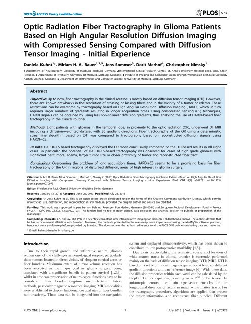
<strong>Optic</strong> <strong>Radiation</strong> <strong>Fiber</strong> <strong>Tractography</strong> <strong>in</strong> <strong>Glioma</strong> <strong>Patients</strong>Based on High Angular Resolution Diffusion Imag<strong>in</strong>gwith Compressed Sens<strong>in</strong>g Compared with DiffusionTensor Imag<strong>in</strong>g - Initial ExperienceDaniela Kuhnt 1 *, Miriam H. A. Bauer 1,2,5 , Jens Sommer 3 , Dorit Merhof 4 , Christopher Nimsky 11 Department of Neurosurgery, University of Marburg, Marburg, Germany, 2 International Cl<strong>in</strong>ical Research Center, St. Anne’s University Hospital Brno, Brno, CzechRepublic, 3 Department of Psychiatry, University of Marburg, Marburg, Germany, 4 Institute of Imag<strong>in</strong>g and Computer Vision, Rhenish-Westphalian Technical UniversityAachen, Aachen, Germany, 5 Department Of Mathematics and Computer Science, University of Marburg, Marburg, GermanyAbstractObjective: Up to now, fiber tractography <strong>in</strong> the cl<strong>in</strong>ical rout<strong>in</strong>e is mostly based on diffusion tensor imag<strong>in</strong>g (DTI). However,there are known drawbacks <strong>in</strong> the resolution of cross<strong>in</strong>g or kiss<strong>in</strong>g fibers and <strong>in</strong> the vic<strong>in</strong>ity of a tumor or edema. Theserestrictions can be overcome by tractography based on High Angular Resolution Diffusion Imag<strong>in</strong>g (HARDI) which <strong>in</strong> turnrequires larger numbers of gradients result<strong>in</strong>g <strong>in</strong> longer acquisition times. Us<strong>in</strong>g compressed sens<strong>in</strong>g (CS) techniques,HARDI signals can be obta<strong>in</strong>ed by us<strong>in</strong>g less non-coll<strong>in</strong>ear diffusion gradients, thus enabl<strong>in</strong>g the use of HARDI-based fibertractography <strong>in</strong> the cl<strong>in</strong>ical rout<strong>in</strong>e.Methods: Eight patients with gliomas <strong>in</strong> the temporal lobe, <strong>in</strong> proximity to the optic radiation (OR), underwent 3T MRI<strong>in</strong>clud<strong>in</strong>g a diffusion-weighted dataset with 30 gradient directions. <strong>Fiber</strong> tractography of the OR us<strong>in</strong>g a determ<strong>in</strong>isticstreaml<strong>in</strong>e algorithm based on DTI was compared to tractography based on reconstructed diffusion signals us<strong>in</strong>gHARDI+CS.Results: HARDI+CS based tractography displayed the OR more conclusively compared to the DTI-based results <strong>in</strong> all eightcases. In particular, the potential of HARDI+CS-based tractography was observed for cases of high grade gliomas withsignificant peritumoral edema, larger tumor size or closer proximity of tumor and reconstructed fiber tract.Conclusions: Overcom<strong>in</strong>g the problem of long acquisition times, HARDI+CS seems to be a promis<strong>in</strong>g basis for fibertractography of the OR <strong>in</strong> regions of disturbed diffusion, areas of high <strong>in</strong>terest <strong>in</strong> glioma surgery.Citation: Kuhnt D, Bauer MHA, Sommer J, Merhof D, Nimsky C (2013) <strong>Optic</strong> <strong>Radiation</strong> <strong>Fiber</strong> <strong>Tractography</strong> <strong>in</strong> <strong>Glioma</strong> <strong>Patients</strong> Based on High Angular ResolutionDiffusion Imag<strong>in</strong>g with Compressed Sens<strong>in</strong>g Compared with Diffusion Tensor Imag<strong>in</strong>g - Initial Experience. PLoS ONE 8(7): e70973. doi:10.1371/journal.pone.0070973Editor: Friedemann Paul, Charité University Medic<strong>in</strong>e Berl<strong>in</strong>, GermanyReceived January 13, 2013; Accepted June 26, 2013; Published July 26, 2013Copyright: ß 2013 Kuhnt et al. This is an open-access article distributed under the terms of the Creative Commons Attribution License, which permitsunrestricted use, distribution, and reproduction <strong>in</strong> any medium, provided the orig<strong>in</strong>al author and source are credited.Fund<strong>in</strong>g: This work was supported <strong>in</strong> part by von Behr<strong>in</strong>g Roentgen Foundation, Germany (58-0044) and European Regional Development Fund – ProjectFNUSA – ICRC (No. CZ.1.05/1.1.00/02.0123). The funders had no role <strong>in</strong> study design, data collection and analysis, decision to publish, or preparation of themanuscript.Compet<strong>in</strong>g Interests: Ch. Nimsky, MD, PhD is a scientific consultant <strong>in</strong>for <strong>in</strong>traoperative imag<strong>in</strong>g for Bra<strong>in</strong>Lab (Feldkirchen,Germany). The authors declare thathe has no commercial affiliation with Bra<strong>in</strong>Lab. Moreover, the methods presented <strong>in</strong> the manuscript were implemented us<strong>in</strong>g the MedAlyVis software platform,hence not on any software platform provided by Bra<strong>in</strong>Lab. This does not alter the authors’ adherence to all the PLOS ONE policies on shar<strong>in</strong>g data and materials.* E-mail: kuhntd@med.uni-marburg.deIntroductionDue to their rapid growth and <strong>in</strong>filtrative nature, gliomasrema<strong>in</strong> one of the challenges <strong>in</strong> neurological surgery, particularlythose tumors located <strong>in</strong> direct vic<strong>in</strong>ity of eloquent cortical areas orfiber bundles. Maximum extent of tumor volume resection hasbeen accepted as the major goal <strong>in</strong> glioma surgery, be<strong>in</strong>gassociated with a significant benefit <strong>in</strong> patient survival [1,2,3],whilst <strong>in</strong> any case preservation of neurological functions have to beconsidered. Thus, besides long-time used electrostimulationmethods, particular magnetic resonance imag<strong>in</strong>g (MRI)-modalitieswere established to display functional cortical sites or fiber bundlesnon-<strong>in</strong>vasively. These data can be <strong>in</strong>tegrated <strong>in</strong>to the navigationsystem and displayed <strong>in</strong>traoperatively, which has been shown tocontribute to low postoperative morbidity [4,5].Due to its practicability, the estimated course and location ofwhite matter tracts <strong>in</strong> cl<strong>in</strong>ical practice is currently performedma<strong>in</strong>ly on the basis of diffusion tensor imag<strong>in</strong>g (DTI)-MRI. DTI isbased on a set of diffusion images acquired for at least six differentgradient directions and one reference image [6]. With these data,the diffusion properties with<strong>in</strong> each voxel can be calculated by theStejskal Tanner equation, result<strong>in</strong>g <strong>in</strong> a 2 nd order tensor. Inanisotropic tensors, the ma<strong>in</strong> eigenvector encodes for thelongitud<strong>in</strong>al direction of axons <strong>in</strong> major white matter tracts. Forthe tractography procedure, algorithms are applied that processthe tensor <strong>in</strong>formation and reconstruct fiber bundles. DifferentPLOS ONE | www.plosone.org 1 July 2013 | Volume 8 | Issue 7 | e70973
<strong>Optic</strong> <strong>Radiation</strong> <strong>Tractography</strong> with DTI vs. HARDIalgorithms have already been implemented and <strong>in</strong>vestigated so far[7,8,9]. However, due to the restricted 2 nd order tensor model,multiple fiber populations with<strong>in</strong> a voxel cannot be resolvedadequately. This causes complications to resolve cross<strong>in</strong>g-, kiss<strong>in</strong>g-,diverg<strong>in</strong>g or highly curved fibers. Another problem appear<strong>in</strong>g withDTI-based fiber tractography is the resolution of fibers <strong>in</strong> areas ofdisturbed diffusion, for example due to tumor or peritumoraledema [10,11,12].To overcome these drawbacks, alternative diffusion imag<strong>in</strong>gand reconstruction schemes denoted as High Angular ResolutionDiffusion Imag<strong>in</strong>g (HARDI) [13] have become <strong>in</strong>creas<strong>in</strong>glyrelevant. A three-dimensional orientation distribution function isherewith <strong>in</strong>terpolated as a steady function on the sphere, forexample by spherical harmonics. However, the cl<strong>in</strong>ical use ofHARDI is limited due to larger number of diffusion-encod<strong>in</strong>ggradients (rang<strong>in</strong>g from 60 to 100), result<strong>in</strong>g <strong>in</strong> long acquisitiontimes [14,15,16]. A solution for this restriction is a sparserepresentation of signals, which is provided for example by theuse of spherical ridgelets, as proposed by Michailovich et al.[17,18,19].Us<strong>in</strong>g this protocol, HARDI data can be represented us<strong>in</strong>g arelatively small number of diffusion-encod<strong>in</strong>g gradients, thusenhanc<strong>in</strong>g the feasibility of HARDI-based fiber tractography <strong>in</strong>the cl<strong>in</strong>ical practice. This is implied <strong>in</strong> compressed sens<strong>in</strong>gtechniques (CS) [17,20,21,22].To <strong>in</strong>vestigate the feasibility of HARDI+CS for the reconstructionof the optic radiation (OR) <strong>in</strong> proximity of gliomas <strong>in</strong> thetemporal lobe with associated disturbed diffusion properties, weperformed fiber tractography based on DTI and HARDI+CS foreight patients. To our knowledge, this is the first cl<strong>in</strong>icalapplication and comparison of DTI-, and HARDI+CS-basedfiber tractography for the OR, show<strong>in</strong>g the potential of HARDIwith respect to challeng<strong>in</strong>g <strong>in</strong>tra-voxel fiber distributions.Materials and Methods<strong>Patients</strong>The study was conducted as a prospective case series withretrospective data analysis after approval by the ethics commissionof the Philipps-University of Marburg, Germany. All patients gavetheir written <strong>in</strong>formed consent to participate <strong>in</strong> the study. The<strong>in</strong>formed consent form was also approved by the ethicscommission, Philipps-University of Marburg, Germany. Eightpatients with gliomas, all located <strong>in</strong> the left temporal lobe, were<strong>in</strong>cluded <strong>in</strong> our study. Mean patient age was 54.8 years. Threefemale patients and five male patients participated. To comparetumor size <strong>in</strong>clud<strong>in</strong>g peritumoral edema and their <strong>in</strong>fluence on thefiber reconstruction, all tumors were manually segmented acrossall slices. In case the tumor showed contrast-enhancement, bothT1+gadol<strong>in</strong>ium-, and T2-weighted tumor outl<strong>in</strong>es were segmented.Otherwise, only T2-weighted tumor outl<strong>in</strong>es were segmented.Tumor volumes are given <strong>in</strong> [cm 3 ]. Furthermore the m<strong>in</strong>imumdistance from the tumor outl<strong>in</strong>es to the fiber object, based onHARDI+CS was calculated <strong>in</strong> [mm]. All tumors were located <strong>in</strong>close proximity to the reconstructed OR with less than 20 mm (seeTable 1).MRIMR images were acquired at a 3T MRI (Tim Trio, Siemens,Erlangen, Germany) preoperatively, <strong>in</strong>clud<strong>in</strong>g T1-weighted <strong>3D</strong>images (<strong>3D</strong>-Magnetization Prepared Rapid Gradient Echo(MPRAGE)): repetition time (TR) 1900 ms, echo time (TE)2.26 ms, field of view (FoV) 256 mm, matrix 2566256, slicethickness 1 mm, 176 slices, sagittal). The protocol for diffusionimag<strong>in</strong>g was TR 7800 ms, TE 90 ms, FoV 256 mm, matrix1286128, slice thickness 2 mm, numbers of excitation (NEX) = 1,b = 1000 s/mm 2 , 30 non-coll<strong>in</strong>ear diffusion-encod<strong>in</strong>g gradients,voxel size of 26262 mm 3 . This same diffusion images were thebase for DTI-based fiber tractography and HARDI+CS-basedfiber tractography. Acquisition of diffusion imag<strong>in</strong>g data took5 m<strong>in</strong>utes per patient.<strong>Fiber</strong> tractographyEvaluation was performed us<strong>in</strong>g the image analysis platformMedAlyVis (Medical Analysis and Visualization) [23] for both,DTI-based and advanced fiber tractography us<strong>in</strong>g HARDI signalsderived from sparsely sampled diffusion data (HARDI+CS). ForDTI-based fiber tractography, a tensor deflection approach withfractional anistotropy (FA) thresholds of 0.18–0.2 was applied. Fortractography based on HARDI+CS, a determ<strong>in</strong>istic multidirectionalorientation distribution function (ODF) track<strong>in</strong>g was applied[12] with the L-<strong>in</strong>dex [24] as anisotropy measure, set to 0.03(maximum angle: 90 degrees, maximum steps: 500, stepsize: 1).For both, DTI-, and HARDI+CS-based fiber reconstruction,the same manually segmented region around the lateral geniculatenucleus (LGN) was used as seed region. The visual cortex was thenused as <strong>in</strong>clude region, result<strong>in</strong>g <strong>in</strong> a fiber object pass<strong>in</strong>g throughthe LGN and term<strong>in</strong>at<strong>in</strong>g <strong>in</strong> the visual cortex (Brodman areas 17–19). These ROIs were determ<strong>in</strong>ed by experienced exam<strong>in</strong>ers (oversix years of expertise), one neurosurgeon and one neurologist.Results – Case SeriesCase 1A 74 year old male patient presented with a temporal tumorwith slight and diffuse contrast enhancement <strong>in</strong> T1-weighted MRI.T2-weighted MR-signal however revealed mass of approximately(app.) 35 cm 3 (Table 1).For both DTI and HARDI+CS-based fiber tractography, wefound a fiber bundle, reach<strong>in</strong>g from LGN to the visual cortex.However, DTI-based fibers were slender and diffusely orientated.HARDI+CS-based tractography, however displayed a more solidfiber bundle, represent<strong>in</strong>g the OR without significant loop<strong>in</strong>g ofMeyer’s loop (ML). Still, all three parts of the OR <strong>in</strong> relation to theventricular system are reproducible with both methods (Figure 1,row 1).Case 2A 65 year-old female patient <strong>in</strong>itially presented with aphasia.T1-weighted MRI scans showed a contrast enhanc<strong>in</strong>g tumor <strong>in</strong>the left temporal lobe of app. 11 cm 3 with central necrosis,however with a strong peritumoral edema (Table 1).In this case, on the basis of DTI no fiber bundle pass<strong>in</strong>g throughLGN and term<strong>in</strong>at<strong>in</strong>g <strong>in</strong> the visual cortex was reliably traced.Only one s<strong>in</strong>gle fiber was found, which can hardly be assigneddef<strong>in</strong>itely to one of the three parts of the OR. This is opposed toHARDI+CS-based fiber tractography, which displayed the ORwith its three bundles <strong>in</strong>clud<strong>in</strong>g the ML, form<strong>in</strong>g a pronouncedloop <strong>in</strong> anterolateral direction (Figure 1, row 2).Case 3HARDI+CS-based fiber tractography of this 41 year-old malepatient with a relatively small anaplastic astrocytoma (WHO III) <strong>in</strong>the left temporal lobe, displayed a fiber bundle from LGN to thecalcar<strong>in</strong>e sulcus. In this case, the ML does not loop significantly.However, tractography results also <strong>in</strong>clude some diffusely runn<strong>in</strong>gfibers, rather not belong<strong>in</strong>g to the OR (<strong>in</strong> anterior temporo-mesialdirection and direct<strong>in</strong>g to the corpus callosum). S<strong>in</strong>gle-ROI DTI-PLOS ONE | www.plosone.org 2 July 2013 | Volume 8 | Issue 7 | e70973
<strong>Optic</strong> <strong>Radiation</strong> <strong>Tractography</strong> with DTI vs. HARDITable 1. Patient Collective.no age gender lesionm<strong>in</strong>imum distance tumor/HARDI+CS-fiber [mm]tumor volume T1+Gd/T2 [cm 3 ]1 73 m Anaplastic oligodendroglioma WHO III 10.0 11.2/35.22 65 f Glioblastoma multiforme WHO IV 18.5 10.7/68.33 41 m Anaplastic astrocytoma WHO III 16.6 -/13.74 52 m Glioblastoma multiforme WHO IV 10.2 10.2/16.05 61 f Anaplastic astrocytoma WHO III 11.5 1.7/41.86 35 m Diffuse Astrocytome WHO II 11.3 -/16.27 45 m Glioblastoma multiforme WHO IV 7.6 23.7/91.18 66 f Glioblastoma multiforme WHO IV 10.0 44.25/118doi:10.1371/journal.pone.0070973.t001based fiber track<strong>in</strong>g results were <strong>in</strong>conclusively, with only one fiberstart<strong>in</strong>g from LGN and term<strong>in</strong>at<strong>in</strong>g <strong>in</strong> the visual cortex, mostlikely belong<strong>in</strong>g to the central bundle of the OR (Figure 1, row 3).Case 4A 52 year-old female patient was admitted with a GBM (WHOIV) of app. 10 cm 3 and little peritumoral edema. Tumorlocalization is <strong>in</strong> close proximity to the OR with only app.10 mm m<strong>in</strong>imum distance. Results of DTI-, and HARDI+CSbasedfiber tractography vary significantly. With HARDI+CS, theOR is reliably displayed. The ML shows a sharp loop <strong>in</strong>anterolateral direction. By contrast, us<strong>in</strong>g the same start<strong>in</strong>g ROIaround the LGN, DTI provides a slight fiber bundle, most likelyshow<strong>in</strong>g the central bundle of the OR. The ML is not tracedsufficiently (Figure 1 (row 4)).Case 5A 61 year-old female patient was admitted with MRI scans of asmall temporal tumor on contrast enhanced T1-weighted images,however T2-weighted images revealed a hyper<strong>in</strong>tense signal ofapp. 42 cm 3 , closely located to the OR, given by tractographyresults (Table 1).DTI-based track<strong>in</strong>g results displayed one fiber, which can eachbe assigned to the central, superior and <strong>in</strong>ferior bundle of the OR.However, the result rema<strong>in</strong>s unconv<strong>in</strong>c<strong>in</strong>g. Compared to theHARDI+CS-based fiber bundle and contrary to the DTI-basedfibers, the OR here is given as a solid bundle, particularly the MLwith a sharp loop (Figure 1, row 5).Case 6In a case of a 35 year-old male patient, only T2-weighted MRIimages showed a small signal alteration <strong>in</strong> the lateral temporal lobewithout contrast enhancement <strong>in</strong> T1-weighted images. Boths<strong>in</strong>gle-ROI tractography results displayed the OR well withcentral, superior and <strong>in</strong>ferior fibers, form<strong>in</strong>g ML (Figure 2).However, HARDI+CS-based tractography showed a more solidand dense fiber bundle compared to DTI-based track<strong>in</strong>g.Particularly the ML, loop<strong>in</strong>g slightly <strong>in</strong> anterolateral direction, isdisplayed more conv<strong>in</strong>c<strong>in</strong>gly by HARDI+CS-based tractography(Figure 1, row 6).Case 7In this case of a 45 year old male patient with a left temporalGBM (WHO IV) accompanied with a large peritumoral edema(app. 24 and 92 cm 3 ), tractography results based on DTI displayedsolitary fibers, connect<strong>in</strong>g LGN and visual cortex. However, fiberswere not safely traced around the temporal horn of the lateralventricle. The HARDI+CS-based results displayed a solid ORobject,show<strong>in</strong>g the three bundles of the OR <strong>in</strong> their coursearound the temporal horn (Figure 1, row 7). Displayed with<strong>in</strong> aT1-weighted <strong>3D</strong> image model it can furthermore be seen, that thefiber bundle is significantly elevated due to the large tumor(Figure 2).Case 8A 66 year old female patient presented with aphasia andhemiparesis. This corresponded to a large temporal tumor onMRI, <strong>in</strong> close vic<strong>in</strong>ity to the HARDI+CS-based fiber-bundle(Table 1). This object reliably displayed the OR as a compoundbundle from LGN to the visual cortex. On the basis of DTI, theOR was not displayed conv<strong>in</strong>c<strong>in</strong>gly, as aga<strong>in</strong> only one s<strong>in</strong>gle fibercan be traced from LGN to the visual cortex (Figure 1, row 8).Summary<strong>Tractography</strong> results based on HARDI+CS display the ORmore conv<strong>in</strong>c<strong>in</strong>gly compared with DTI-based fiber objects <strong>in</strong> alleight cases. Overall, HARDI+CS-based objects conta<strong>in</strong> morefibers and the OR is given as a more compound object. Thus, <strong>in</strong>six out of eight cases, HARDI+CS-based fiber tractography results<strong>in</strong> significantly improved fiber objects (patients 2–5, 7, 8)compared with DTI. Even <strong>in</strong> two cases (patients 2, 8), DTI basedfiber tractography fails to generate an OR-object at all.The ML is given <strong>in</strong> four cases via HARDI-based tractography,<strong>in</strong> which it can not be reliably displayed by DTI (patients 2, 4, 7,8). In all 6 cases (patients 2–5, 7, 8), <strong>in</strong> which HARDI+CSdelivered much better tractography results, patients suffered fromhigh-grade tumors that were also associated with significant edema<strong>in</strong> 4 cases (patients 2, 5, 7, 8). Also closer location of tumor andOR (<strong>in</strong> patients 4, 5, 7, 8) resulted <strong>in</strong> significantly improved resultsvia HARDI+CS compared with DTI.Discussion<strong>Fiber</strong> tractography of the OR and applications <strong>in</strong> cl<strong>in</strong>icalpracticeThe classical descriptions of the visual pathway, particularly theOR are based on dissection studies by Leuret and Gratiolet [25] <strong>in</strong>1839 or Meyer [26] <strong>in</strong> 1907. These were revised and complementedwith f<strong>in</strong>d<strong>in</strong>gs from newer studies [27,28], mostly based ondissection techniques by Kl<strong>in</strong>gler et al. [29]. Basically, the ORconsists of three bundles – anterior, central and posterior bundles(otherwise named <strong>in</strong>ferior, central and superior bundles). Start<strong>in</strong>gPLOS ONE | www.plosone.org 3 July 2013 | Volume 8 | Issue 7 | e70973
<strong>Optic</strong> <strong>Radiation</strong> <strong>Tractography</strong> with DTI vs. HARDIFigure 1. <strong>Fiber</strong> tractography results presented for each patient(patients 1–8 accord<strong>in</strong>g rows 1–8) based on DTI (column 1),<strong>Slicer</strong> 4 (column 2), and based on HARDI+CS (column 4) with<strong>in</strong>MedAlyVis. Overlay of DTI-based (red) and HARDI+CS-based tractography(green) (r = right; l = left; a = anterior; p = posterior).doi:10.1371/journal.pone.0070973.g001at the LGN, they pass through the temporal stem to term<strong>in</strong>ate <strong>in</strong>the calcar<strong>in</strong>e sulcus. In the deep white matter of the <strong>in</strong>feriorlimit<strong>in</strong>g sulcus of the temporal lobe, they cover the superior andlateral wall of the lateral ventricle’s temporal horn. Meyer [26]described the anterior bundle as the most anterior extent of theOR, loop<strong>in</strong>g the roof of the temporal horn beh<strong>in</strong>d the anteriorcommissure, now called ML. The central bundle crosses the roofof the temporal horn without form<strong>in</strong>g an anterolateral loop. Theposterior bundle <strong>in</strong>stead is thought to run straight <strong>in</strong> posteriordirection from the LGN <strong>in</strong> the lateral wall of the ventricle.However, the temporal stem conta<strong>in</strong>s multiple fiber bundles, e.g.the unc<strong>in</strong>ate fascicle or the <strong>in</strong>ferior occipito-frontal fascicle, so thatthe more recent studies found these fascicles hard to accuratelydel<strong>in</strong>eate from each other [30]. In this way, the visual pathwaysstill accounts for the neuroanatomically most complex fiberbundles [31].Navigation systems are widely used among neurosurgicaloperat<strong>in</strong>g theatres, show<strong>in</strong>g outl<strong>in</strong>es of pre-operatively segmentedrisk structures and targets <strong>in</strong> the microscope heads-up display.This requires a previous registration process of physical space andimage space [32]. Besides merely anatomical MR images,Figure 2. 3-D models of T1-weighted MR-images with overlayof HARDI+CS-(red), and DTI-(green)-based tractography of theoptic radiation presented as hulled fiber objects. A+A9: Casepresentation (case 7): Case of a large temporo-mesial GBM of a 45 yearoldmale patient, show<strong>in</strong>g the marked differences of HARDI+CS-, andDTI-based fiber objects <strong>in</strong> case of a large, high-grade tumor. A: axialoblique view. A9: Axial oblique view. Tumor manually segmented <strong>in</strong>yellow. B+B9: Case presentation (case 6): 35 year-old male patient withsmall diffuse astrocytoma. Less remarkable differences of tractographyresults <strong>in</strong> case of this smaller, low-grade tumor. B: Left sagittal obliqueview. B9: Axial oblique view. Tumor manually segmented <strong>in</strong> yellow.doi:10.1371/journal.pone.0070973.g002PLOS ONE | www.plosone.org 4 July 2013 | Volume 8 | Issue 7 | e70973
<strong>Optic</strong> <strong>Radiation</strong> <strong>Tractography</strong> with DTI vs. HARDIfunctional data have also been <strong>in</strong>tegrated <strong>in</strong> recent years, nowdesignated as multimodality navigation. This concept <strong>in</strong>cludes thedisplay of eloquent cortical sites (given for example by fMRI [33]),metabolic data (given for example by magnetic resonancespectroscopy imag<strong>in</strong>g [34], s<strong>in</strong>gle photon emission computedtomography) or major white matter tracts computed with fibertractography.The by far most frequently used basis for fiber tractography <strong>in</strong>the cl<strong>in</strong>ical practice is DTI, which is still developed further<strong>in</strong>clud<strong>in</strong>g the processes of data acquisition, image process<strong>in</strong>g andanalysis [35]. The pr<strong>in</strong>ciple DTI is based on the assumption thatdiffusion is faster alongside white matter tracts than perpendicularto the fiber bundle direction [6]. Although consist<strong>in</strong>g of one b0-image and at least six non-coll<strong>in</strong>ear diffusion images, today a totalof 30 gradient directions <strong>in</strong> a diffusion dataset has been proposedfor acceptable tractography results of neuroanatomically complexfiber bundles and is thus the basel<strong>in</strong>e standard for a DTI dataset[36]. One major restriction of the DTI technique is the<strong>in</strong>capability to resolve complex <strong>in</strong>tra-voxel diffusion profiles suchas cross<strong>in</strong>g, kiss<strong>in</strong>g or diverg<strong>in</strong>g fibers. However, this is of special<strong>in</strong>terest for tractography results of neuroanatomically complexfiber bundles like for example the visual pathways, particularly theOR <strong>in</strong> its course through the temporal lobe. So far, several groupshave already succeeded <strong>in</strong> a DTI-based reconstruction of ML andOR [27,37]. However their quantitative results (e.g. measureddistances from temporal lobe structures like the temporal horn orthe temporal pole) varied significantly [27,38,39]. A simiilarobservation was made <strong>in</strong> studies us<strong>in</strong>g pre-, and postoperativevisual field deficits for validation [37,39]. A major drawback is the<strong>in</strong>capability of DTI to resolve fibers <strong>in</strong> areas of disturbed diffusionreliably, which is most commonly the case <strong>in</strong> neurosurgical patientcollectives. Apart from DTI-tractography of the OR <strong>in</strong> temporallobe epilepsy [37], <strong>in</strong> which there is no structural change of thewhite matter, fiber tractography is of major <strong>in</strong>terest for temporalgliomas as highly <strong>in</strong>vasive and <strong>in</strong>filtrative tumors are hard todel<strong>in</strong>eate from the surround<strong>in</strong>g healthy bra<strong>in</strong> parenchyma.Although DTI-based tractography results have been shown tocontribute to a low postoperative morbidity when <strong>in</strong>tegrated <strong>in</strong> thenavigation system [4,5], comparable to electrostimulation methods[40,41], methods to provide even higher patient safety are stillunder <strong>in</strong>tense <strong>in</strong>vestigation:Apart from alternative approaches [42] or even the applicationof <strong>in</strong>traoperative DTI [43] us<strong>in</strong>g high-field <strong>in</strong>traoperative MRIsystems, several approaches for improvement of the DTItractographyprocedure itself have been published. Regard<strong>in</strong>gthe OR, there has to be mentioned fiber track<strong>in</strong>g from multipleseed volumes as proposed by Wu et al. [44] or Tao et al. [45],variations on the number of directional motion prob<strong>in</strong>g gradients[38] or tractography algorithms, for example the advanced fastmarch<strong>in</strong>g algorithm by Staempfli et al. [46] among others.Despite these <strong>in</strong>novations, the previously described <strong>in</strong>tr<strong>in</strong>sicdrawbacks of the 2 nd order tensor model rema<strong>in</strong>. These can beovercome by us<strong>in</strong>g advanced diffusion imag<strong>in</strong>g and reconstructionschemes based on HARDI. Us<strong>in</strong>g this approach, multiple<strong>in</strong>travoxel fiber orientations can be resolved. However, acquisitionof HARDI data sets requires a significantly higher number ofdiffusion gradients, rang<strong>in</strong>g from 60 to 100, with associated dataacquisition times of up to 25 m<strong>in</strong>utes as opposed to approximately4 m<strong>in</strong>utes for DTI (on 3T MRI-systems). In this way, cl<strong>in</strong>icalapplications of HARDI are still rare although frequently used fortheoretical neuroimag<strong>in</strong>g e.g. by Frey et al. [47].The disadvantage of <strong>in</strong>creased acquisition times has beenalleviated by HARDI+CS, which is based on the theory of sparserepresentation. Thus, CS enables the reconstruction of HARDIsignals from as low as 20 diffusion gradients, although with a lowreconstruction error of approximately 1%. In this way, ofconventional diffusion weighted data set, which is also used forDTI-based fiber tractography can be used for reconstruction [17].Apart from this, current research also addresses higherpracticability for HARDI-based fiber reconstruction <strong>in</strong> cl<strong>in</strong>icalapplications. Due to the <strong>in</strong>creased diffusion <strong>in</strong>formation providedby HARDI, naïve fiber track<strong>in</strong>g approaches based on HARDIdata are computationally expensive. Several frameworks havebeen proposed, for example by Prckowska et al. [48] us<strong>in</strong>g a fusedDTI/HARDI visualization or Reisert et al. [49].Particularly for reconstruction of neuroanatomically complexfiber bundles, a high impact of advanced diffusion models as basefor fiber tractography is likely. We <strong>in</strong>vestigated HARDI+CS’spossible advantages over DTI on the example of the OR <strong>in</strong> aneurosurgical patient collective, all patients suffer<strong>in</strong>g from gliomas<strong>in</strong> the temporal lobe with their associated more complex whitematter architecture.Interpretation of our tractography resultsThe basis for <strong>in</strong>terpretation of our results are classical and morerecently published dissection studies also mentioned above[27,28].In all eight cases, tractography results based on HARDI+CSdisplay the OR better compared with DTI-based fiber objects.HARDI+CS-based objects generally display more fibers, thusdisplay<strong>in</strong>g the OR as a solid tract <strong>in</strong>clud<strong>in</strong>g its three differentbundles. This suggests, accord<strong>in</strong>g to the recent literature that DTIbasedfiber tractography generally underdeterm<strong>in</strong>es the extent of afiber bundle [50].In six out of eight cases however, HARDI+CS-based fibertractography results <strong>in</strong> significantly improved fiber objects(patients 2–5, 7, 8) compared with DTI. In these cases, thes<strong>in</strong>gle-ROI DTI-based results only give slender fiber bundles(patients 3–5, 7) or even do not display the OR conv<strong>in</strong>c<strong>in</strong>gly at all(patients 2, 8).These obvious advantages of HARDI+CS-based fiber tractographyare particularly associated with the follow<strong>in</strong>g factors:– tumor size/size of peritumoral edema– tumor localization/distance from the OR– tumor histopathology– tumor morphology <strong>in</strong> MRIThus, <strong>in</strong> those cases for which HARDI+CS delivered significantlybetter results, we found a high-grade <strong>in</strong>vasive tumor <strong>in</strong> sixcases (patients 2–5, 7, 8), mostly associated with a significantperitumoral edema (4 cases: patients 2, 5, 7, 8). Only one of thesetumors (patient 3) did not show significant contrast enhancementon the T1-weighted MRI. Furthermore, the tumors were located<strong>in</strong> direct vic<strong>in</strong>ity to the OR based on HARDI+CS with less than15 mm <strong>in</strong> four cases (patients 4, 5, 7, 8). Compared to this, s<strong>in</strong>gle-ROI DTI-based fiber tractography resulted <strong>in</strong> acceptable objects<strong>in</strong> two patients (1 and 6). Here, both tumors showed no contrastenhancement <strong>in</strong> T1-weighted MRI, suggest<strong>in</strong>g less <strong>in</strong>vasivebehaviour, which was confirmed by histopathology (oligodendroglioma,diffuse astrocytoma). Furthermore, these tumors werecomparatively small accord<strong>in</strong>g to manual segmentation (35 cm 3and 16 cm 3 ).Compar<strong>in</strong>g our DTI-based tractography results of the OR withthose of other study groups, we can conclude that our results seemto be of lower quality. However, there are several explanations forthis: Most DTI-based tractography results were performed <strong>in</strong>healthy bra<strong>in</strong> [38,44,46,51] or, if used <strong>in</strong> neurosurgical practice, <strong>in</strong>PLOS ONE | www.plosone.org 5 July 2013 | Volume 8 | Issue 7 | e70973
<strong>Optic</strong> <strong>Radiation</strong> <strong>Tractography</strong> with DTI vs. HARDItemporal lobe epilepsy without any structural changes <strong>in</strong> MRI[37]. In other cases, multi-ROI approaches were used with seedvolumes placed around the LGN and alongside the course the OR[38,44,46,51]. Furthermore, the FA-threshold for DTI-basedtractography varies significantly or is not mentioned. In somecases a threshold as low as 0.15 [45] was described, which can leadto false estimation of the created object’s volume.To obta<strong>in</strong> comparable results for DTI-, and HARDI+CS-basedfiber tractography, we used FA-thresholds of 0.18–0.2 and onlyone identical ROI around the LGN as start for the tractography.Potentials and drawbacks of HARDI+CS-based fibertractographyAs shown, we found obvious advantages of HARDI+CS-basedfiber tractography over DTI-based tractography <strong>in</strong> bra<strong>in</strong> whitematter areas of disturbed diffusion properties. This can be ofparticular neurosurgical <strong>in</strong>terest <strong>in</strong> high-grade glioma surgery ofthe temporal lobe or also for low-grade gliomas which are closelylocated to the OR, as DTI-based tractography of the OR seems tobe of m<strong>in</strong>or quality <strong>in</strong> these cases.Of course, results of fiber tractography differ for the various<strong>in</strong>tracerebral fiber bundles, so that our described f<strong>in</strong>d<strong>in</strong>gs shouldbe <strong>in</strong>vestigated and ensured for other neurosurgically relevantpathways as well, particularly for those with a neuroanatomicallycomplex course. So far, this was already done for the languageassociatedpathways <strong>in</strong> a glioma patient collective [52]. Here, wefound similar results <strong>in</strong> terms of improved tractography based onHARDI+CS, particularly <strong>in</strong> cases of closely located larger<strong>in</strong>traaxial high-grade tumors associated with <strong>in</strong>creased peritumoraledema.However, certa<strong>in</strong> drawbacks of the HARDI+CS-based fiberreconstruction have to be mentioned. Although us<strong>in</strong>g the samediffusion dataset with 30 non-coll<strong>in</strong>ear gradients, time forHARDI+CS-based fiber tractography (<strong>in</strong>clud<strong>in</strong>g the calculationof ODFs and the fiber track<strong>in</strong>g procedure itself) is significantlylonger with 45 m<strong>in</strong>utes, compared with 5 m<strong>in</strong>utes for DTI-basedtractography. In this way, cl<strong>in</strong>ical practicability is still reduced,requir<strong>in</strong>g larger effort <strong>in</strong> time and personnel. Furthermore, thesoftware platform MedAlyVis, on which HARDI+CS is implemented,is not commercially available and strongly emphazisesscientific applications. We found, that the teach-<strong>in</strong> period issignificantly longer compared with DTI-tractography applicationson cl<strong>in</strong>ically orientated and commercially used navigation systems.OutlookReferences1. Lacroix M, Abi-Said D, Fourney DR, Gokaslan ZL, Shi W, et al. (2001) Amultivariate analysis of 416 patients with glioblastoma multiforme: prognosis,extent of resection, and survival. J Neurosurg 95: 190–198.2. McGirt MJ, Chaichana KL, Gath<strong>in</strong>ji M, Attenello FJ, Than K, et al. (2009)Independent association of extent of resection with survival <strong>in</strong> patients withmalignant bra<strong>in</strong> astrocytoma. J Neurosurg 110: 156–162.3. Sanai N, Polley MY, McDermott MW, Parsa AT, Berger MS (2011) An extentof resection threshold for newly diagnosed glioblastomas. J Neurosurg 115: 3–8.For optimization, we <strong>in</strong>tend to vary parameters (e.g. gantry tilt,voxel size, repetitions) of the diffusion imag<strong>in</strong>g sequence based forthe fiber reconstruction via DTI-, and HARDI+CS. This will firstbe <strong>in</strong>vestigated with a software phantom, provid<strong>in</strong>g a ground truthfiber bundle to objectively compare the reconstructed fibers.Subsequently, the most promis<strong>in</strong>g sequence parameters will beevaluated <strong>in</strong> a collective of healthy subjects. Furthermore,optimization of tractography data for fibers with neuroanatomicallycomplex course such the language-associated pathways, theoptic radiation or the limbic system will be <strong>in</strong>vestigated via use ofdifferent tractography algorithms applied also to the HARDI dataand compared with the tensor deflection algorithm. For future<strong>in</strong>vestigations, HARDI+CS-based reconstruction the mentionedfiber bundles should be rout<strong>in</strong>ely <strong>in</strong>tegrated <strong>in</strong>to the navigationsystem besides conventional DTI-based tractography to supportour hypothesis and evaluate the cl<strong>in</strong>ical impact on postoperativemorbidity. In this context, it should be the aim to applyHARDI+CS <strong>in</strong> an open source software platform e.g. <strong>Slicer</strong> [53].To implement the fiber object obta<strong>in</strong>ed via HARDI+CS <strong>in</strong> the<strong>in</strong>traoperatively used navigation system, a b<strong>in</strong>ary mask of thederived fiber bundle can be used for visualization. The <strong>in</strong>traoperativelydisplayed data should be compared and validated us<strong>in</strong>gthe pre-, and postoperative neurological-, and neuropsychologicalexam<strong>in</strong>ation and <strong>in</strong>traoperative electrostimulation methods, particularlysubcortical stimulation. Furthermore, <strong>in</strong> case of thelanguage-associated pathways, awake surgery can be considered <strong>in</strong>selected cases.To provide an <strong>in</strong>traoperative fiber-estimation with compensationfor bra<strong>in</strong>shift, we suggest match<strong>in</strong>g preoperatively obta<strong>in</strong>edHARDI+CS fibers with <strong>in</strong>traoperative MRI us<strong>in</strong>g non-l<strong>in</strong>earregistration or sophisticated pattern recognition techniques [54].Alternatively, non l<strong>in</strong>ear registration techniques can also beapplied to other <strong>in</strong>traoperative imag<strong>in</strong>g methods like <strong>3D</strong>ultrasound, provid<strong>in</strong>g multimodal <strong>in</strong>formation <strong>in</strong>traoperatively[55,56].ConclusionsWith our prospectively conducted case series on eight patients,we can clearly show the potential of HARDI+CS-based fibertractography compared with conventionally used DTI-based fiberreconstruction. HARDI+CS thus offers high resolution fibertractography, even for neuroanatomically complex fiber bundleslike the OR and <strong>in</strong> areas of disturbed diffusion patterns, howeverus<strong>in</strong>g low data acquisition times required for cl<strong>in</strong>ical use. In thisway, a neurosurgical major <strong>in</strong>terest focuses on glioma surgery ofthe temporal lobe, as <strong>in</strong> these cases peritumoral diffusion patternshave to be considered disturbed <strong>in</strong> any case. HARDI+CS seems tobe a promis<strong>in</strong>g approach, comb<strong>in</strong><strong>in</strong>g the advantages of HARDI’sestimation of multiple <strong>in</strong>travoxel fiber populations <strong>in</strong> the temporalstem with the cl<strong>in</strong>ical feasibility of rout<strong>in</strong>ely used DTI image dataacquisition <strong>in</strong> presurgical practice.AcknowledgmentsWe thank Joerg W. Bartsch PhD, Department of Neurosurgery, UniversityMarburg for proofread<strong>in</strong>g the manuscript.Author ContributionsConceived and designed the experiments: DK. Performed the experiments:DK MB. Analyzed the data: DK MB DM JS CN. Wrote the paper: DKMB.4. Kuhnt D, Bauer MH, Becker A, Merhof D, Zolal A, et al. (2011) Intraoperativevisualization of fiber track<strong>in</strong>g based reconstruction of language pathways <strong>in</strong>glioma surgery. Neurosurgery.5. Nimsky C, Ganslandt O, Merhof D, Sorensen AG, Fahlbusch R (2006)Intraoperative visualization of the pyramidal tract by diffusion-tensor-imag<strong>in</strong>gbasedfiber track<strong>in</strong>g. Neuroimage 30: 1219–1229.6. Basser PJ, Mattiello J, LeBihan D (1994) MR diffusion tensor spectroscopy andimag<strong>in</strong>g. Biophys J 66: 259–267.7. Basser PJ, Pajevic S, Pierpaoli C, Duda J, Aldroubi A (2000) In vivo fibertractography us<strong>in</strong>g DT-MRI data. Magn Reson Med 44: 625–632.PLOS ONE | www.plosone.org 6 July 2013 | Volume 8 | Issue 7 | e70973
<strong>Optic</strong> <strong>Radiation</strong> <strong>Tractography</strong> with DTI vs. HARDI8. Friman O, Farneback G, West<strong>in</strong> CF (2006) A Bayesian approach for stochasticwhite matter tractography. IEEE Trans Med Imag<strong>in</strong>g 25: 965–978.9. Mori S, van Zijl PC (2002) <strong>Fiber</strong> track<strong>in</strong>g: pr<strong>in</strong>ciples and strategies - a technicalreview. NMR Biomed 15: 468–480.10. Alexander DC, Barker GJ, Arridge SR (2002) Detection and model<strong>in</strong>g of non-Gaussian apparent diffusion coefficient profiles <strong>in</strong> human bra<strong>in</strong> data. MagnReson Med 48: 331–340.11. Frank LR (2001) Anisotropy <strong>in</strong> high angular resolution diffusion-weighted MRI.Magn Reson Med 45: 935–939.12. Descoteaux M, Deriche R, Knosche TR, Anwander A (2009) Determ<strong>in</strong>istic andprobabilistic tractography based on complex fibre orientation distributions.IEEE Trans Med Imag<strong>in</strong>g 28: 269–286.13. Behrens TE, Berg HJ, Jbabdi S, Rushworth MF, Woolrich MW (2007)Probabilistic diffusion tractography with multiple fibre orientations: What canwe ga<strong>in</strong>? Neuroimage 34: 144–155.14. Tuch DS, Reese TG, Wiegell MR, Makris N, Belliveau JW, et al. (2002) Highangular resolution diffusion imag<strong>in</strong>g reveals <strong>in</strong>travoxel white matter fiberheterogeneity. Magn Reson Med 48: 577–582.15. Frank LR (2002) Characterization of anisotropy <strong>in</strong> high angular resolutiondiffusion-weighted MRI. Magn Reson Med 47: 1083–1099.16. Anderson AW (2005) Measurement of fiber orientation distributions us<strong>in</strong>g highangular resolution diffusion imag<strong>in</strong>g. Magn Reson Med 54: 1194–1206.17. Michailovich O, Rathi Y (2010) Fast and accurate reconstruction of HARDIdata us<strong>in</strong>g compressed sens<strong>in</strong>g. Med Image Comput Comput Assist Interv 13:607–614.18. Michailovich O, Rathi Y (2010) On approximation of orientation distributionsby means of spherical ridgelets. IEEE Trans Image Process 19: 461–477.19. Michailovich O, Rathi Y, Dolui S (2011) Spatially regularized compressedsens<strong>in</strong>g for high angular resolution diffusion imag<strong>in</strong>g. IEEE Trans Med Imag<strong>in</strong>g30: 1100–1115.20. Candes E, Romberg J (2006) Quantitative robust uncer ta<strong>in</strong>ty pr<strong>in</strong>ciples andoptimally sparse decompositions. Foundations of Computional Mathematics 6:227–254.21. Candes E, Romberg J, Tao T (2006) Robust uncerta<strong>in</strong>ty pr<strong>in</strong>ciples: Exactsignalreconstrruction from highly <strong>in</strong>comlete frequency <strong>in</strong>formation. IEEE Transactionsand Information Theory 52: 489–509.22. Donoho D (2006) Compressed sens<strong>in</strong>g. IEEE Transactions and InformationTheory 52: 1289–1306.23. Merhof D (2007) Reconstruction and visualization of neuronal pathways fromdiffusion tensor data. Erlangen, Germany: University of Erlangen-Nuremberg.24. Landgraf P, Richter M, Merhof D (2011) Anisotropy of HARDI DiffusionProfiles based on the L2-Norm. Bildverarbeitung für die Mediz<strong>in</strong> (BVM).25. Leuret F, Gratiolet L-P (1839) Anatomie Comparee du Syste’me Nerveux. J-BBaille’re et fils II.26. Meyer A (1907) The connections of the occipital lobes and present status of thecerebral visual affections. Trans Assoc Am Phys 22: 7–23.27. Sherbondy AJ, Dougherty RF, Napel S, Wandell BA (2008) Identify<strong>in</strong>g thehuman optic radiation us<strong>in</strong>g diffusion imag<strong>in</strong>g and fiber tractography. J Vis 8: 1211–11.28. Parraga RG, Ribas GC, Well<strong>in</strong>g LC, Alves RV, de Oliveira E (2012)Microsurgical anatomy of the optic radiation and related fibers <strong>in</strong> 3-dimensionalimages. Neurosurgery 71: ons160–172.29. Kl<strong>in</strong>gler J (1935) Development of a macroscoptic preparation of the bra<strong>in</strong>through the process of freez<strong>in</strong>g (<strong>in</strong> German). Schweiz Arch Neurol Psychiatr:247–256.30. S<strong>in</strong>coff EH, Tan Y, Abdulrauf SI (2004) White matter fiber dissection of theoptic radiations of the temporal lobe and implications for surgical approaches tothe temporal horn. J Neurosurg 101: 739–746.31. Ture U, Yasargil MG, Friedman AH, Al-Mefty O (2000) <strong>Fiber</strong> dissectiontechnique: lateral aspect of the bra<strong>in</strong>. Neurosurgery 47: 417–426; discussion426–417.32. Roberts DW, Strohbehn JW, Hatch JF, Murray W, Kettenberger H (1986) Aframeless stereotaxic <strong>in</strong>tegration of computerized tomographic imag<strong>in</strong>g and theoperat<strong>in</strong>g microscope. J Neurosurg 65: 545–549.33. Gasser T, Sandalcioglu E, Schoch B, Gizewski E, Forst<strong>in</strong>g M, et al. (2005)Functional magnetic resonance imag<strong>in</strong>g <strong>in</strong> anesthetized patients: a relevant steptoward real-time <strong>in</strong>traoperative functional neuroimag<strong>in</strong>g. Neurosurgery 57: 94–99; discussion 94–99.34. Stadlbauer A, Prante O, Nimsky C, Salomonowitz E, Buchfelder M, et al. (2008)Metabolic imag<strong>in</strong>g of cerebral gliomas: spatial correlation of changes <strong>in</strong> O-(2-18F-fluoroethyl)-L-tyros<strong>in</strong>e PET and proton magnetic resonance spectroscopicimag<strong>in</strong>g. J Nucl Med 49: 721–729.35. Assaf Y, Pasternak O (2008) Diffusion tensor imag<strong>in</strong>g (DTI)-based white mattermapp<strong>in</strong>g <strong>in</strong> bra<strong>in</strong> research: a review. J Mol Neurosci 34: 51–61.36. Jones DK (2004) The effect of gradient sampl<strong>in</strong>g schemes on measures derivedfrom diffusion tensor MRI: a Monte Carlo study. Magn Reson Med 51: 807–815.37. Chen X, Weigel D, Ganslandt O, Fahlbusch R, Buchfelder M, et al. (2007)Diffusion tensor-based fiber track<strong>in</strong>g and <strong>in</strong>traoperative neuronavigation for theresection of a bra<strong>in</strong>stem cavernous angioma. Surg Neurol 68: 285–291;discussion 291.38. Yamamoto A, Miki Y, Urayama S, Fushimi Y, Okada T, et al. (2007) Diffusiontensor fiber tractography of the optic radiation: analysis with 6-, 12-, 40-, and 81-directional motion-prob<strong>in</strong>g gradients, a prelim<strong>in</strong>ary study. AJNRAm J Neuroradiol 28: 92–96.39. Nilsson D, Starck G, Ljungberg M, Ribbel<strong>in</strong> S, Jonsson L, et al. (2007)Intersubject variability <strong>in</strong> the anterior extent of the optic radiation assessed bytractography. Epilepsy Res 77: 11–16.40. Duffau H, Denvil D, Lopes M, Gaspar<strong>in</strong>i F, Cohen L, et al. (2002)Intraoperative mapp<strong>in</strong>g of the cortical areas <strong>in</strong>volved <strong>in</strong> multiplication andsubtraction: an electrostimulation study <strong>in</strong> a patient with a left parietal glioma.J Neurol Neurosurg Psychiatry 73: 733–738.41. Berger MS (1996) M<strong>in</strong>imalism through <strong>in</strong>traoperative functional mapp<strong>in</strong>g. Cl<strong>in</strong>Neurosurg 43: 324–337.42. Merhof D, Meister M, B<strong>in</strong>gol E, Nimsky C, Gre<strong>in</strong>er G (2009) Isosurface-basedgeneration of hulls encompass<strong>in</strong>g neuronal pathways. Stereotact FunctNeurosurg 87: 50–60.43. Nimsky C, Ganslandt O, Hastreiter P, Wang R, Benner T, et al. (2007)Preoperative and <strong>in</strong>traoperative diffusion tensor imag<strong>in</strong>g-based fiber track<strong>in</strong>g <strong>in</strong>glioma surgery. Neurosurgery 61: 178–185; discussion 186.44. Wu W, Rigolo L, O’Donnell LJ, Norton I, Shriver S, et al. (2012) Visualpathway study us<strong>in</strong>g <strong>in</strong> vivo diffusion tensor imag<strong>in</strong>g tractography tocomplement classic anatomy. Neurosurgery 70: 145–156; discussion 156.45. Tao XF, Wang ZQ, Gong WQ, Jiang QJ, Shi ZR (2009) A new study ondiffusion tensor imag<strong>in</strong>g of the whole visual pathway fiber bundle and cl<strong>in</strong>icalapplication. Ch<strong>in</strong> Med J (Engl) 122: 178–182.46. Staempfli P, Rienmueller A, Reischauer C, Valavanis A, Boesiger P, et al. (2007)Reconstruction of the human visual system based on DTI fiber track<strong>in</strong>g. J MagnReson Imag<strong>in</strong>g 26: 886–893.47. Frey S, Campbell JS, Pike GB, Petrides M (2008) Dissociat<strong>in</strong>g the humanlanguage pathways with high angular resolution diffusion fiber tractography.J Neurosci 28: 11435–11444.48. Prckovska V, Peeters TH, van Almsick M, Romeny B, Vilanova A (2011) FusedDTI/HARDI visualization. IEEE Trans Vis Comput Graph 17: 1407–1419.49. Reisert M, Mader I, Anastasopoulos C, Weigel M, Schnell S, et al. (2011) Globalfiber reconstruction becomes practical. Neuroimage 54: 955–962.50. K<strong>in</strong>oshita M, Yamada K, Hashimoto N, Kato A, Izumoto S, et al. (2005) <strong>Fiber</strong>track<strong>in</strong>gdoes not accurately estimate size of fiber bundle <strong>in</strong> pathologicalcondition: <strong>in</strong>itial neurosurgical experience us<strong>in</strong>g neuronavigation and subcorticalwhite matter stimulation. Neuroimage 25: 424–429.51. Wang F, Sun T, Li XG, Liu NJ (2008) Diffusion tensor tractography of thetemporal stem on the <strong>in</strong>ferior limit<strong>in</strong>g sulcus. J Neurosurg 108: 775–781.52. Kuhnt D, Bauer MH, Egger J, Richter M, Kapur T, et al. (2013) <strong>Fiber</strong>tractography based on diffusion tensor imag<strong>in</strong>g compared with high-angularresolutiondiffusion imag<strong>in</strong>g with compressed sens<strong>in</strong>g: <strong>in</strong>itial experience.Neurosurgery 72 Suppl 1: 165–175.53. The <strong>Slicer</strong> Community (2012). <strong>3D</strong> <strong>Slicer</strong>. Available: http:/www.slicer.org/.Accessed 2012 Aug 12.54. Archip N, Clatz O, Whalen S, Kacher D, Fedorov A, et al. (2007) Non-rigidalignment of pre-operative MRI, fMRI, and DT-MRI with <strong>in</strong>tra-operative MRIfor enhanced visualization and navigation <strong>in</strong> image-guided neurosurgery.Neuroimage 35: 609–624.55. Cao A, Thompson RC, Dumpuri P, Dawant BM, Galloway RL, et al. (2008)Laser range scann<strong>in</strong>g for image-guided neurosurgery: <strong>in</strong>vestigation of image-tophysicalspace registrations. Med Phys 35: 1593–1605.56. Miga MI, S<strong>in</strong>ha TK, Cash DM, Galloway RL, Weil RJ (2003) Cortical surfaceregistration for image-guided neurosurgery us<strong>in</strong>g laser-range scann<strong>in</strong>g. IEEETrans Med Imag<strong>in</strong>g 22: 973–985.PLOS ONE | www.plosone.org 7 July 2013 | Volume 8 | Issue 7 | e70973


