PDF File - Mahidol University
PDF File - Mahidol University
PDF File - Mahidol University
Create successful ePaper yourself
Turn your PDF publications into a flip-book with our unique Google optimized e-Paper software.
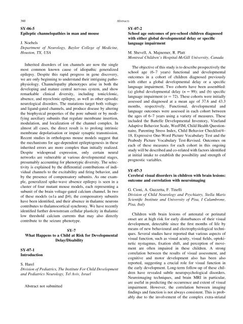
360<br />
Abstracts<br />
SY-06-5<br />
Epileptic channelopathies in man and mouse<br />
J. Noebels<br />
Department of Neurology, Baylor College of Medicine,<br />
Houston, TX, USA<br />
Inherited disorders of ion channels are now the single<br />
most common known cause of idiopathic generalized<br />
epilepsy. Despite this rapid progress in gene discovery,<br />
we are only beginning to understand their intriguing pathophysiology.<br />
Channelopathy phenotypes arise in both the<br />
developing and mature central nervous system, and show<br />
remarkable clinical diversity, including tonic/clonic,<br />
absence, and myoclonic epilepsy, as well as other episodic<br />
neurological disorders. The mutations target both voltageand<br />
ligand-gated channels, and produce disease by altering<br />
the biophysical properties of the pore subunit or by modifying<br />
auxiliary subunits that regulate membrane insertion,<br />
modulation, and localization of the channel complex. In<br />
almost all cases, the direct result is to prolong intrinsic<br />
membrane depolarisation or impair synaptic transmission.<br />
Recent studies in orthologous mouse models suggest that<br />
the mechanisms for age-dependent epileptogenesis in these<br />
inherited errors are more complex than initially realized.<br />
Despite widespread expression, only certain neural<br />
networks are vulnerable at various developmental stages,<br />
presumably accounting for phenotypic diversity. The selectivity<br />
is explained by the differential contributions of individual<br />
channels to the excitability and firing behavior, and<br />
by the presence of compensatory subunits. As one example,<br />
generalized spike-wave absence epilepsy is seen in a<br />
cluster of four mutant mouse models, each representing a<br />
subunit of the brain voltage-gated calcium channel. In two<br />
of these models (a1a and b4), the compensatory subunits<br />
have been identified, and their absence in thalamic neurons<br />
contributes to thalamocortical synchrony. We have recently<br />
identified further downstream cellular plasticity in thalamic<br />
low threshold calcium currents that may also directly<br />
contribute to the seizure phenotype.<br />
SY-7<br />
What Happens to a Child at Risk for Developmental<br />
Delay/Disability<br />
SY-07-1<br />
Introduction<br />
S. Harel<br />
Division of Pediatrics, The Institute For Child Development<br />
and Pediatrics Neurology, Tel Aviv, Israel<br />
Abstract not submitted<br />
SY-07-2<br />
School age outcomes of pre-school children diagnosed<br />
with either global developmental delay or specific<br />
language impairment<br />
M. Shevell, A. Majnemer, R. Platt<br />
Montreal Children’s Hospital-McGill <strong>University</strong>, Canada<br />
The objective of this study is to describe prospectively the<br />
school age (6–7 years) functional and developmental<br />
outcomes in a cohort of children diagnosed previously<br />
with either a global developmental delay or a specific<br />
language impairment. Two cohorts have been assembled:<br />
(a) global developmental delay (n ¼ 99); and (b) specific<br />
language impairment (n ¼ 72). These cohorts were initially<br />
assessed and diagnosed at a mean age of 37.6 and 43.3<br />
months, respectively. Functional, developmental and<br />
language outcomes were assessed in each cohort between<br />
the ages of 6–7 years using a variety of measures. These<br />
included the Battelle Developmental Inventory, Vineland<br />
Adaptive Behavior Scale, WeeFIM, Child Health Questionnaire,<br />
Parenting Stress Index, Child Behavior Checklist/4–<br />
18, Expressive One-Word Picture Vocabulary Test and the<br />
Peabody Picture Vocabulary Test-Revised. Outcomes on<br />
each of these measures for each cohort in this ongoing<br />
study will be described and co-related with factors identified<br />
at initial intake to establish the possibility and strength of<br />
prognostic variables.<br />
SY-07-3<br />
Cerebral visual disorders in children with brain lesions:<br />
outcome and correlation with neuroimaging<br />
G. Cioni, A. Guzzetta, F. Tinelli<br />
Division of Child Neurology and Psychiatry, Stella Maris<br />
Scientific Institute and <strong>University</strong> of Pisa, I Calambrone,<br />
Pisa, Italy<br />
Children with brain lesions of antenatal or perinatal<br />
onset are at high risk for early disturbances of their visual<br />
development, detectable since the first months of life by<br />
means of new behavioural and electrophysiological techniques.<br />
Several studies have reported that various aspects of<br />
visual function, such as visual acuity, visual fields, optokinetic<br />
nystagmus, fixation shift, and perception of movement<br />
are often impaired in these children. A strong<br />
correlation between the results of visual assessment, and<br />
cognitive and motor development also has been also<br />
reported, suggesting a crucial role for visual function in<br />
the early development. Long-term follow-up of these children<br />
have revealed subtle neuropsychological disorders.<br />
Neuroimaging techniques, and brain MRI in particular,<br />
are useful in predicting the occurrence and extent of visual<br />
impairment. However, the correlation between imaging<br />
findings and function is not always consistent. This is probably<br />
due to the involvement of the complex extra-striatal


