PDF File - Mahidol University
PDF File - Mahidol University
PDF File - Mahidol University

Create successful ePaper yourself
Turn your PDF publications into a flip-book with our unique Google optimized e-Paper software.
350<br />
Abstracts<br />
patterns of behavior, interests, and activities. MRI studies in<br />
autism have found a variety of alterations including a<br />
decrease of size of various brain structures. In functional<br />
neuroimaging studies such as functional MRI (fMRI),<br />
magnetic resonance spectroscopy (MRS), positron emission<br />
spectroscopy (PET) and single photon emission computed<br />
tomography (SPECT), various abnormalities have been<br />
reported. SPECT has found a decrease of regional cerebral<br />
blood flow (rCBF) in the frontal and temporal regions and the<br />
left cerebral hemisphere, and has shown the relationship<br />
between rCBF and symptoms. PET has found metabolic<br />
abnormalities in frontal monoaminergic neuron systems,<br />
and a decrease of glucose metabolic activity in anterior<br />
cingulate gyrus. MRS has found energy metabolism abnormality<br />
in the frontal lobe using 31P-MRS, and abnormality in<br />
medial temporal and lateral frontal regions using 1 H-MRS.<br />
fMRI has found a lower activity in the amygdala during face<br />
recognition test and in the right fusiform gyrus during face<br />
discrimination test. These structural and functional imaging<br />
findings suggest that the anatomic abnormalities are multiple<br />
and not located, and are more likely to be the results of<br />
abnormal development of connections of undefined<br />
disturbed neural networks for complex information processing<br />
involving the frontal cerebral cortex, limbic system, and<br />
posterior fosse brain structures. In this presentation,<br />
previously reported findings will be discussed in relations<br />
with possible mechanism of symptom occurrence in autism.<br />
TU-5B<br />
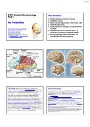
ADHD: current concepts and neurobiology<br />
M. Denckla<br />
The Kennedy Krieger Institute, Baltimore, MD, USA<br />
Aided by advances in cognitive neuroscience and neuroimaging,<br />
the emerging current concept of ADHD is that it<br />
represents a group of deficits in self-control; affected in<br />
some combination are motor control, cognitive control, and<br />
emotional control. So frequent is the comorbidity with<br />
ADHD of Developmental Motor Coordination Disorder<br />
that this combination mirrors Gillberg’s DAMP syndrome.<br />
Neuroimaging implicates parallel underlying circuits<br />
comprised of frontal, striatal, and cerebellar regions, with<br />
consensus based on MRI measurements that somewhat smaller<br />
total brain volumes (3–5% reductions) are of multifocal<br />
(not diffuse) origin, mostly small frontal and striatal hypoplasia.<br />
This topic update on ADHD will also review diagnosis,<br />
behavioral genetics, prognosis (especially genderdiscrepant),<br />
neurotransmitters (with implications for genetics<br />
and pharmacotherapeutics) and non-pharmacological<br />
therapies. The emphasis, however, will be on the following:<br />
implications of motor localization clues; neurocognitive<br />
elucidation that attention allocation (not attention per se) is<br />
deficient; and evidence from in vivo MR neuroimaging, both<br />
anatomic MRI and fMRI, that frontal-striatal-cerebellar<br />
circuits deserve scrutiny in relation to ADHD.<br />
SYMPOSIUM<br />
SY-1<br />
Pathogenesis and Prevention of Prenatal and Perinatal<br />
Brain Damage<br />
SY-01-1<br />
Structural and functional studies on cadherins, synaptic<br />
adhesion molecules<br />
W. Shan, D.R. Colman<br />
Fishberg Research Center for Neurobiology, The Mount<br />
Sinai School of Medicine, New York, New York, USA<br />
The ability of the mammalian CNS to perform complex<br />
informative processes and highly cognitive functions is<br />
based on the precise and specifically determined connectivity<br />
of different neuronal cell types at synapses. Recently studies<br />
implicate that cadherins, calcium-dependent cell adhesion<br />
molecules, are involved in synaptic formation, specifying<br />
connections among different synapses and functional modulation<br />
during early development. The molecular mechanisms<br />
by which the classic cadherin mediate adhesion are beginning<br />
to be elucidated. We have identified certain key residues<br />
at the interface of N-cadherin molecules that participate in<br />
cell-cell adhesion formation. In addition, two different, but<br />
conserved cadherins are highly likely to form hetero-interaction<br />
at adherent junction sites, which may increase the<br />
diversity of synaptic connections. Furthermore, synaptic<br />
modulation could be activated in either physiological or<br />
pathological conditions, indicating a role for cadherins in<br />
synaptic plasticity. Taken together, our data provide insights<br />
into the mechanisms of formation of adherens junctions,<br />
synaptic targeting, and modulation of synaptic activity.<br />
SY-01-2<br />
Basic mechanisms and prevention of prenatal brain<br />
damage<br />
P. Evrard<br />
Service de Neurologie Pediatrique et des Maladies Metaboliques,<br />
Faculte de Medicine Xavier-Bichat, Hospital<br />
Robert-Debre, Paris, France<br />
Abstract not submitted<br />
SY-01-3<br />
Neurophysilogical analysis of periventricular<br />
leukomalacia in preterm infants<br />
A. Okumura, K. Watanabe, F. Hayakawa, T. Kato<br />
Department of Pediatrics, Nagoya <strong>University</strong> Graduate<br />
School of Medicine, Nagoya, Japan<br />
PVL is an important cause of cerebral palsy in preterm<br />
infants. Serial EEG recordings are not only of great prognostic<br />
value but also useful to determine the timing of brain