Silicon carbide radiation detector for harsh environments - Nuclear ...
Silicon carbide radiation detector for harsh environments - Nuclear ...
Silicon carbide radiation detector for harsh environments - Nuclear ...

You also want an ePaper? Increase the reach of your titles
YUMPU automatically turns print PDFs into web optimized ePapers that Google loves.
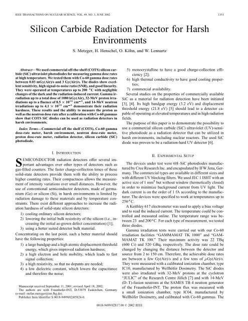
IEEE TRANSACTIONS ON NUCLEAR SCIENCE, VOL. 49, NO. 3, JUNE 2002 1351<br />
<strong>Silicon</strong> Carbide Radiation Detector <strong>for</strong> Harsh<br />
Environments<br />
S. Metzger, H. Henschel, O. Köhn, and W. Lennartz<br />
Abstract—We used commercial off-the-shelf (COTS) silicon <strong>carbide</strong><br />
(SiC) ultraviolet photodiodes <strong>for</strong> measuring gamma dose rates<br />
at high temperature. We tested them with Co-60 gamma dose rates<br />
between 0.03 mGy(Air)/s and 3 Gy(Air)/s. The diodes show excellent<br />
sensitivity, high signal-to-noise ratio (SNR), and good linearity.<br />
They were operated at temperatures up to 200 C with negligible<br />
changes of the dark and the <strong>radiation</strong>-induced current. Gamma ir<strong>radiation</strong><br />
up to a total dose of 1080 kGy(Air), 32-MeV proton ir<strong>radiation</strong>s<br />
up to a fluence of 8.5 10 12 cm 2 , and 14-MeV neutron<br />
ir<strong>radiation</strong>s up to 4.1 10 12 cm 2 demonstrate their <strong>radiation</strong><br />
hardness. These results and the ability to measure the proton as<br />
well as the neutron dose rate after a calibration with Co-60 gammas<br />
show that COTS SiC diodes can be used as <strong>radiation</strong> <strong>detector</strong>s in<br />
<strong>harsh</strong> <strong>environments</strong>.<br />
Index Terms—Commercial off the shelf (COTS), Co-60 gamma<br />
dose-rate meter, <strong>harsh</strong> environment, neutron dose-rate meter,<br />
proton dose-rate meter, <strong>radiation</strong> <strong>detector</strong>, silicon <strong>carbide</strong> (SiC)<br />
photodiode.<br />
5) monocrystalline to have a good charge-collection efficiency<br />
[2];<br />
6) high thermal conductivity to have good cooling properties;<br />
7) commercial availability.<br />
Several studies on the properties of commercially available<br />
SiC as a material <strong>for</strong> <strong>radiation</strong> detection have been initiated<br />
[3], [4]. Its high bandgap energy (3.2 eV) and displacement<br />
threshold energy (21.8 eV) [5] should lead to a <strong>detector</strong> capable<br />
of operating at elevated temperatures and in high <strong>radiation</strong><br />
fields.<br />
The purpose of this paper is to demonstrate the possibility to<br />
use a commercial silicon <strong>carbide</strong> (SiC) ultraviolet (UV)-sensitive<br />
photodiode as a <strong>radiation</strong> <strong>detector</strong> that can be utilized in<br />
<strong>harsh</strong> <strong>environments</strong>, including nuclear reactors. The used SiC<br />
diode was proven to be a <strong>radiation</strong>-hard UV <strong>detector</strong> [6].<br />
I. INTRODUCTION<br />
SEMICONDUCTOR <strong>radiation</strong> <strong>detector</strong>s offer several important<br />
advantages over other types of <strong>detector</strong>s such as<br />
gas-filled counters. The faster charge-collection times of these<br />
solid-state <strong>detector</strong>s provide them with the ability to process<br />
higher counting rates. Their compactness allows the measurement<br />
of intensity variations over small distances. However, the<br />
use of conventional semiconductor <strong>detector</strong>s, made of germanium<br />
(Ge) or silicon (Si), in <strong>harsh</strong> <strong>environments</strong> is limited by<br />
<strong>radiation</strong> damage to these materials and by temperature constraints.<br />
There exist different approaches to increase the <strong>radiation</strong><br />
hardness of solid-state silicon <strong>detector</strong>s:<br />
1) cooling ordinary silicon <strong>detector</strong>s;<br />
2) lowering the initial bulk resistivity of the silicon (i.e., increasing<br />
the initial as-grown defect concentration) [1];<br />
3) using a better suited <strong>detector</strong> bulk material.<br />
Concentrating on the last point, such a better material should<br />
have the following properties:<br />
1) a large bandgap and a high atomic displacement threshold<br />
energy, which gives improved <strong>radiation</strong> hardness;<br />
2) a high electron and hole mobility, which leads to fast<br />
signal collection;<br />
3) a high resistivity, so that no dopants are needed;<br />
4) a low dielectric constant, which lowers the capacitance<br />
and there<strong>for</strong>e the noise;<br />
Manuscript received September 11, 2001; revised April 18, 2002.<br />
The authors are with Fraunhofer-INT, D-53879 Euskirchen, Germany<br />
(e-mail: stefan.metzger@int.fhg.de).<br />
Publisher Item Identifier S 0018-9499(02)05824-0.<br />
II. EXPERIMENTAL SETUP<br />
The devices under test were 6H–SiC photodiodes manufactured<br />
by Cree Research Inc. and encapsulated by IFW Jena, Germany.<br />
The commercial types are available in different sizes and<br />
with different UV blocking filters. We used JEC 1 ISHT with an<br />
active size of 1 mm but without window (hermetically closed)<br />
in order to minimize background current from UV light. The<br />
dark current is on the order of 1 fA according to the manufacturer.<br />
The devices were specified to work at temperatures up to<br />
250 C.<br />
A Keithley 617 electrometer was used to apply a bias voltage<br />
and to read the induced current. The temperature could be controlled<br />
and measured online. The temperature range was between<br />
23 and 200 C. For each type of measurement, we tested<br />
three diodes.<br />
Gamma ir<strong>radiation</strong> tests were carried out with our Co-60<br />
ir<strong>radiation</strong> facilities “GAMMAMAT TK 1000” and “GAM-<br />
MAMAT TK 100.” Their maximum activity was 22 TBq<br />
(600 Ci) and 520 GBq, respectively. The dose rate could be<br />
changed by changing the distance between the <strong>detector</strong> and<br />
source from 2 to 150 cm. There<strong>for</strong>e, the achievable dose rates<br />
are between a few Gy(Air)/s and a few tens of Gy(Air)/s.<br />
They were measured with a calibrated ionization chamber, type<br />
IC10, manufactured by Wellhöfer Dosimetry. The SiC diodes<br />
were also irradiated with 32-MeV protons at the cyclotron<br />
“JULIC” of the Research Centre Jülich [7] and with 14-MeV<br />
(D–T)-fusion neutrons at the SAMES TB 4 neutron generator<br />
of the Fraunhofer-INT. The proton flux was measured with<br />
a small ionization chamber, type IC04, manufactured by<br />
Wellhöfer Dosimetry, and calibrated with Co-60 gammas. The<br />
0018-9499/02$17.00 © 2002 IEEE
1352 IEEE TRANSACTIONS ON NUCLEAR SCIENCE, VOL. 49, NO. 3, JUNE 2002<br />
Fig. 1. Dark current–voltage characteristic of a JEC 1 ISHT SiC diode at room<br />
temperature.<br />
neutron flux during the ir<strong>radiation</strong> was monitored with two<br />
calibrated uranium-238 fission chambers, type FC4A, from<br />
Centronic Inc.<br />
Fig. 2. Increase of the gamma-induced current in a SiC UV-sensitive<br />
photodiode with increasing bias voltage at room temperature. The gamma dose<br />
rate was 30 mGy/s.<br />
III. RESULTS<br />
A. The Effects of Bias Voltage<br />
Initial characterization of the SiC diodes was per<strong>for</strong>med be<strong>for</strong>e<br />
<strong>radiation</strong> testing of the <strong>detector</strong>s. Fig. 1 shows the dark current–voltage<br />
characteristic. The dark current is only in the range<br />
of 10–50 fA <strong>for</strong> bias voltages below 20 V. As a consequence, the<br />
signal-to-noise ratio (SNR) during ir<strong>radiation</strong> will be high, even<br />
with low signal amplitudes.<br />
The active layer thickness of a diode is proportional to the<br />
square root of the applied (reverse) bias voltage [8]. As an external<br />
voltage is applied to the diode, the active region grows<br />
until the maximum active layer thickness is reached. At this<br />
point, the diode is said to be fully depleted and the applied bias<br />
voltage is referred to as the depletion voltage. Further increase<br />
in the applied voltage cannot result in any more growth of the<br />
active layer thickness.<br />
Fig. 2 shows the effect of the applied (reverse) bias voltage on<br />
the signal current at a gamma dose rate of about 30 mGy(Air)/s.<br />
The gamma-induced current increases with increasing reverse<br />
bias voltage. This can be attributed to the widening of the<br />
diode’s active layer because <strong>for</strong> the same dose rate, more<br />
ionization is produced in a bigger volume.<br />
As an outcome of the initial characterization, the bias voltage<br />
was fixed at 20 V <strong>for</strong> all further <strong>radiation</strong> testing because at this<br />
point, the SNR is nearly optimal. At lower voltages, the SNR<br />
should be higher because of the smaller dark current, but these<br />
values (below 20 fA) are stable only after 5–10 min of settling<br />
time after any change of any experimental parameter (voltage<br />
change, beam on/off, etc.).<br />
B. Response to Radiation<br />
Fig. 3 shows the gamma-induced current versus time at room<br />
temperature of the SiC diode. The device under test (DUT) is<br />
exposed with a dose rate of 30 mGy(Air)/s <strong>for</strong> six successive<br />
<strong>radiation</strong> periods. A short time is required be<strong>for</strong>e the current<br />
Fig. 3. Response of a SiC diode to six successive gamma ir<strong>radiation</strong>s of 15 s<br />
duration at room temperature. The dose rate was 30 mGy(Air)/s. The applied<br />
bias voltage was 20 V.<br />
reaches a stable value. This is partly because it takes about 0.3<br />
s to move the gamma source into or out of the shielding container.<br />
It was found that the settling time (time to reach 95% of<br />
the steady-state value) is less than 4 s. This is suitable <strong>for</strong> applications<br />
with slowly changing dose rates.<br />
Fig. 4 shows a plot of gamma-induced current of a SiC photodiode<br />
versus gamma dose rate. The gamma-induced current<br />
is linear in the log-log plot over nearly six orders of magnitude.<br />
Corresponding values <strong>for</strong> proton- (deduced from Fig. 11) and<br />
neutron-induced current (from Fig. 12) are also plotted in this<br />
figure to demonstrate that SiC diodes can detect and measure<br />
different types of <strong>radiation</strong> after a calibration at a Co-60 gamma<br />
source.<br />
From these measurements, we could deduce a lower limit of<br />
the <strong>detector</strong>’s sensitivity (collected charge per absorbed dose),<br />
which was 1.2 nC/Gy(Air) at 3 Gy(Air)/s, whereas the lower<br />
limit of the SNR is 25 at 28 Gy(Air)/s. This is shown in Fig. 5,<br />
where the temporal response of the diode <strong>for</strong> the above-mentioned<br />
dose rates at room temperature is shown.
METZGER et al.: SILICON CARBIDE RADIATION DETECTOR 1353<br />
Fig. 4. Gamma-induced current of a SiC photodiode as a function of gamma<br />
dose rate at room temperature, with an applied bias of 20 V.<br />
Fig. 6. Effect of temperature on the dark current of a SiC diode be<strong>for</strong>e<br />
ir<strong>radiation</strong> and after a total dose of 20 kGy(Air). The applied bias voltage was<br />
20 V.<br />
Fig. 5. Temporal response of a SiC diode at two different dose rates, showing<br />
the high SNR. The applied bias voltage was 20 V.<br />
C. Temperature Effects<br />
Additional experiments were per<strong>for</strong>med to study the <strong>detector</strong><br />
response with changing temperature. First, the change of the<br />
dark current with temperature was studied be<strong>for</strong>e ir<strong>radiation</strong><br />
and after a total dose of 20 kGy(Air). There<strong>for</strong>e, the diode was<br />
heated to 250 C and the dark current was monitored while it<br />
was cooling down to room temperature. This measurement was<br />
made with a bias voltage of 20 V. The result <strong>for</strong> the temperature<br />
range between 30 and 150 C is shown in Fig. 6. There is only<br />
a small change <strong>for</strong> temperatures below 100 C, but at higher<br />
temperatures, the dark current increases exponentially. So if<br />
one wants to measure dose rates at temperatures above 100 C,<br />
the use of this <strong>detector</strong> is limited to applications where either<br />
small temperature variations or high dose rates, e.g., 0.01<br />
Gy/s, occur. Over the measured temperature range, the dark<br />
current rose about 50 fA after a dose of 20 kGy(Air).<br />
Secondly, the linearity of the <strong>detector</strong> response was measured<br />
be<strong>for</strong>e ir<strong>radiation</strong> and after a total dose of 20 kGy(Air). These<br />
tests were done at five constant temperatures between 23 and<br />
200 C. Results are shown in Fig. 7. No significant changes were<br />
observed.<br />
Fig. 7. Effect of temperature on the linearity of the <strong>detector</strong> response after a<br />
total gamma dose of 20 kGy(Air). The applied bias voltage was 20 V.<br />
These results demonstrate that the response of a SiC diode<br />
to gamma <strong>radiation</strong> is nearly unperturbed by temperature in the<br />
studied range. This could be expected due to the wide bandgap<br />
energy of SiC.<br />
D. Radiation Hardness<br />
The <strong>radiation</strong> hardness of SiC diodes was studied with<br />
two types of <strong>radiation</strong>. The tests were per<strong>for</strong>med at room<br />
temperature and with an applied bias voltage of 20 V. At first,<br />
they were irradiated with Co-60 gammas up to a total dose of<br />
870 kGy(Air). During that test, the gamma-induced current<br />
only decreased by 10% and seemed to reach a plateau after a<br />
total dose of 500 kGy(Air). This is shown in Fig. 8.<br />
Secondly, the gamma-induced increase of leakage current<br />
was studied. In Fig. 9, two ir<strong>radiation</strong>s are shown: one from<br />
zero to a total dose of 10 kGy(Air) and one from zero to<br />
870 kGy(Air). The dark current increased from 0.013 to 0.2 pA<br />
during the first ir<strong>radiation</strong> and from 0.007 to 1.4 pA during the<br />
second one. Furthermore, it can be seen that the <strong>radiation</strong>-induced<br />
increase of the leakage current after 10 kGy(Air) rapidly
1354 IEEE TRANSACTIONS ON NUCLEAR SCIENCE, VOL. 49, NO. 3, JUNE 2002<br />
Fig. 8. Decrease of the gamma-induced current of a SiC diode during gamma<br />
ir<strong>radiation</strong> with an applied bias voltage of 20 V.<br />
Fig. 10. Linearity of the <strong>detector</strong> response be<strong>for</strong>e and after several total dose<br />
values. The applied bias was 20 V, and the temperature was 23 C.<br />
Fig. 9. Increase of dark current during two gamma ir<strong>radiation</strong>s up to 10 and<br />
870 kGy(Air), respectively. The applied bias was 20 V.<br />
(within 2000 s) anneals to preir<strong>radiation</strong> levels, whereas after<br />
the second (longer) ir<strong>radiation</strong>, no annealing was observed.<br />
In a third step, the total dose effect on the linearity of the <strong>detector</strong><br />
response was studied. The results are shown in Fig. 10. As<br />
can be seen, there is no major effect of the total dose on the linearity<br />
of the <strong>detector</strong> response after 1080 kGy(Air). This could<br />
be expected after the results from the previous <strong>radiation</strong> hardness<br />
tests [slight decrease of the signal amplitude (see Fig. 8)<br />
and slight increase of the leakage current (see Fig. 9)].<br />
In [6], JEC 0.1 SiC diodes (same type but only 0.1 mm active<br />
area) were irradiated with 32-MeV protons. This is shown<br />
in Fig. 11. During the time intervals I 1, 2, and 3 (shaded in<br />
Fig. 11), their UV sensitivity was measured in order to test their<br />
<strong>radiation</strong> hardness as a UV <strong>detector</strong> used in proton <strong>environments</strong>.<br />
During that ir<strong>radiation</strong>, the proton-induced current was<br />
monitored. It nicely follows the proton flux up to a fluence of<br />
8.5 10 cm (except <strong>for</strong> the time intervals I 1, 2, and 3 when<br />
the UV light was on).<br />
The SiC diodes were also irradiated with 14-MeV neutrons.<br />
The neutron-induced current in the SiC diode was measured online.<br />
The behavior of its signal amplitude should be an indication<br />
Fig. 11. The 32-MeV proton flux and proton-induced current in a SiC diode<br />
JEC0.1 at room temperature.<br />
<strong>for</strong> the effect of neutron <strong>radiation</strong> on the <strong>radiation</strong> sensitivity of<br />
a SiC diode. The results are shown in Fig. 12 up to a total neutron<br />
fluence of 4.1 10 cm . The ir<strong>radiation</strong> was interrupted<br />
every 30 min <strong>for</strong> 5 min to study the effects of neutrons on the<br />
leakage current.<br />
Up to a total neutron fluence of about 2.2 10 cm (after<br />
12 500 s), the signal amplitude follows the temporal behavior of<br />
the neutron flux identically. After that fluence, the signal began<br />
to decrease by about 20% until the end of the ir<strong>radiation</strong>, while<br />
the neutron flux was nearly constant. In Fig. 12, there is no evidence<br />
that 14-MeV neutron <strong>radiation</strong> has an effect on the dark<br />
current, at least up to a total fluence of 4.1 10 cm .<br />
So SiC diodes of this type can be used as <strong>radiation</strong> <strong>detector</strong>s<br />
up to a total gamma dose of 1000 kGy(Air), to a 32-MeV proton<br />
fluence of nearly 10 cm , or to a 14-MeV neutron fluence of<br />
about 4.1 10 cm without major degradation. This could<br />
be expected because of the high displacement energy in SiC.<br />
E. Radiation Dosimetry<br />
The good agreement between particle flux (proton as well as<br />
neutron) and particle-induced current brought up the following
METZGER et al.: SILICON CARBIDE RADIATION DETECTOR 1355<br />
Fig. 12. The 14-MeV neutron flux and neutron-induced current in a SiC diode<br />
at room temperature. The applied bias was 20 V.<br />
question: Is it possible to use SiC diodes also as particle <strong>radiation</strong><br />
dosimeter based on a calibration with Co-60 gammas?<br />
A proton-induced current of 32 nA (ten times the current<br />
from Fig. 11) at a flux of 9 10 cm s is also induced<br />
by a Co-60 gamma dose rate of 25.5 Gy(Air). From these two<br />
values, a proton energy loss in SiC of 17.7 MeV cm g can<br />
be derived. This is in good agreement with published data of<br />
15.8 MeV cm g [9].<br />
A neutron-induced current of about 20 pA at 2.5 10<br />
cm s (see Fig. 12) corresponds to a Co-60 gamma dose<br />
rate of about 5 mGy(Air)/s. The division of both values results<br />
in a fluence dose conversion factor of 2 10 Gy cm ,<br />
which is in good agreement with a derived kerma factor of 1.88<br />
10 Gy cm <strong>for</strong> SiC (published kerma factors are 1.30<br />
10 Gy cm <strong>for</strong> Si and 2.46 10 Gy cm <strong>for</strong> C [10]).<br />
This strongly supports the opinion of using SiC diodes as <strong>detector</strong>s<br />
<strong>for</strong> any kind of <strong>radiation</strong>. After calibration at a Co-60<br />
gamma source, it is possible to measure the ionizing dose applied<br />
by protons and neutrons.<br />
IV. CONCLUSION<br />
The possibility of using a commercial SiC UV photodiode<br />
as a <strong>radiation</strong> <strong>detector</strong> has been investigated. We found the following.<br />
1) The small dark current in combination with a large <strong>radiation</strong>-induced<br />
current (sensitivity) gives a high SNR.<br />
2) The <strong>detector</strong> response is linear over five orders of magnitude<br />
of gamma dose rate.<br />
3) No evidence of temperature effects on the per<strong>for</strong>mance<br />
of the SiC <strong>detector</strong> was found <strong>for</strong> temperatures below<br />
200 C.<br />
4) The <strong>detector</strong> per<strong>for</strong>mance is nearly unchanged up to a<br />
total gamma dose of 1000 kGy(Air), a 32-MeV proton<br />
fluence of 8.5 10 cm , or a 14-MeV neutron fluence<br />
of 4.1 10 cm .<br />
5) After a calibration with Co-60 gammas, it can be used as<br />
a proton or a neutron <strong>radiation</strong> dosimeter.<br />
These results demonstrate the potential of SiC COTS photodiodes<br />
as <strong>radiation</strong> (ionizing and particle) <strong>detector</strong>s <strong>for</strong> use in<br />
applications where intense <strong>radiation</strong> fields and high temperatures<br />
are expected.<br />
Last but not least, in [6], it was shown that their initial function<br />
as a UV <strong>detector</strong> can also withstand <strong>harsh</strong> <strong>environments</strong>.<br />
REFERENCES<br />
[1] Z. Li, V. Eremin, I. Ilyashenko, A. Ivanov, and E. Verbitskaya, “Investigation<br />
of epitaxial silicon layers as a material <strong>for</strong> <strong>radiation</strong> hardened<br />
silicon <strong>detector</strong>s,” IEEE Trans. Nucl. Sci., vol. 45, no. 3, pp. 585–590,<br />
1998.<br />
[2] M. Schlögl and B. E. Fischer, “Investigation of the detection efficiency<br />
of polycrystalline diamond <strong>detector</strong>s with a heavy ion microprobe,” in<br />
Proc. RADECS 1999, Saumur, France, 1999, pp. 132–135.<br />
[3] F. H. Rudy, A. R. Dulloo, S. Seshadri, C. D. Brandt, and J. G. Seidel,<br />
“Development of a silicon <strong>carbide</strong> semiconductor neutron <strong>detector</strong> <strong>for</strong><br />
monitoring thermal neutron fluxes,” Westinghouse Science and Technology,<br />
96-9TK1-NUSIC-R1, 1996.<br />
[4] A. R. Dulloo, F. H. Rudy, J. G. Seidel, S. Seshadri, and L. B. Rowland,<br />
“Development of a silicon <strong>carbide</strong> <strong>radiation</strong> <strong>detector</strong>,” IEEE Trans. Nucl.<br />
Sci., vol. 45, pp. 536–541, June 1998.<br />
[5] A. L. Barry, B. Lehmann, D. Fritsch, and D. Bräunig, “Energy dependence<br />
of electron damage and displacement threshold energy in 6H silicon<br />
<strong>carbide</strong>,” IEEE Trans. Nucl. Sci., vol. 38, pp. 1111–1115, Dec.<br />
1991.<br />
[6] S. Metzger, H. Henschel, O. Köhn, and W. Lennartz, “Radiation effects<br />
in ultraviolet sensitive SiC photodiodes,” in Proc. RADECS 1999,<br />
Saumur, France, 1999, pp. 457–460.<br />
[7] S. Metzger, H. G. Böge, W. Bräutigam, R. Brings, N. Gad, H. Henschel,<br />
O. Köhn, W. Lennartz, and H. J. Probst, “Low energy proton testing of<br />
space electronics at ‘JULIC’,” in Proc. RADECS 1999, Saumur, France,<br />
1999, pp. 163–166.<br />
[8] S. M. Sze, Physics of Semiconductor Devices, 2nd ed. New York:<br />
Wiley, 1969.<br />
[9] J. F. Janni, “Proton range-energy tables, 1 keV–10 GeV. Part 2, Elements,”<br />
Atomic Data <strong>Nuclear</strong> Data Tables, vol. 27, no. 4–5, 1982.<br />
[10] R. S. Caswell, J. J. Coyne, and M. L. Randolph, “Kerma factors <strong>for</strong> tissue<br />
compositions, compounds and mixtures,” in Basic Physical Data <strong>for</strong><br />
Neutron Dosimetry. Luxembourg: CEC, 1976, vol. EUR 5629e, pp.<br />
69–76.