NTEGRA spectra: Cutting edge of molecular sciences - NT-MDT
NTEGRA spectra: Cutting edge of molecular sciences - NT-MDT
NTEGRA spectra: Cutting edge of molecular sciences - NT-MDT

You also want an ePaper? Increase the reach of your titles
YUMPU automatically turns print PDFs into web optimized ePapers that Google loves.
<strong><strong>NT</strong>EGRA</strong> <strong>spectra</strong>:<br />
<strong>Cutting</strong> <strong>edge</strong> <strong>of</strong> <strong>molecular</strong> <strong>sciences</strong>
<strong><strong>NT</strong>EGRA</strong> Spectra for non-transparent samples<br />
High Aperture AFM<br />
The only commercial AFM on the market<br />
integrated with 100x high NA objective in<br />
upright geometry<br />
With technical questions contact:<br />
Pavel Dorozhkin, dorozhkin@ntmdt.ru<br />
Vasiliy Gavrilyuk, gavrilyuk@ntmdt.ru
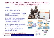
Fully functional “AFM+3D confocal Raman microscope” system<br />
for non-transparent samples<br />
Optomechanical unit<br />
(excitation, confocal and spectrometer modules)<br />
High NA objective<br />
High Aperture AFM
AFM with 100x 0.7 NA objective in upright configuration –<br />
for non-transparent samples<br />
CCD video camera<br />
Probe<br />
deflectometer<br />
Optical AFM head<br />
(100x,0.7 NA<br />
objective inside)<br />
Imaging optics<br />
Beam splitter 2<br />
Beam splitter 1<br />
AFM probe<br />
Excitation light<br />
Scattered light<br />
Laser<br />
deflectometer<br />
objective<br />
Laser<br />
input&scanning<br />
module<br />
XYZ scanner<br />
Sample<br />
The only commercial AFM in the world integrated with 100x high<br />
NA objective in upright configuration !!!
Laser input & scanning module<br />
To video camera<br />
6<br />
Dichroic<br />
mirror<br />
2<br />
5<br />
Angle scanning<br />
7 mirror<br />
3<br />
1<br />
100x, 0.7 NA<br />
objective<br />
AFM probe<br />
8<br />
From / to<br />
confocal module<br />
Light input-output & scanning module:<br />
2D confocal microscope regime by scanning mirror 7<br />
Fine focus adjustment <strong>of</strong> the excitation beam by moving lens 3<br />
Adjustable height and orientation <strong>of</strong> the entrance aperture (mirror 8)<br />
Dichroic mirror 6 allows the sample & probe observation with highest optical resolution
Laser input & scanning module<br />
Laser input & scanning module allows:<br />
- Scan excitation beam along field <strong>of</strong> view <strong>of</strong> 100x objective (up to<br />
100 µm) and get 2D confocal image<br />
- Precisely position laser beam at any sample point (for example at<br />
the apex <strong>of</strong> AFM tip) and fix it there - thanks to closed-loop<br />
operation <strong>of</strong> the scanning mirror #7<br />
This is a crucial option for TERS experiment
Integration with standard (Solar-TII) spectrometer<br />
Optomechanical unit (excitation &<br />
confocal modules, spectrometer)<br />
Laser input/scanning module<br />
High Aperture AFM<br />
Fully functional “AFM + optical microscope + 3D confocal Raman<br />
microscope” system for non-transparent samples<br />
Scanning by both sample and laser beam
<strong><strong>NT</strong>EGRA</strong> can be integrated with other spectrometers<br />
Thanks to its open geometry, <strong>NT</strong>- <strong>MDT</strong> <strong><strong>NT</strong>EGRA</strong> system can be integrated with<br />
most spectrographs available on the market to provide fully functional “AFM +<br />
optical microscope + confocal Raman microscope” system
High Aperture Head – various realizations<br />
High Aperture head units<br />
Units<br />
STM<br />
AFM<br />
670<br />
AFM<br />
830<br />
1<br />
Objective unit<br />
**<br />
**<br />
**<br />
2<br />
STM probe unit<br />
**<br />
3<br />
AFM probe holder unit<br />
**<br />
**<br />
4<br />
Optical microscope with LED illuminator<br />
*<br />
*<br />
*<br />
5<br />
Light input-output system<br />
*<br />
*<br />
*<br />
6<br />
AFM control system, 670 nm laser<br />
**<br />
7<br />
AFM control system, 830 nm laser<br />
**<br />
8<br />
Objective Z-piezodrive<br />
*<br />
*<br />
*<br />
9<br />
Piezodriven steering mirror<br />
*<br />
*<br />
*
High Aperture Head: AFM setup<br />
Objective holder unit:<br />
Fine XYZ positioning<br />
Mitutoyo 100x M PlanApo<br />
objective<br />
– NA = 0.7, F = 2 mm<br />
– 6 mm working distance.<br />
– 0.4 μm optical resolution<br />
Infinity corrected optics<br />
Bright Field and Dark Field<br />
objectives available
High Aperture Head – STM setup<br />
Objective holder unit:<br />
Fine XYZ positioning<br />
Mitutoyo 100x M PlanApo<br />
objective<br />
– NA = 0.7, F = 2 mm<br />
– 6 mm working distance.<br />
– 0.4 μm optical resolution<br />
Infinity corrected optics<br />
Bright Field and Dark Field<br />
objectives available<br />
High aperture head: STM setup
Optical AFM (100x objective) with thermohead<br />
TV camera + objective<br />
N.B. The system is currently<br />
in development status<br />
light coupling<br />
and scanning unit<br />
AFM with 100x,<br />
0.7 NA objective<br />
thermohead<br />
Temperature range: -30-60°C . Heat up time
Simultaneous imaging and AFM scanning<br />
Atomic Force Microscopy:<br />
Observe the sample surface during<br />
the scanning process<br />
The high NA objective enables<br />
sample imaging even under the probe<br />
tip<br />
Available for both standard contact<br />
and semi contact AFM modes<br />
Probe tip<br />
AFM topography and optical image <strong>of</strong> a<br />
rectangular 5 μm crater on the sample<br />
surface
Simultaneous imaging and AFM scanning<br />
Cantilever<br />
1 µm height letters are<br />
readable – thanks to<br />
100x objective<br />
(see next slide for AFM)<br />
Black spot at the apex <strong>of</strong><br />
cantilever is the exact<br />
point there the tip<br />
touches substrate !!!<br />
AFM probe over a structured Si substrate. View through 0.7NA 100x objective<br />
Apex <strong>of</strong> opaque Si tip looks transparent on the image!<br />
This unique observation is due to high aperture (0.7 NA) <strong>of</strong> the imaging objective
Simultaneous imaging and AFM scanning<br />
1 µm height letters (see previous slide) can now be resolved<br />
with ultimate nanometer-scale resolution <strong>of</strong> AFM<br />
Topography 12.6x12.6 µm 2 Topography 12.6x12.6 µm 2<br />
AFM-image from the same sample area as on previous slide
Simultaneous imaging and AFM scanning<br />
µm –scale features are perfectly resolved –<br />
thanks to the 100x & 0.7 NA objective<br />
Cantilever<br />
1.5 µm width electrode<br />
AFM probe (Nanosensors, AdvancedTEC series) over a Si<br />
substrate with metal electrodes.<br />
View through 0.7NA 100x objective<br />
Apex <strong>of</strong> opaque Si tip looks transparent on the image!<br />
This unique observation is due to high aperture<br />
(0.7 NA) <strong>of</strong> the imaging objective.<br />
AFM image<br />
under the tip, 6x6 µm<br />
Black spot at the apex <strong>of</strong><br />
cantilever is the exact<br />
point there the tip<br />
touches substrate !!!
Ready for TERS experiments on non-transparent<br />
samples<br />
Laser scanning module allows to position laser beam precisely at the apex <strong>of</strong> AFM<br />
tip and fix it there - thanks to closed-loop operation <strong>of</strong> the scanning mirror<br />
Cantilever<br />
AFM probe on Si substrate. Laser spot (
AFM + two types <strong>of</strong> confocal microscopy<br />
AFM topography<br />
__ 1μm<br />
__ 1μm<br />
Confocal<br />
Raman, 520 cm -1<br />
Scanning by<br />
stage<br />
Si/SiO 2<br />
grating<br />
Confocal Raman,<br />
520 cm -1<br />
Scanning by mirror !
Sample rough positioning is performed with 10x<br />
(or any other magnification) objective<br />
It takes ~5 seconds to<br />
exchange objectives<br />
1x1 mm 2<br />
10x objective module<br />
Cantilever<br />
100x100 µm 2<br />
100x objective + AFM<br />
Objectives are placed with a few µm<br />
precision to ensure staying exactly<br />
on the same sample place<br />
Optical images <strong>of</strong> patterned Si substrate
High Aperture Head – main Values<br />
1. Fully functional 3D confocal microscope for non-transparent<br />
(and transparent) samples<br />
2. Two types confocal microscopy in one instrument:<br />
scanning by sample & scanning by beam<br />
3. AFM probe directly under 100x 0.7 NA objective !<br />
- AFM scanning simultaneously with sample imaging (at nearly<br />
highest possible resolution <strong>of</strong> 0.4 µm !) and confocal scanning<br />
- Best suited for TERS and other tip-enhanced experiments<br />
- Good AFM performance (Z-noise ~0.04 nm RMS, preliminary)<br />
The only commercial AFM on the market integrated with 100x<br />
high NA objective in upright geometry