Synthesis of biocompatible surfaces by nanotechnology methods
Synthesis of biocompatible surfaces by nanotechnology methods
Synthesis of biocompatible surfaces by nanotechnology methods

Create successful ePaper yourself
Turn your PDF publications into a flip-book with our unique Google optimized e-Paper software.
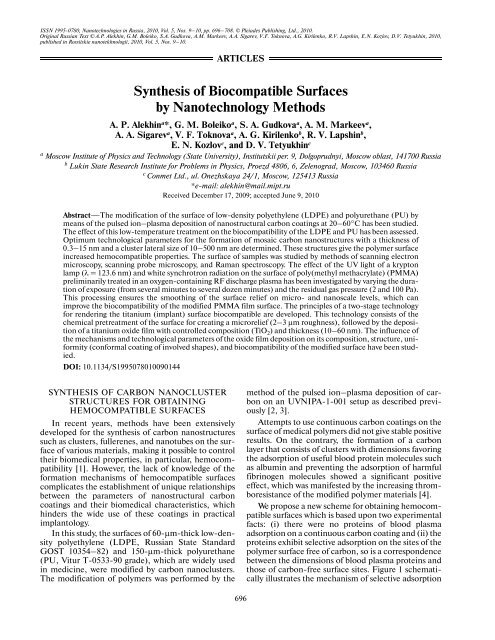
SYNTHESIS OF BIOCOMPATIBLE SURFACES BY NANOTECHNOLOGY METHODS 697Albumin(1)Fibrinogen(2)Carbon cluster(3)K ads K desK ads K desK ads K desK ads Kdes1<strong>of</strong> albumin molecules in competition with fibrinogenmolecules.As can be seen from Fig. 1a, both albumin andfibrinogen molecules adsorb on the initial polymersurface. Fibrinogen molecules, which are 4–5 timesgreater than albumin molecules (~50 nm against~9 nm, respectively), are, for thermodynamic reasons,almost irreversibly adsorbed so that they stick to thesubstrate with their maximum surface area. In contrast,part <strong>of</strong> the modified surface (Fig. 1b) is occupied<strong>by</strong> carbon clusters so that fibrinogen molecules have(a)(b)Fig. 1. Schematic diagram illustrating the adsorption–desorption<strong>of</strong> (1) albumin and (2) fibrinogen molecules on the(a) initial and (b) modified polymer surface containingcarbon clusters (3). Arrows indicate the relative rate constants<strong>of</strong> adsorption (K ads ) and desorption (K des ) for albuminand fibrinogen.32no area to settle and stick, whereas significantlysmaller albumin molecules can readily adsorb on carbon-freesurface sites. Practical implementation <strong>of</strong>this adsorption scheme would improve the hemocompatibility<strong>of</strong> polymer <strong>surfaces</strong> and eliminate the formation<strong>of</strong> thrombuses.In order to provide conditions for the selective(competitive) adsorption <strong>of</strong> albumin and fibrinogenmolecules as depicted in Fig. 1, it is necessary to producethe nanostructurization <strong>of</strong> the substrate surface<strong>by</strong> carbon clusters so that the dimensions <strong>of</strong> open(adsorption-accessible) surface regions between theclusters are be on the order <strong>of</strong> several units to severaldozen nanometers.As is known, the ion–plasma deposition <strong>of</strong> nanostructuralcarbon coatings in a pulsed rather than continuousregime makes it possible to quite simply controlthe degree <strong>of</strong> carbon supersaturation (Δg) in thegas phase and, hence, the mechanism <strong>of</strong> initial growthstages [5]. Indeed, as low Δg values, the carbon filmgrowth may proceed via the layered or two-dimensional(2D) mechanism <strong>of</strong> nucleation and growth. Theprobability <strong>of</strong> this process is characterized <strong>by</strong> the isobaric–isothermalpotential ΔG 2 ~ 1/Δg. As the Δgvalue increases, the mechanism can change from 2Dto 3D, in which case ΔG 3 ~ 1/Δg 2 (power dependence)and the conditions are more nonequilibrium. This circumstanceimplies a greater possibility that the carbon-coatedsurface morphology will be influenced <strong>by</strong>the varying conditions <strong>of</strong> the technological process. Inaddition, the pulsed regime facilitates the removal <strong>of</strong>heat from the substrate surface, since the period <strong>of</strong>time between pulses is an order <strong>of</strong> magnitude greater(as characterized <strong>by</strong> the <strong>of</strong>f/on ratio) than the pulseduration. The parameters <strong>of</strong> this technological processcan be optimized <strong>by</strong> studying the surface morphologyand introducing changes in the process so as to obtainthe desired surface relief.Figure 2 schematically illustrates the dynamics <strong>of</strong>film nucleation and growth according to the Volmer–Weber (island growth) mechanism. At the stage <strong>of</strong> car-(a) (b) (c) (d)Fig. 2. Schematic diagram <strong>of</strong> the island nucleation and film growth mechanism: (a) nucleation and growth <strong>of</strong> carbon clusterswithout mutual interactions; (b) lateral growth and island formation; (c) coalesce <strong>of</strong> islands at a sufficiently large surface coverageand the formation <strong>of</strong> a network with electrically continuous structure capable <strong>of</strong> conducting electric current (percolation threshold);(d) formation <strong>of</strong> a continuous film.NANOTECHNOLOGIES IN RUSSIA Vol. 5 Nos. 9–10 2010
698ALEKHIN et al.bon coating formation, it is necessary to determine theso-called percolation threshold (see Fig. 2c), where<strong>by</strong>a carbon network that forms an electrically continuousstructure capable <strong>of</strong> conducting electric current isformed, typically when the substrate is covered up to70–80%. Why is determining the percolation thresholdso important? Because this point at the stage <strong>of</strong>carbon deposition corresponds to the formation <strong>of</strong> amosaic carbon nanostructure with cluster dimensionsin a nanometer range. This situation leads to a changein the physicochemical and biomedical properties,thus making it possible to control the hemocompatibility<strong>of</strong> the carbon-modified surface [7, 8]. It is highlyprobable that the percolation threshold determines thefinal stage <strong>of</strong> formation <strong>of</strong> a hemocompatible mosaiccluster structure on the substrate surface. In this study,the percolation threshold was determined as a function<strong>of</strong> the number N and the repetition frequency f <strong>of</strong>pulses <strong>of</strong> the carbon plasma generator. The thickness<strong>of</strong> the deposited carbon coating varied within 0.3–15 nm. The proposed technology was experimentallyimplemented and patented [6].DETERMINING THE TECHNOLOGICALPARAMETERS AND PHYSICALAND BIOMEDICAL PROPERTIESOF PLASMA-DEPOSITED CARBONNANOSTRUCTURESWe have experimentally determined the optimumtechnological parameters for the process <strong>of</strong> LDPE andPU modification <strong>by</strong> carbon clusters to reach a percolationthreshold as the pulse repetition frequency f =1 Hz and their number N = 50, which corresponded toan effective carbon coating thickness <strong>of</strong> ~7.5 nm. Thesurface morphology <strong>of</strong> samples was studied <strong>by</strong> scanningelectron microscopy (SEM) and scanning probemicroscopy (SPM).The SEM measurements were performed on aCamscan 4 (Cambridge Instruments) microscopewith a typical magnification <strong>of</strong> ×1000–5000, whichcorresponds to the size <strong>of</strong> visible fields on a level <strong>of</strong>several tens <strong>of</strong> microns. By studying the characteristicfeatures <strong>of</strong> morphology, it is possible to evaluate theoptimum technological parameters, introduce thenecessary corrections, and obtain the surface reliefnecessary for further research. It was established thatthe relief <strong>of</strong> modified polymer <strong>surfaces</strong> was significantlydependent on the number N <strong>of</strong> carbon plasmapulses and their repetition frequency f. The dynamics<strong>of</strong> variation <strong>of</strong> the surface relief exhibited the followingtrends. At a small amount <strong>of</strong> the deposited material(N = 2–10), the necessary surface morphology (correspondingto Fig. 2c) is not formed. In this case, thecarbon-modified polymer surface has a globular structure,i.e., exhibits separate clusters with dimensionswithin 100–200 nm. Large values <strong>of</strong> the pulse repetitionfrequency and number ( f ≥ 10 Hz, N ≥ 100) correspondedto the formation <strong>of</strong> structures representingeither fibrils (3000–4000 nm long) or continuouschains. The most favorable regimes were those with f =0.3–1.0 Hz and N = 30–50.Direct evidence for the formation <strong>of</strong> a carbon clusterstructure on the LDPE surface was provided <strong>by</strong> theresults <strong>of</strong> SPM investigations in air at room temperature.These measurements were performed on aSolver TM Pro M (NT-MDT Company) instrumentoperating in a tapping mode with 2D and 3D imaging.The cantilever had an elasticity coefficient <strong>of</strong> about20 N/m and a resonance frequency <strong>of</strong> 131.851 kHz;the point probe had a tip radius <strong>of</strong> several nanometers.The minimum spatial resolution <strong>of</strong> the microscope, asevaluated <strong>by</strong> the size <strong>of</strong> the minimum visible elements<strong>of</strong> the sample surface, was about 10 nm.Figures 3 and 4 present typical 2D images obtained<strong>by</strong> an SPM examination <strong>of</strong> LDPE and PU modified atvarious repetition frequencies ( f = 0.1–1.0 Hz) andnumbers (N = 30–50) <strong>of</strong> carbon plasma pulses. As thepulse repetition frequency was increased from 0.1 to1.0 Hz and/or their number was increased from 2 to50, the size <strong>of</strong> the deposited carbon clusters or theiraggregates, as well as the degree <strong>of</strong> surface coverage <strong>by</strong>carbon, increased to 70–80%. This led to the formation<strong>of</strong> a mosaic carbon network with an electricallyconducting structure.Thus, based on our results, it was concluded thatthe optimum pulse repetition frequency and carbondeposition velocity for the formation <strong>of</strong> the desiredcluster structure on the polymer surface are 0.3–1 Hzand 0.1–0.2 nm/s, respectively.Additional information on the structure <strong>of</strong> modifiedcarbon coatings on polymer substrates wasobtained <strong>by</strong> mathematically processing their Ramanspectra [9]. An analysis <strong>of</strong> parameters such as theRaman shift and the heights and widths <strong>of</strong> model spectrumcomponents (Fig. 5), which were determinedusing the results <strong>of</strong> Tamba et al. [10], allowed us tomake the following conclusions concerning the structure<strong>of</strong> layers formed on polymer substrates. It was evidentthat both sp 2 and sp 3 hybridization is present incarbon deposits. According to the general principles <strong>of</strong>thermodynamics, structural elements with the sp 2 type<strong>of</strong> hybridization form small-size clusters. Of all thepossible clusters <strong>of</strong> this kind, the most probable aresix-member rings, which can be either separate orconnected into sets <strong>of</strong> several rings. These rings maypossess angular misorientation and can be shaped likeirregular hexagons. These clusters are bound <strong>by</strong> elements<strong>of</strong> the sp 3 hybridization and can be chaoticallyoriented relative to each other.Medico-technical investigations were performed atthe Research Institute <strong>of</strong> Transplantology and ArtificialOrgans (Ministry <strong>of</strong> Public Health <strong>of</strong> the RussianFederation, Moscow), which involved (i) an analysis<strong>of</strong> the thrombocyte adhesion with allowance for theirmorphological characteristics and (ii) a determinationNANOTECHNOLOGIES IN RUSSIA Vol. 5 Nos. 9–10 2010
SYNTHESIS OF BIOCOMPATIBLE SURFACES BY NANOTECHNOLOGY METHODS 699(a)40(a)55nm4003002001000 100 200 300(b)400 nm4003002001000 100 200 300(c)400 nm4000nm400nm40nmnm0.80.60.40.20 0.2 0.4 0.6(b)0.80.80.60.40.20 0.2 0.4 0.6(c)0.80.8nm045nm0453002001000 100 200 300 400nm0nmFig. 3. 2D images obtained <strong>by</strong> SPM examination <strong>of</strong> LDPEsurface modified <strong>by</strong> N = 30–50 pulses <strong>of</strong> carbon plasma ata repetition frequency <strong>of</strong> f = (a) 0.1, (b) 0.3, and (c) 1.0 Hz.nm0.60.40.2nm00 0.2 0.4 0.6 0.8nmFig. 4. 2D images obtained <strong>by</strong> a SPM examination <strong>of</strong> thePU surface modified <strong>by</strong> N = 30–50 pulses <strong>of</strong> carbonplasma at a repetition frequency <strong>of</strong> f = (a) 0.1, (b) 0.3, and(c) 1.0 Hz.<strong>of</strong> the degree <strong>of</strong> activation <strong>of</strong> the complement systembased on the general hemolytic activity <strong>of</strong> the complementin human blood serum. The results showed (seetable) that the proposed modification <strong>of</strong> the polymersurface <strong>by</strong> carbon leads to a decrease in the thrombocyte(platelet) adhesion and a drop in the rate constantK ind <strong>of</strong> induced activation <strong>of</strong> the complementsystem. These characteristic were calculated using thetimes <strong>of</strong> the half-lysis <strong>of</strong> sensitized goat erythrocytes<strong>by</strong> human blood serum before and after incubationwith a sample. The surface is classified as hemocompatible,provided that its K ind does not exceed 1.5. Itshould be noted that the initial polymer <strong>surfaces</strong> hadK ind = 5.1 and 5.5 for LDPE and PU, respectively. Ananalysis <strong>of</strong> data in the table shows that the proposedmodification significantly improves the hemocompatibility<strong>of</strong> the polymer surface at an affective carbonfilm thickness <strong>of</strong> 5–12 nm and cluster dimensionswithin 50–120 nm.NANOTECHNOLOGIES IN RUSSIA Vol. 5 Nos. 9–10 2010
700ALEKHIN et al.Intensite, rel. units30201008001000 1200 1400 1600 1800Wavenumber, cm −1Fig. 5. Analysis <strong>of</strong> the Raman spectrum <strong>of</strong> a carbon coatingshowing general line envelope and its components correspondingto sp 2 hybridization, sp 3 hybridization, and misorientedstates, respectively.NANOSTRUCTURAL MODIFICATIONOF POLY(METHYL METHACRYLATE)SURFACE BY A COMBINATIONOF PLASMACHEMICAL TREATMENTAND VACUUM ULTRAVIOLET IRRADIATIONPoly(methyl methacrylate) (PMMA) is now widelyused in practical transplantology as a base material forvarious implants [11].Plasmachemical Processing <strong>of</strong> PMMA SurfacePreviously, Vasilets et al. [12, 13] showed that plasmachemicalprocessing and vacuum ultraviolet (VUV)irradiation modify the properties <strong>of</strong> near-surface layers<strong>of</strong> medical polymers. The destruction <strong>of</strong> polymers andthe formation <strong>of</strong> active chemical radicals and speciesunder these conditions make it possible to control thebiocompatibility <strong>of</strong> the polymers. In particular, themodification <strong>of</strong> the near-surface layers <strong>of</strong> a polymerleads to a change in its nanoroughness. In this context,we have studied the effect <strong>of</strong> plasmachemical treatmentin an oxygen-containing medium at a small partialpressure (~2 Pa) followed <strong>by</strong> VUV irradiation onthe PMMA surface roughness in a nanometer range.The initial PMMA film with a molecular mass <strong>of</strong>8 × 10 4 and a thickness <strong>of</strong> about 0.8 μm was deposited<strong>by</strong> centrifuging onto a polished Si(100) single crystalsurface (Fig. 6a) and then nanostructured <strong>by</strong> beingtreated for 20 s in an RF (13.56 MHz) plasma. Theparameters <strong>of</strong> the surface roughness and geometry <strong>of</strong>the surface features (nanohills and nanograins) onPMMA films were studied <strong>by</strong> SPM in the atomic forcemicroscopy (AFM) mode on a sample surface area <strong>of</strong>1.4 × 1.4 μm 2 . The dimensions <strong>of</strong> the features weredetermined <strong>by</strong> the computer-aided recognition <strong>of</strong> thesurface relief, which was performed using the method<strong>of</strong> feature-oriented scanning in the virtual regime[14].It was established that the plasmachemical treatment<strong>of</strong> a PMMA film led to the formation <strong>of</strong> flat nanograins(Fig. 6b) with average lateral dimensions <strong>of</strong>about 66 nm, an average height <strong>of</strong> about 1.8 nm, andan average distance <strong>of</strong> about 104 nm between adjacentgrains. The number <strong>of</strong> nanograins per unit surface areawas about 122 μm –2 . The proposed nanostructuretechnology ensures that these results can be easilyreproduced.The formation <strong>of</strong> surface nanograins can beexplained <strong>by</strong> local changes in the PMMA film density.These local changes are related to the presence <strong>of</strong>inhomogeneities in most polymers, primarily globules/grainsand lamellae, as well as coils, nodes, twists,entanglements, etc. <strong>of</strong> molecular chains, which agreeswith the results <strong>of</strong> other investigations [15, 16]. In thecourse <strong>of</strong>PMMA treatment in oxygen-containing plasma,the polymer is subject to significant chemical modification,including the formation <strong>of</strong> polar functionalgroups [17, 128], carbonate (O 2 C=O), and carbonyl(C=O) groups. In addition, there are significantchanges in the binding energies <strong>of</strong> carbon atoms invarious molecular groups. The content <strong>of</strong> oxygen inthe near-surface layer exhibits a significant increaseduring the first seconds <strong>of</strong> treatment; then the growthslows down and eventually oxygen rather slowly penetratesin depth <strong>of</strong> the material [17].Treatment <strong>of</strong> PMMA Film Surface<strong>by</strong> VUV RadiationPlasma-treated PMMA samples were exposed tothe radiation <strong>of</strong> krypton lamps with a characteristicwavelength <strong>of</strong> λ = 123.6 nm in vacuum at a residualThe dependence that the cluster size, the rate constant (K ads ) <strong>of</strong> blood plasma protein adsorption, and the rate constant K ind<strong>of</strong> induced activation <strong>of</strong> the complement system has on the effective thickness <strong>of</strong> the carbon-containing layer on LDPE surfaceCarbon filmthickness, nmClustersize, nmK ads , ml/(mg s)K indLDPE Vitur LDPE Vitur12 Surface coverage >80% 0.25 0.1 4.3 ± 0.5 3.5 ± 0.4NANOTECHNOLOGIES IN RUSSIA Vol. 5 Nos. 9–10 2010
SYNTHESIS OF BIOCOMPATIBLE SURFACES BY NANOTECHNOLOGY METHODS 701nm12001000800600400200(a)0 200 400 600 800 1000 1200nmnm2.22.01.81.61.41.21.00.80.60.40.2012001000800600400200(b)0 200 400 600 800 1000 1200nm87654nm3210Fig. 6. AFM images <strong>of</strong> PMMA films: (a) initial film on silicon substrate; (b) nanostructured surface upon treatment in an oxygencontainingRF plasma.pressure <strong>of</strong> 2 and 100 Pa for various periods <strong>of</strong> time(0.5, 1, 2, 5, 10, and 20 min) on a setup described indetail elsewhere [3].The photon energy at λ = 123.6 is about 10 eV,which is large enough to excite polymer molecules andbreak chemical bonds such as C–C, C–H, C–CH 2 ,and C–O [15]. Photolysis leads to the formation <strong>of</strong>low-molecular-mass gaseous products such as CO,CO 2 , H 2 , H 2 O, and CH 4 , as well as to high-molecularmassgaseous products such as methyl formate(HCOOCH 3 ), formaldehyde (CH 2 =O), methanol(CH 3 –OH), and methyl methacrylate[CH 2 =C(CH 3 )–COOCH 3 ] [19]. In addition, exposureto VUV leads to the formation <strong>of</strong> intermolecularcross-links. Thus, the VUV-induced smoothing <strong>of</strong>PMMA nanorelief is due to the joint action <strong>of</strong> severalprocesses, including the photoetching <strong>of</strong> the polymer,the redeposition <strong>of</strong> the gaseous products <strong>of</strong> photolysis,and the formation <strong>of</strong> intermolecular cross-links. Thedegree <strong>of</strong> smoothing (at a fixed radiation intensity) isdetermined primarily <strong>by</strong> the treatment duration.An analysis <strong>of</strong> our results showed that even 2-minVUV irradiation produced clearly detectable smoothing<strong>of</strong> the roughnesses on the nanostructured PMMAsurface. After 10-min exposure, the average roughnesssize decreased <strong>by</strong> a factor <strong>of</strong> 2.6–3; the average lateralsize <strong>of</strong> nanograins was reduced <strong>by</strong> half, and their averageheight decreased <strong>by</strong> a factor <strong>of</strong> 15–18. Thus, 10-min VUV treatment rendered the PMMA film surfacepractically smooth.MODIFICATION OF PMMA SURFACEBY SYNCHROTRON RADIATIONIt was also <strong>of</strong> interest to study the modification <strong>of</strong>near-surface layers <strong>of</strong> polymers <strong>by</strong> irradiation in thehard VUV (λ = 50–100 nm) and s<strong>of</strong>t X-ray (λ = 10–50 nm) ranges. The experiments on polymer modificationin the short-wavelength spectral range wereperformed using a small synchrotron ring <strong>of</strong> the KurchatovSource <strong>of</strong> Synchrotron Radiation (Moscow).However, since it is still a difficult task to separate thecoherent collimated beams <strong>of</strong> various wavelengthsfrom the entire synchrotron spectrum, the samples <strong>of</strong>PMMA films were exposed to a beam <strong>of</strong> “white” synchrotronradiation (SR). The white beam representedSR in the entire spectrum with λ = 5–400 nm.The irradiation <strong>of</strong> PMMA <strong>by</strong> high-energy photons<strong>of</strong> the white SR beam leads to the formation <strong>of</strong> radicals(via the Norrish type I reaction) <strong>by</strong> rupturing chemicalbonds (C–C, C–H, C=O) with different energies andforming simple products such H 2 , CO, CO 2 , and CH 4 .At the same time, long-wavelength photons producethe reverse effect (cross linking). As a result, exposureto white SR is accompanied <strong>by</strong> the destruction <strong>of</strong>PMMA and the subsequent cross linking <strong>of</strong> the products.Figure 7 shows AFM images <strong>of</strong> the surface relief <strong>of</strong>PMMA films, which show evidence for self-organizationand a decrease in the size <strong>of</strong> nanostructured particles.An explanation <strong>of</strong> this phenomenon can bebased on the fact that polymers containing tertiarycarbon atoms and possessing low enthalpy <strong>of</strong> polymerizationare subject to depolymerization (thermodestruction).In addition, this is accompanied <strong>by</strong> UVradiation-inducedphotodestruction in the presence <strong>of</strong>C=O, which decreases the strength <strong>of</strong> bonds in thepolymer backbone. Both processes lead to polymerdestruction with a quantitative yield <strong>of</strong> monomers[15]. Thus the exposure <strong>of</strong> PMMA to the white SRbeam leads to the appearance <strong>of</strong> structured monomerson the sample surface.NANOTECHNOLOGIES IN RUSSIA Vol. 5 Nos. 9–10 2010
702ALEKHIN et al.µmµm4.54.03.53.02.52.01.51.00.54.54.03.53.02.52.01.51.00.50 0.5 1.0 1.5 2.0 2.5 3.0 3.5 4.0 4.5(b)0 0.5 1.0 1.5 2.0 2.5 3.0 3.5 4.0 4.5(c)nm4.5124.0103.53.082.562.01.541.020.500 0.5 1.0 1.5 2.0 2.5 3.0 3.5 4.0 4.5μmFig. 7. AFM images <strong>of</strong> PMMA films exposed to white SRbeam for (a) 1 min, (b) 5 min, and (c) 17 min.µm(a)In order to check for the biocompatibility <strong>of</strong> radiation-modified<strong>surfaces</strong>, we have studied their hydrophilic–hydrophobicbalance characteristics. Figure 86050403020100nm20151050nmMSR, nm76100905804WettingVUV 703SR6021Roughness 50400 5 10 15 20 25t, minFig. 8. Plots <strong>of</strong> the mean-square roughness (MSR) size and(wetting) contact angle on PMMA surface versus VUV andSR exposure duration t.Contact angle, degshows plots <strong>of</strong> the mean-square roughness (MSR) sizeand (wetting) contact angle on the PMMA surfaceversus VUV and SR exposure duration. As can be seen,the MSR ceases to depend on the exposure time afterabout 10-min irradiation and is determined <strong>by</strong> theparameters (wavelength and intensity) <strong>of</strong> the radiationsource. In the case <strong>of</strong> VUV, the contact angle change iscorrelated with the MSR variation on the PMMA surface.Using the exposure to SR, it is possible to simultaneouslymodify both the surface topography (roughness)and its chemical composition <strong>by</strong> varying only theexposure time. Note that the surface wetting dependsmostly on the chemical composition. These resultsindicate that SR affects the polymer surface roughnessto a much greater extent than VUV radiation does.CREATION OF A BIOACTIVE SURFACEWITH ACCELERATED OSTEOINTEGRATIONABILITY BY THE ATOMIC LAYERDEPOSITION METHODMost dental implants throughout the world aremade <strong>of</strong> commercially available Grade 4 titanium.The most favorable type <strong>of</strong> interaction between theimplant and natural bone tissue is commonly acceptedto be osteointegration, which involves a complex <strong>of</strong>physiological reactions directly dependent on the surfacemorphology and chemical composition <strong>of</strong> theimplant [20].A developed microrelief on the titanium surface withroughness R a ~ 1.4–1.7 is conventionally formed <strong>by</strong> two<strong>methods</strong>: (i) sandblasting <strong>by</strong> corundum (Al 2 O 3 ) particleswith dimensions ranging from several tens to severalhundred <strong>of</strong> microns and (ii) etching with variousappropriate acid mixtures [21]. In addition to obeyingmicrorelief requirements, the implant surface must bewettable (hydrophilic), which is ensured <strong>by</strong> giving it anecessary chemical state [22].It was recently demonstrated [23] that modifyingthe surface <strong>of</strong> titanium <strong>by</strong> anatase—one <strong>of</strong> the mostNANOTECHNOLOGIES IN RUSSIA Vol. 5 Nos. 9–10 2010
SYNTHESIS OF BIOCOMPATIBLE SURFACES BY NANOTECHNOLOGY METHODS 703μm9.58.57.56.55.54.53.52.51.50.5−0.5−1.5−2.5−3.5−4.5−5.5−6.5500600 700 800 900 1000μmFig. 9. Roughness pr<strong>of</strong>ile <strong>of</strong> the titanium surface upon sand-blasting and chemical etchingwidely occurring forms <strong>of</strong> titanium dioxide—leads toa significant improvement <strong>of</strong> the bioactivity <strong>of</strong>implants. In view <strong>of</strong> the above data, it was decided tocreate a new combined technology for the formation<strong>of</strong> <strong>biocompatible</strong> <strong>surfaces</strong> <strong>of</strong> titanium for medicalapplications.Developing Integrated Technology<strong>of</strong> Implant Surface ModificationThe samples <strong>of</strong> titanium were washed in an ultrasonicbath with isopropyl alcohol (special puritygrade) at 50°C; then they were rinsed in ultrahighpuritywater and etched in various inorganic acids. Thequantitative compositions <strong>of</strong> etchants and the treatmentconditions and duration were experimentallyselected with respect to two criteria: (i) obtaining amicrorelief with MSR ~ 2–3 μm and (ii) ensuring acontact angle for water within 0°–5°.The topology <strong>of</strong> the etched titanium surface wascharacterized using an Alpha-Step prophilometer(Tencor Instruments), which showed that etched samplespossess a developed relief with a maximum roughnessheight <strong>of</strong> Z max = 13.4 μm and an average roughness<strong>of</strong> R a = 2.61 μm (Fig. 9).Figure 10 shows the influence that the etching timehas on the surface topography and contact angle variation,which was studied using a KSV Instrumentssetup. The insets show SEM images <strong>of</strong> the sample surfaceupon etching for various periods <strong>of</strong> times. As canbe seen, a minimum contact angle (~4°) and mostdeveloped surface microrelief are observed upon 30-setching.SEM data confirmed the facts that (i) the unevenedges <strong>of</strong> surface roughnesses are smoothed <strong>by</strong> acidetching and (ii) active centers <strong>of</strong> selective etchingappear on the metal surface, which lead to the formation<strong>of</strong> a fine porous structure. The surface porositywas studied <strong>by</strong> SPM on a Solver Pro M (NT-MDTCompany) instrument operating in a tapping mode.The maximum probed area size was 11.5 × 11.5 μm.the lateral pore dimensions were within 2–4 μm(Fig. 11).However, the etching also led to a significant scatter<strong>of</strong> contact angles, which varied within 5°–90° in someregions. The surface chemical composition <strong>of</strong> thesesamples was studied <strong>by</strong> Auger electron spectroscopy(AES) (Perkin-Elmer AES instrument). The results <strong>of</strong>this analysis showed that the surface <strong>of</strong> samples exhibitinghydrophobic properties upon etching was characterized<strong>by</strong> the presence <strong>of</strong> Ti–C bonds, which wasevidence for the presence <strong>of</strong> carbon on the surface <strong>of</strong>titanium (Fig. 12). It was an increased content <strong>of</strong>hydrocarbons on the metal surface which impartedhydrophobic properties to the material surface.Thus, the proposed method allowed a developedmicrorelief to be obtained on the surface <strong>of</strong> titanium.However, the formation <strong>of</strong> a carbidelike layer on theetched surface hindered a stable hydrophilic state overthe entire surface <strong>of</strong> a titanium sample (Fig. 12). Forthis reason it was decided to stabilize the chemicalcomposition <strong>of</strong> a titanium surface with developedmicrorelief using atomic layer deposition (ALD).NANOTECHNOLOGIES IN RUSSIA Vol. 5 Nos. 9–10 2010
704ALEKHIN et al.3 μm 3 μm 3 μmWetting angle, deg1086420 10 20 30 40 50 60 70Etching time, sFig. 10. Plot <strong>of</strong> the contact angle versus etching time. Insets show SEM images <strong>of</strong> the sample surface morphology at the indicatedpoints.(a)μmμm101.61.4861.21.00.8420.60.40.200 2 4 6 8 10 μm(b)μm1.60.80108642106840 2Fig. 11. (a) 2D and (b) 3D images obtained <strong>by</strong> AFM <strong>of</strong> atitanium surface etched in an acid mixture.µmµmWhy did we select the ALD method? Because it<strong>of</strong>fers the following unique possibilities [24–26]:(i) Obtaining regular, highly stoichiometric soliddeposits with the desired crystal structure on a substrateat low temperatures (300–500 K).(ii) Performing phase transformations <strong>by</strong>passinghigh energy barriers related to nucleation, thus ensuringa conformal coating within 0.5–1.0 nm.One distinctive feature <strong>of</strong> the ALD technique is thepulsed supply <strong>of</strong> reactants to the substrate surface.Between these pulses, the reactor is either purged withan inert gas or evacuated. Correctly choosing the processparameters yields conditions where only a chemisorbedlayer <strong>of</strong> one reactant is left on the substrate toreact with another reactant that is supplied with thenext pulse. This process type is sometimes referred toas self-saturation or self-control [27]. Multiplyrepeated reaction cycles make the layer-<strong>by</strong>-layergrowth <strong>of</strong> a thin film <strong>of</strong> a preset composition possible,the total thickness <strong>of</strong> which is controlled <strong>by</strong> merelychanging the number n <strong>of</strong> repeated reaction cycles.In this study we used a Sunale-R150 Model verticalACO-type reactor (Picosum Oy) in which titaniumduioxide was deposited using ethoxytitanium [97%Ti(OC 2 H 5 ) 4 ] and water. Since the saturated vaporpressure <strong>of</strong> Ti(OC 2 H 5 ) 4 is low, it was supplied from asource heated to 150°C. The substrate temperatureduring deposition was 250°C.An ellipsometric analysis <strong>of</strong> these titanium dioxidefilms showed that their refractive index was 2.48 andthe standard deviation <strong>of</strong> film thickness relative to thesubstrate surface was within 1.8%. The deposition rateNANOTECHNOLOGIES IN RUSSIA Vol. 5 Nos. 9–10 2010
SYNTHESIS OF BIOCOMPATIBLE SURFACES BY NANOTECHNOLOGY METHODS 705Min:−1664Max:1308dN(E)Ti−C202 214 226 238 250 262Energy, eV274 286Fig. 12. AES spectrum <strong>of</strong> the acid-etched surface <strong>of</strong> titanium in the region <strong>of</strong> poor wetting (where the contact angle varied within5°–90°).was 0.039 nm per cycle. We have prepared sampleswith film thicknesses <strong>of</strong> 8, 24, and 48 nm, which wereobtained using n = 200, 600, and 1200 reaction cycles,respectively.In addition to high precision in maintaining presetfilm thicknesses, the ALD method also ensures uniqueresults with respect to the uniform (conformal) coating<strong>of</strong> large developed <strong>surfaces</strong> (including those <strong>of</strong>complicated shapes), which is achieved <strong>by</strong> virtue <strong>of</strong> thefact that the reaction proceeds every time in a singlelayer chemisorbed on the substrate surface. We haveexperimentally checked for this on test structuresobtained <strong>by</strong> the ion-beam processing <strong>of</strong> a silicon platein which trapezoid-pr<strong>of</strong>iled grooves with a base width<strong>of</strong> ~2 μm and a depth <strong>of</strong> ~4 μm were formed and subsequentlycoated <strong>by</strong> titanium dioxide using the ALDmethod. As can be seen from Fig. 13, the TiO 2 layerthickness is virtually the same over the entire surfacewith a complicated relief.Characterization <strong>of</strong> TiO 2 Films Obtained<strong>by</strong> the ALD Method on TitaniumThe TiO 2 layers on titanium substrates were studied<strong>by</strong> Fourier transform IR spectroscopy (FTIR)(Perkin-Elmer Spectrum 100 spectrometer) and X-ray diffraction (XRD) (Rigaku Ultima IY universaldiffractometer), which showed that both the thicknessand structure <strong>of</strong> TiO 2 layers depend on thenumber <strong>of</strong> reaction cycles. Figure 14 shows theFTIR spectra measured in the reflection modebefore (curve 1) and after (curves 2–4) TiO 2 deposition<strong>by</strong> the ALD method. As can be seen, the spectra<strong>of</strong> both the titanium substrate (curve 1) and TiO 2layer deposited for 200 cycles (curve 2) display abroad absorption band at 700–1000 cm –1 , whichcorresponds to amorphous titanium dioxide [9]. Anincrease in the number <strong>of</strong> cycles to n > 600 leads tothe appearance <strong>of</strong> a sharp peak at 870 cm –1 , whichcorresponds to a crystalline TiO 2 with an anatasestructure (curves 3 and 4). According to the FTIRdata, similar changes from the amorphous to polycrystallineanatase structure were observed for thefilms grown <strong>by</strong> the ALD on silicon substrates.The appearance <strong>of</strong> a polycrystalline anatase structurein the layer grown <strong>by</strong> ALD with increasing number<strong>of</strong> reaction cycles was also confirmed <strong>by</strong> the XRDdata (Fig. 15).LayerTiO 2Bottomlayer2 μmFig. 13. SEM image <strong>of</strong> the surface <strong>of</strong> silicon with a trapezoidalgroove formed using ion-beam processing followed<strong>by</strong> TiO 2 deposition <strong>by</strong> the ALD method.NANOTECHNOLOGIES IN RUSSIA Vol. 5 Nos. 9–10 2010
706ALEKHIN et al.R, %90801703260450401500 1400 1300 1200 1100 1000 900 800 700 600 500Wavenumber, cm −1Fig. 14. FTIR spectra <strong>of</strong> TiO 2 films on polished titanium substrates: (1) initial Ti substrate; (2–4) TiO 2 layers grown <strong>by</strong> the ALDmethod using n = 200, 600, and 1200 reaction cycles, respectively.Measurements <strong>of</strong> the wetting contact angle on thesubstrates with titanium dioxide coatings obtained <strong>by</strong>the ALD with n > 600 on acid-etched titanium <strong>surfaces</strong>showed easily reproducible results with the contactangle within 0°–5°.Relative intensity, %10090200 cycles80 600 cycles7060501200 cyclesAnatase40302010020 24 28 32 36 40 44 48 52 56TiO 2Ti(101) AnataseFig. 15. XRD patterns obtained for TiO 2 coating formedon titanium <strong>by</strong> the ALD for n = (1) 200, (2) 600, and(3) 1200 reaction cycles. .3212θ, degThe biocompatibility <strong>of</strong> modified titanium sampleswas assessed in experiments performed according tostandard method [28] with an evaluation <strong>of</strong> the ability<strong>of</strong> MC3T3-E1 osteoblast cells to proliferate, adhere,and differentiate. The ability to differentiate was eval-Alkaline phosphatase activity,ng/(mg protein)/min (%)1601401201002020202001 2 3 4Fig. 16. Histogram <strong>of</strong> the alkaline phosphatase activity onmedical titanium samples prepared <strong>by</strong> different <strong>methods</strong>:(1) initial Ti surface; (2) ALD <strong>of</strong> TiO 2 (n = 1500);(3) 1% HF acid etching followed <strong>by</strong> the ALD <strong>of</strong> TiO 2 (n =1600); (4) HCl/H 2 SO 4 acid etching followed <strong>by</strong> the ALD<strong>of</strong> TiO 2 (n = 1600).NANOTECHNOLOGIES IN RUSSIA Vol. 5 Nos. 9–10 2010