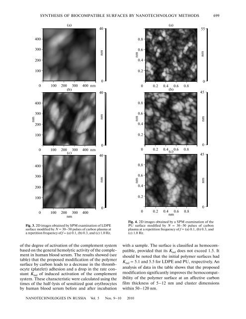
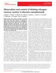
SYNTHESIS OF BIOCOMPATIBLE SURFACES BY NANOTECHNOLOGY METHODS 699(a)40(a)55nm4003002001000 100 200 300(b)400 nm4003002001000 100 200 300(c)400 nm4000nm400nm40nmnm0.80.60.40.20 0.2 0.4 0.6(b)0.80.80.60.40.20 0.2 0.4 0.6(c)0.80.8nm045nm0453002001000 100 200 300 400nm0nmFig. 3. 2D images obtained <strong>by</strong> SPM examination <strong>of</strong> LDPEsurface modified <strong>by</strong> N = 30–50 pulses <strong>of</strong> carbon plasma ata repetition frequency <strong>of</strong> f = (a) 0.1, (b) 0.3, and (c) 1.0 Hz.nm0.60.40.2nm00 0.2 0.4 0.6 0.8nmFig. 4. 2D images obtained <strong>by</strong> a SPM examination <strong>of</strong> thePU surface modified <strong>by</strong> N = 30–50 pulses <strong>of</strong> carbonplasma at a repetition frequency <strong>of</strong> f = (a) 0.1, (b) 0.3, and(c) 1.0 Hz.<strong>of</strong> the degree <strong>of</strong> activation <strong>of</strong> the complement systembased on the general hemolytic activity <strong>of</strong> the complementin human blood serum. The results showed (seetable) that the proposed modification <strong>of</strong> the polymersurface <strong>by</strong> carbon leads to a decrease in the thrombocyte(platelet) adhesion and a drop in the rate constantK ind <strong>of</strong> induced activation <strong>of</strong> the complementsystem. These characteristic were calculated using thetimes <strong>of</strong> the half-lysis <strong>of</strong> sensitized goat erythrocytes<strong>by</strong> human blood serum before and after incubationwith a sample. The surface is classified as hemocompatible,provided that its K ind does not exceed 1.5. Itshould be noted that the initial polymer <strong>surfaces</strong> hadK ind = 5.1 and 5.5 for LDPE and PU, respectively. Ananalysis <strong>of</strong> data in the table shows that the proposedmodification significantly improves the hemocompatibility<strong>of</strong> the polymer surface at an affective carbonfilm thickness <strong>of</strong> 5–12 nm and cluster dimensionswithin 50–120 nm.NANOTECHNOLOGIES IN RUSSIA Vol. 5 Nos. 9–10 2010
700ALEKHIN et al.Intensite, rel. units30201008001000 1200 1400 1600 1800Wavenumber, cm −1Fig. 5. Analysis <strong>of</strong> the Raman spectrum <strong>of</strong> a carbon coatingshowing general line envelope and its components correspondingto sp 2 hybridization, sp 3 hybridization, and misorientedstates, respectively.NANOSTRUCTURAL MODIFICATIONOF POLY(METHYL METHACRYLATE)SURFACE BY A COMBINATIONOF PLASMACHEMICAL TREATMENTAND VACUUM ULTRAVIOLET IRRADIATIONPoly(methyl methacrylate) (PMMA) is now widelyused in practical transplantology as a base material forvarious implants [11].Plasmachemical Processing <strong>of</strong> PMMA SurfacePreviously, Vasilets et al. [12, 13] showed that plasmachemicalprocessing and vacuum ultraviolet (VUV)irradiation modify the properties <strong>of</strong> near-surface layers<strong>of</strong> medical polymers. The destruction <strong>of</strong> polymers andthe formation <strong>of</strong> active chemical radicals and speciesunder these conditions make it possible to control thebiocompatibility <strong>of</strong> the polymers. In particular, themodification <strong>of</strong> the near-surface layers <strong>of</strong> a polymerleads to a change in its nanoroughness. In this context,we have studied the effect <strong>of</strong> plasmachemical treatmentin an oxygen-containing medium at a small partialpressure (~2 Pa) followed <strong>by</strong> VUV irradiation onthe PMMA surface roughness in a nanometer range.The initial PMMA film with a molecular mass <strong>of</strong>8 × 10 4 and a thickness <strong>of</strong> about 0.8 μm was deposited<strong>by</strong> centrifuging onto a polished Si(100) single crystalsurface (Fig. 6a) and then nanostructured <strong>by</strong> beingtreated for 20 s in an RF (13.56 MHz) plasma. Theparameters <strong>of</strong> the surface roughness and geometry <strong>of</strong>the surface features (nanohills and nanograins) onPMMA films were studied <strong>by</strong> SPM in the atomic forcemicroscopy (AFM) mode on a sample surface area <strong>of</strong>1.4 × 1.4 μm 2 . The dimensions <strong>of</strong> the features weredetermined <strong>by</strong> the computer-aided recognition <strong>of</strong> thesurface relief, which was performed using the method<strong>of</strong> feature-oriented scanning in the virtual regime[14].It was established that the plasmachemical treatment<strong>of</strong> a PMMA film led to the formation <strong>of</strong> flat nanograins(Fig. 6b) with average lateral dimensions <strong>of</strong>about 66 nm, an average height <strong>of</strong> about 1.8 nm, andan average distance <strong>of</strong> about 104 nm between adjacentgrains. The number <strong>of</strong> nanograins per unit surface areawas about 122 μm –2 . The proposed nanostructuretechnology ensures that these results can be easilyreproduced.The formation <strong>of</strong> surface nanograins can beexplained <strong>by</strong> local changes in the PMMA film density.These local changes are related to the presence <strong>of</strong>inhomogeneities in most polymers, primarily globules/grainsand lamellae, as well as coils, nodes, twists,entanglements, etc. <strong>of</strong> molecular chains, which agreeswith the results <strong>of</strong> other investigations [15, 16]. In thecourse <strong>of</strong>PMMA treatment in oxygen-containing plasma,the polymer is subject to significant chemical modification,including the formation <strong>of</strong> polar functionalgroups [17, 128], carbonate (O 2 C=O), and carbonyl(C=O) groups. In addition, there are significantchanges in the binding energies <strong>of</strong> carbon atoms invarious molecular groups. The content <strong>of</strong> oxygen inthe near-surface layer exhibits a significant increaseduring the first seconds <strong>of</strong> treatment; then the growthslows down and eventually oxygen rather slowly penetratesin depth <strong>of</strong> the material [17].Treatment <strong>of</strong> PMMA Film Surface<strong>by</strong> VUV RadiationPlasma-treated PMMA samples were exposed tothe radiation <strong>of</strong> krypton lamps with a characteristicwavelength <strong>of</strong> λ = 123.6 nm in vacuum at a residualThe dependence that the cluster size, the rate constant (K ads ) <strong>of</strong> blood plasma protein adsorption, and the rate constant K ind<strong>of</strong> induced activation <strong>of</strong> the complement system has on the effective thickness <strong>of</strong> the carbon-containing layer on LDPE surfaceCarbon filmthickness, nmClustersize, nmK ads , ml/(mg s)K indLDPE Vitur LDPE Vitur12 Surface coverage >80% 0.25 0.1 4.3 ± 0.5 3.5 ± 0.4NANOTECHNOLOGIES IN RUSSIA Vol. 5 Nos. 9–10 2010