Download the PDF copy - Bruker
Download the PDF copy - Bruker
Download the PDF copy - Bruker

You also want an ePaper? Increase the reach of your titles
YUMPU automatically turns print PDFs into web optimized ePapers that Google loves.
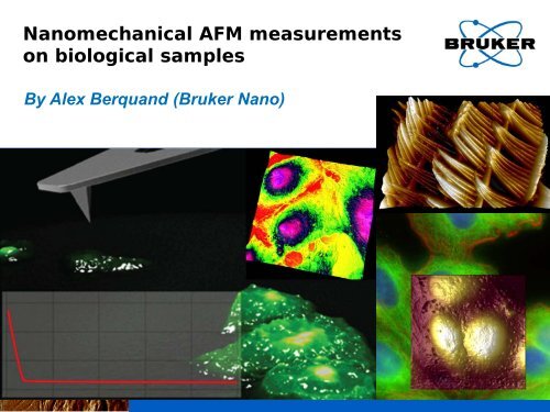
Nanomechanical AFM measurements<br />
on biological samples<br />
By Alex Berquand (<strong>Bruker</strong> Nano)
What’s behind “cell mechanics” and why is it<br />
so important in biology<br />
Complexity of signal transduction in cells<br />
B<br />
A<br />
C<br />
• A, B and C have different stiffness and contain various molecules.<br />
• Those molecules are associated to <strong>the</strong> inner components of <strong>the</strong> cell.<br />
• They can regulate normal functions or lead to imbalance/disease.<br />
• Identifying A, B and C and <strong>the</strong>ir components is of great importance.<br />
1/22/2013 BRUKER CONFIDENTIAL<br />
2
What’s behind “cell mechanics” and why is it<br />
so important in biology<br />
Stiffness in Cell differentiation<br />
• Nature of <strong>the</strong> scaffold:<br />
(same stem cells)<br />
or<br />
1 kPa 0.1 kPa 0.01 kPa<br />
or<br />
or<br />
or<br />
different cell types<br />
• Tilt:<br />
(same stem cells and same substrate)<br />
or or or or<br />
0º 5º 10º<br />
different cell types<br />
• AFM and fluorescence micros<strong>copy</strong> can be used.<br />
1/22/2013 BRUKER CONFIDENTIAL<br />
3
Concrete example<br />
Cancer: why is sensing differences in elasticity<br />
important<br />
• Cancer cells are usually softer than <strong>the</strong>ir counterparts, especially in <strong>the</strong><br />
case of bladder (Lekka et al. 1999), prostate (Faria et al. 2008), breast<br />
(Cross et al. 2007), cartilage (Darling et al. 2007), blood (Rosenbluth et al.<br />
2006) and ovarian (Sharma et al. 2011) tissues.<br />
• Some organs are not permeable to antibiotics.<br />
• Cancer surgery is often highly risky.<br />
• Identifying differences in elasticity at an early stage is required. Possible by<br />
AFM on <strong>the</strong> corresponding cells lines.<br />
- Lekka M. and Laidler P., Nat. Nanotechnol. 72 (2009) 72-73.<br />
- Faria E. C. et al., Analyst 133 (2008) 1498-500.<br />
- Cross S.E. et al. Nat. Nanotechnol. 2 (2007) 780-783.<br />
- Rosenbluth M.J. et al., Biophys. J. 90 (2006) 2994-3003.<br />
- Sharma S. et al., Nanomed. Nanotechnol. Biol. Med. (2011) under press.<br />
1/22/2013 BRUKER CONFIDENTIAL<br />
4
Usual tools to probe cell mechanics<br />
Major techniques<br />
• Micropipette aspiration (HochMuth, 2000).<br />
• Acoustic wave micros<strong>copy</strong> (Hildebrand 2001).<br />
• Bio-imprinting (Dickert et al. 2002).<br />
• Optical tweezers / optical traps (Dao et al. 2003).<br />
• AFM (Force Spectros<strong>copy</strong>) (Rotsch et al 2000).<br />
- HochMuth R.M., J.Biomech 33 (2000) 15-22.<br />
- Hildebrand J.A. et al., PNAS 78 (1981) 1656-1660.<br />
- Dickert J.L. et al., Anal. Chem. 74 (2002) 1302-1306.<br />
- Dao M. et al., Mech. Phys. Sol. 51 (2003) 2259-2280.<br />
- Rotsch C. et al, Biophys J. 78 (2000) 520-535.<br />
1/22/2013 BRUKER CONFIDENTIAL<br />
5
Principle of AFM<br />
Optical detection system<br />
Different feedbacks for different AFM modes<br />
1/22/2013 BRUKER CONFIDENTIAL<br />
6
AFM Resolution<br />
Compared to o<strong>the</strong>r micros<strong>copy</strong> techniques<br />
0.1nm 1nm 10nm 100nm 1µm 10µm 100µm<br />
AFM<br />
light micros<strong>copy</strong><br />
EM<br />
atoms<br />
molecules<br />
bacterioΦ chloroplasts<br />
bacteria<br />
cells
Combining AFM to Fluorescence<br />
2 techniques in 1 tool<br />
AFM<br />
Optics<br />
1/22/2013 BRUKER CONFIDENTIAL
Combining AFM to IOM<br />
Compatibility with various<br />
optical techniques<br />
AFM + …<br />
Sample courtesy<br />
INRA Nantes<br />
Sample<br />
courtesy Zeiss<br />
Sample<br />
courtesy<br />
NIH<br />
Sample courtesy<br />
SWMC Stanford<br />
Sample courtesy Nat.<br />
Sample courtesy Univ. of<br />
Univ. Singapore<br />
New South Wales<br />
1/22/2013 BRUKER CONFIDENTIAL<br />
9
Combining AFM to fluorescence<br />
Automatic Overlay (MIRO)<br />
1) Capture 3 sample images at 3 different locations.<br />
2) Capture 1 tip image.<br />
3) Capture a background image and overlay it with an AFM image.<br />
1) Import optical image<br />
into Nanoscope<br />
2) Target a location for<br />
<strong>the</strong> AFM scan<br />
3) Overlay optical and<br />
AFM images<br />
1/22/2013 BRUKER CONFIDENTIAL<br />
10
Combining AFM to fluorescence<br />
Automatic Overlay (MIRO)<br />
Live Hela cells<br />
1/22/2013 BRUKER CONFIDENTIAL<br />
11
Force Spectros<strong>copy</strong><br />
Get access to stiffness and adhesion<br />
2<br />
force (nN)<br />
1<br />
0<br />
-1<br />
0<br />
distance (nm)<br />
500<br />
1/22/2013 BRUKER CONFIDENTIAL<br />
12
Contact <strong>the</strong>ories in AFM<br />
Different models / samples<br />
Hertz DMT JKR<br />
Sneddon<br />
MD<br />
(Most adapted for<br />
biological samples)<br />
Standard for Peak Force QNM
FV/Fluo Applications in Biology<br />
CSK disrupting agents<br />
• Nocodazole can disrupt Tubulin CSK.<br />
• Latrunculin can disrupt Actin CSK.<br />
• Tested on 2 cell types in fluorescence, AFM imaging, AFM FV.<br />
Hela cells<br />
OS cells
FV/Fluo Applications in Biology<br />
CSK disrupting agents<br />
tubulin<br />
Effect of <strong>the</strong> 2 drugs:<br />
1) decay of fluorescence<br />
2) loss of tracking<br />
actin<br />
3) change in elasticity<br />
Nocodazole<br />
Latrunculin<br />
z<br />
z<br />
0<br />
x<br />
x<br />
Actin plays a much more important role in cell rigidity<br />
than tubulin.<br />
Too slow (low res.) + not enough information.
Popular AFM techniques<br />
Are <strong>the</strong>y quantitative<br />
• Force measurements: Force Volume<br />
• Is it quantitative<br />
• What does it provide<br />
• Imaging: Tapping mode<br />
• Is it quantitative<br />
• What does it provide<br />
ϕ<br />
ϕ<br />
Not Quantitative!<br />
1/22/2013 BRUKER CONFIDENTIAL<br />
16
FV to slow to probe biological processes<br />
True for most of <strong>the</strong>m<br />
• Examples:<br />
• Diffusion of proteins: 10 -5 -10 -9 cm 2 .s -1 .<br />
• Polymerization of microtubules: sec-min.<br />
• Protein translocation to nucleus (can be probed by fluorescence): 30<br />
min to 1 h.<br />
• Cell migration: µm/sec-µm/h.<br />
• Cell division: 20 min to 3 days.<br />
• Ideally: less than 10 min per AFM image. Impossible with FV or loss of<br />
resolution.<br />
• Need for acceptable resolution to compare AFM and Fluorescence data (for<br />
instance: increasing number of STED publications).<br />
1/22/2013 BRUKER CONFIDENTIAL<br />
17
Resolution is important for biologists<br />
Example: STED<br />
80<br />
70<br />
60<br />
50<br />
40<br />
30<br />
20<br />
10<br />
0<br />
2007-2008 2009-2010 2011-2012<br />
• Number of STED publications in Biology/Medicine research.<br />
• AFM offers similar spatial resolution but is much slower.<br />
• Need for higher resolution, higher force control and faster imaging to<br />
be complementary to fluorescence techniques.<br />
1/22/2013 BRUKER CONFIDENTIAL<br />
18
Need for a new characterization<br />
technique<br />
Peak Force Tapping and Peak Force QNM<br />
PFT is based on ScanAsyst (fully Automated AFM)<br />
Works with most standard AFM probes in <strong>the</strong> standard AFM cantilever<br />
holders.<br />
Z piezo is driven with sinusoidal waveform<br />
(not a triangle as in force-distance curves).<br />
Z drive frequency is 1-2 kHz. Z drive amplitude is fixed at typical<br />
value of 150 nm (300 nm peak-to-peak)<br />
Vertical motion of probe produces force-distance plots as it taps on<br />
<strong>the</strong> sample.<br />
Imaging feedback is based on <strong>the</strong> Peak Force of <strong>the</strong> force-distance<br />
curve.<br />
The probe can be calibrated before <strong>the</strong> experiment so that all <strong>the</strong><br />
channels are directly quantitative: PFQNM<br />
1/22/2013 BRUKER CONFIDENTIAL<br />
19
Needed range of Young’s moduli<br />
Example: Human Body<br />
1/22/2013 BRUKER CONFIDENTIAL<br />
20
Overview: PeakForce QNM®<br />
Basic Principle<br />
• PeakForce Tapping is an oscillating<br />
technique that can be used to image a<br />
wide range of samples at a high<br />
resolution.<br />
5nm<br />
• The probe can easily be calibrated<br />
prior to <strong>the</strong> experiment. The technique<br />
is referred to as PeakForce QNM. In<br />
that case, all <strong>the</strong> recorded data are<br />
directly quantitative.<br />
• Peak Force QNM can now be operated<br />
on live cells and can be used to test<br />
changes in morphology and any o<strong>the</strong>r<br />
property in response to various<br />
factors.<br />
1/22/2013<br />
21
Preliminary test on a stiff sample<br />
FV/HMX/QNM comparison on a daphnia<br />
A<br />
A<br />
FV<br />
QNM<br />
A<br />
B<br />
C<br />
100<br />
µm<br />
Bright field image of Daphnia<br />
(20x)<br />
B<br />
• Daphnia scanned in FV, HMX and QNM modes (20 min per image).<br />
• Young’s modulus calculated by using <strong>the</strong> 3 modes.<br />
• QNM offers much higher resolution and information than FV. HMX<br />
returns intermediate results.<br />
1/22/2013 22
Preliminary test on a stiff sample<br />
FV/HMX/QNM comparison on a daphnia<br />
• QNM offers <strong>the</strong> best compromise in terms of resolution, ease-of-use<br />
and delivered information.<br />
1/22/2013 23
Preliminary test on a soft sample.<br />
FV/QNM comparison on a cell<br />
FV (32x32)<br />
QNM (128x128)<br />
• 90x90 µm<br />
• 5 min / image<br />
1/22/2013 24
Preliminary test on a soft sample.<br />
FV/QNM comparison on a cell<br />
Processed area<br />
QNM image + pfc image<br />
Bearing Analysis<br />
FV<br />
QNM<br />
36 curves Export + post-processing<br />
Export + post-processing<br />
144 curves<br />
1/22/2013 25
Preliminary test on a soft sample.<br />
FV/QNM comparison on a cell<br />
Line plot<br />
Indentation<br />
Modify force parameters<br />
Baseline correction<br />
Multiple line plots<br />
Boxcar filter<br />
Run History<br />
FV [Sneddon fit] = 10.9 MPa<br />
QNM (bearing Analysis) [Sneddon fit] = 21.72 MPa<br />
QNM (PFC+export) [Sneddon fit] = 18.42 MPa<br />
• The 2 QNM results are very close.<br />
• The FV result is different from <strong>the</strong> QNM results but <strong>the</strong> number<br />
of data point is also quite different + it’s hard to perform <strong>the</strong><br />
analysis on exactly <strong>the</strong> same area.<br />
• How about an analysis on higher scale (Bio experiment)<br />
1/22/2013 26
FV/QNM accuracy in Biology<br />
Study on glioblastoma<br />
• Isogenic cell line U251 transfected with tumor suppressive factors.<br />
PF error (z: 0-250 pN) Elasticity (z: 0-1.2 MPa) Deformation (z: 0-250 nm)<br />
• 5-6 min per image.<br />
• 128x128 resolution.<br />
• Enough to observe contrast on all mechanical channels.<br />
1/22/2013 BRUKER CONFIDENTIAL<br />
27
FV/QNM accuracy in Biology<br />
Study on glioblastoma<br />
Submitted to JJAP<br />
• Same tendency (cancer cells softer than normal ones) but STD very<br />
different (number of data points: 589 824 for QNM / 9214 for FV; less<br />
than 10 min per image).<br />
• Same remark for cell diffentiation, cell migration, cell remodeling,…<br />
1/22/2013 BRUKER CONFIDENTIAL<br />
28
QNM study on live Hacats<br />
Effect of Glyphosate on Human Skin<br />
• The Human skin is <strong>the</strong> first physiological<br />
barrier against physical and chemical<br />
aggressions.<br />
• Glyphosate (N-(phosphonomethyl)Glycine)<br />
is a broad spectrum systemic herbicide<br />
used to kill weeds. It’s been extensively<br />
used in <strong>the</strong> late 2000’s.<br />
• Its toxicity on lab animals has been clearly<br />
demonstrated but its effects on humans<br />
remain unclear.<br />
1/22/2013<br />
29
Background: Glyphosate<br />
Existing Data in Cytology and Main Challenges<br />
[H 2 O 2 ] marker<br />
• Glyphosate-treated samples show a<br />
higher number of necrotic and apoptotic<br />
cells, and a higher [H 2 O 2 ] than controls.<br />
• Keratinocytes and HaCat cells are<br />
difficult to image by classical AFM<br />
modes. A more reliable technique is<br />
required.<br />
• Need for a faster way to probe <strong>the</strong><br />
oxidative stress. Changes in morphology<br />
and mechanical properties might be<br />
detectable by using PeakForce QNM.<br />
1/22/2013
PeakForce QNM: Much more information<br />
Probe changes in mechanical properties<br />
• Drug treatment induces a significant increase in Young’s modulus (x3)<br />
and a decrease in deformation (x2).<br />
• Changes in mechanical properties can directly be correlated to changes<br />
in morphology.<br />
1/22/2013<br />
31
Journal of Structural Biology<br />
Publication<br />
January 2012<br />
• Data was published in a paper<br />
entitled ‘Glyphosate-induced<br />
stiffening of HaCat keratinocytes,<br />
a Peak Force Tapping study on<br />
living cells’ in Journal of<br />
Structural Biology.<br />
• The System: Bioscope Catalyst<br />
with PeakForce QNM and<br />
operated in fluid<br />
• Authors are Celine Heu, Celine<br />
Elie and Laurence Nicod (FEMTO)<br />
and Alexandre Berquand<br />
(<strong>Bruker</strong> Nano GmbH)
Different Euk. cells: Diatoms<br />
Interest in Industry<br />
• Diatoms are of interest in micro and nanoscale science for<br />
applications ranging from solar cells to optical systems.<br />
• As a living vesicle, <strong>the</strong>y also have potential for drug delivery.<br />
33
PeakForce QNM:<br />
Easier imaging of Live Diatoms in Seawater<br />
5 µm<br />
• Large scale Catalyst images remarkably stable.<br />
• The Perfusing Stage Incubator accessory enables <strong>the</strong> active exchange of medium.<br />
34
PeakForce QNM: Easier Imaging<br />
Mechanical Properties at High Resolution<br />
150 mV<br />
50 MPa<br />
0 0<br />
Peak force Error<br />
Young’s modulus<br />
20 nm<br />
0<br />
Deformation<br />
3d Height + YM skin<br />
• All PeakForce QNM channels are directly quantitative.<br />
• Unprecedented resolution from imaging to modulus.<br />
Pletikapic et al., J.<br />
Phycol 48 (2012) 174.<br />
35
PeakForce QNM: Easier Imaging<br />
First AFM Investigation of <strong>the</strong> Frustule<br />
• The frustule is a filtration wall allowing communication between <strong>the</strong> sea water and <strong>the</strong> inner<br />
part of <strong>the</strong> diatom.<br />
• These are <strong>the</strong> first images of <strong>the</strong> frustle by AFM.<br />
• Additionally, QNM channels provide quantitative mechanical properties, and show nice<br />
contrast between <strong>the</strong> pores, <strong>the</strong> rings & <strong>the</strong> rest of <strong>the</strong> frustule.<br />
36
QNM on Live Bacteria<br />
E. Coli K12<br />
Bacteria with pili 3d-height (pili retracted) YM = 183 kPa<br />
1/22/2013 37
Correlating topography to Force curves<br />
HSDC files<br />
Export + post-processing<br />
1/22/2013 BRUKER CONFIDENTIAL<br />
38
Correlating topography to Force curves<br />
HSDC files<br />
AFM tip<br />
fibril<br />
cell<br />
1 2<br />
Young’s modulus calculated<br />
over very specific areas<br />
Young’s modulus calculated<br />
over <strong>the</strong> entire cell<br />
1/22/2013 BRUKER CONFIDENTIAL<br />
39
Functionalized probes<br />
HSDC files - Detecting specific unbinding events<br />
1/22/2013 BRUKER CONFIDENTIAL<br />
40
Combining QNM with functionalized<br />
probes - Pathogenesis of Malaria<br />
Erythrocyte (Red Blood Cell) Infection<br />
P. falciparum<br />
Anopheles mosquito<br />
Healthy RBC<br />
• Parasites enter bloodstream and<br />
infect RBCs.<br />
• As parasites multiply, <strong>the</strong> RBCs<br />
breaks open and infects more RBCs.<br />
• Infected RBCs are misshapen with<br />
knob-like structures on surface.<br />
Knobs<br />
Infected RBC<br />
1/22/2013<br />
41
Background: Cytoadherence<br />
Erythrocyte (Red Blood Cell) Infection<br />
• P. falciparum infected erythrocytes (IE’s) exhibit cytoadherence<br />
(stick to blood vessel walls /endo<strong>the</strong>lium).<br />
• Prevents IE elimination by <strong>the</strong> spleen and causes vascular blockages.<br />
• Interaction with endo<strong>the</strong>lium surface receptors (eg. CD36).<br />
Adapted from http://cmr.asm.org/cgi/content-nw/full/13/3/439/F1.<br />
Adapted from <strong>the</strong> Plasmodium Genome Resource.<br />
1/22/2013<br />
42
The Biological Question:<br />
Can we map <strong>the</strong> distribution of cytoadherent<br />
molecules to specific cell surface structures<br />
• AFM probes were functionalized with endo<strong>the</strong>lial surface receptor CD36.<br />
• Used PeakForce QNM with functionalized probe to obtain 2D map of <strong>the</strong><br />
distribution of CD36 molecular binding sites on IE.<br />
(CD36)<br />
Adapted from Li et al. (2011) PLoS ONE 6: 1-10.<br />
1/22/2013<br />
43
Molecular Recognition Imaging of IEs<br />
Colocalization of CD36 binding sites with knobs<br />
Topography Adhesion Overlay<br />
• CD36 binding sites (high adhesion) correlate to knob structures (circles).<br />
• Adhesion is a result of specific interactions between CD36 and knobs and<br />
not due to crosstalk between data channels (arrow).<br />
1/22/2013<br />
44
Challenge: Glycocalyx in force measurements<br />
Obstacle for access to <strong>the</strong> membrane<br />
• Exists in bacteria and euk. Cells.<br />
• Idea: use probes with various chemistries<br />
1/22/2013 BRUKER CONFIDENTIAL<br />
45
Application Note #135<br />
Quantitative imaging of living biological samples<br />
by Peak Force QNM Atomic Force Micros<strong>copy</strong><br />
• A comprehensive review of Peak<br />
Force QNM applications on soft<br />
biological samples<br />
• Authors are Dr. Alex Berquand and<br />
Dr. Ben Ohler (<strong>Bruker</strong> Nano Inc.).<br />
• + New App Note with latest<br />
developments and examples to be<br />
released soon.<br />
1/22/2013<br />
46
Contact information<br />
Alexandre Berquand<br />
Life Science Applications Scientist<br />
Alexandre.Berquand@bruker-nano.com<br />
Tel: +49 174 333 94 62<br />
www.bruker.com/en/products/surface-analysis/atomic-force-micros<strong>copy</strong>.html<br />
www.brukerafmprobes.com/<br />
http://nanoscaleworld.bruker-axs.com/<br />
1/22/2013 BRUKER CONFIDENTIAL<br />
47