

Annals of Diagnostic Paediatric Pathology
Annals of Diagnostic Paediatric Pathology
Annals of Diagnostic Paediatric Pathology

Create successful ePaper yourself
Turn your PDF publications into a flip-book with our unique Google optimized e-Paper software.
92<br />
Summary<br />
Liver steatosis <strong>of</strong> severe degree involving 100% <strong>of</strong> liver parenchyma<br />
was found in three patients (No 1, 2, and 5) who<br />
died during ALTE. Moderate steatosis involving approximately<br />
60% <strong>of</strong> liver was present in one biopsy ( No 3). Mild steatosis<br />
affecting 30% and 15% <strong>of</strong> liver parenchyma was found in two<br />
biopsies coming from cases 4 and 6, respectively. In one autopsy<br />
sample liver steatosis involved 10% <strong>of</strong> parenchyma (No<br />
7). Steatosis was macrovacuolar or mixed. The first type was<br />
found in two sequential examinations <strong>of</strong> the same patient (biopsy<br />
– No 4 and autopsy – No 5) and in two autopsies (samples<br />
No 2 and 7). Macrovacuolar steatosis was severe in two cases<br />
(No 2 and 5) and mild in two others (No 4 and 7). Mixed<br />
steatosis was seen in two biopsies (No 3 and 6) and one autopsy<br />
(No 1). Its degeree was mild (sample No 6), moderate<br />
(sample No 3), and severe (sample No 1). None <strong>of</strong> the patients<br />
had pure microvacuolar fatty liver. In six cases distribution<br />
<strong>of</strong> steatotic hepatocytes was diffuse, in one focal (sample<br />
No 4). Inflammation was present in three cases. It was mild<br />
(cases No 3 and 5) or severe (case No 4). Fibrosis was disclosed<br />
in four examined samples derived from three patients:<br />
in two cases its intensity was mild (No 2 and 7) in two severe<br />
(samples No 4 and 5). Liver biopsy samples available for assessment<br />
from the same patient disclosed progression from<br />
precirrhosis to cirrhosis. Data concerning patients and their<br />
liver morphology are presenting in Table 1.<br />
Discussion<br />
It is known that the process <strong>of</strong> mitochondrial fatty acid oxidation<br />
can be divided in two stages. First involves transport<br />
through mitochondrial membrane, the second consists <strong>of</strong> cutting<br />
<strong>of</strong>f two-carbon moieties from fatty acid chain by several<br />
enzymes for long, medium, and short chain fatty acids, respectively<br />
[5, 9, 11]. Fatty liver may develop in both patients<br />
with defects <strong>of</strong> the first stage involving carnitine dependent<br />
transport [2, 3, 12], and with defects <strong>of</strong> the proper beta-oxidation<br />
[2, 4, 6-8, 10, 11]. Baldridge in his study <strong>of</strong> 82 patients<br />
with liver steatosis reported only one child in whom the<br />
LCHAD deficiency was established [1]. Our group comprises<br />
six patients with this diagnosis. Roe and Coates provided general<br />
description <strong>of</strong> liver morphology in LCHAD deficiency<br />
[9]. They pointed lipid accumulation and sometimes accompanying<br />
fibrosis as characteristic features <strong>of</strong> this disorder. Our<br />
findings are consistent with this observation. However, they<br />
have not provided particulars concerning intensity, type, and<br />
distribution <strong>of</strong> steatosis. Gilbert-Barness and Barness have<br />
presented more detailed description <strong>of</strong> liver microscopical<br />
pathology in LCHAD deficient patients [5]. They considered<br />
diffuse or zonal macrovacuolar severe fatty liver to be characteristic<br />
<strong>of</strong> the majority <strong>of</strong> LCHAD-deficient patients. In our<br />
group this type <strong>of</strong> morphological pattern was found in three<br />
patients always when the liver biopsy was performed at the<br />
acute clinical presentation.<br />
In a number <strong>of</strong> described patients with LCHAD deficiency<br />
the liver discloses only focal steatosis <strong>of</strong> mild intensity<br />
[5]. We observed focal fatty changes only in one case out <strong>of</strong><br />
seven (sample No 4). In remaining patients the degree <strong>of</strong> steatosis<br />
was mild to moderate and was almost invariably diffusely<br />
distributed.<br />
Moore described morphology <strong>of</strong> the liver in six months<br />
old girl with LCHAD deficiency [7]. There were mixed steatosis<br />
accompanied by mild cholestasis. Author has not provided<br />
data concerning intensity and localization <strong>of</strong> fatty<br />
changes. In our series <strong>of</strong> seven cases we did not find any case<br />
with the features <strong>of</strong> cholestasis.<br />
Tyni described liver morphology including microscopical<br />
findings <strong>of</strong> 16 patients coming from 12 families, who died<br />
with diagnosis <strong>of</strong> LCHAD deficiency [11]. All patients in her<br />
study had features <strong>of</strong> macrovacuolar fatty liver. In our patients<br />
steatosis was not only macrovacuolar, but also mixed. Tyni<br />
concluded that steatosis was typical for fatty acid beta oxidation<br />
defects, and emphasized that accompanying fibrosis was<br />
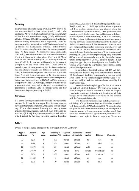
Table 1<br />
Details <strong>of</strong> morphological changes <strong>of</strong> the liver in patients with LCHADD<br />
Type <strong>of</strong><br />
examination<br />
Sample<br />
No<br />
Age<br />
(mo)<br />
Intensity <strong>of</strong><br />
steatosis (%)<br />
Type <strong>of</strong><br />
steatosis<br />
Localization<br />
<strong>of</strong> steatosis<br />
Inflammation<br />
Fibrosis Cholesta<br />
A 3299 4 100 mixed diffuse no no no<br />
A 3289 3 100 macro diffuse no mild no<br />
B 30573 12 60 mixed diffuse mild no no<br />
B 30760 3 30 macro focal severe precirrhosis no<br />
A 2701 6 100 macro diffuse mild cirrhosis no<br />
B 51605 7 15 mixed diffuse no no no<br />
A 3070 6 10 macro diffuse no mild no


