

Annals of Diagnostic Paediatric Pathology
Annals of Diagnostic Paediatric Pathology
Annals of Diagnostic Paediatric Pathology

Create successful ePaper yourself
Turn your PDF publications into a flip-book with our unique Google optimized e-Paper software.
116<br />
Table 1<br />
Total and direct bilirubin level in serum in distinguished groups <strong>of</strong> primary hyperbilirubinemia and with non-specific changes<br />
Disease<br />
Gilbert<br />
Compl.<br />
Incompl.<br />
Number <strong>of</strong> patients Age (years)<br />
10<br />
11<br />
10-17<br />
6-7<br />
Bilirubin (mg%)<br />
Total Average Direct Ave<br />
1,5-4,6 2,56 0,5-0,9 0,62<br />
1,2-5,1 2,48 0,3-1,1 0,95<br />
Dubin-Johnson 5 9-17 1,3-4,7 2,54 0,6-2,0 1,05<br />
Rotor 3 16-17 1,6-4,5 2,10 0,4-0,8 0,55<br />
Non specific 33 7-17 1,2-5,7 2,62 0,2-2,8 1,45<br />
Histology<br />
Five mm thick paraffin sections were stained in a routine manner<br />
with hematoxylin-eosin, azan, silver, PAS, PAS with<br />
diastaze digestion and than reviewed to exclude other disease<br />
than hyperbilirubinemia.<br />
Electron microscopy<br />
Samples were fixed in 2.5% glutaraldehyde in cacodylate<br />
buffer at pH 7.3, postfixed in 2% OsO 4,<br />
dehydrated in alcohols<br />
and embedded in Epon 812. Thick sections stained with toluidine<br />
blue were analysed under light microscope. Ultrathin<br />
sections stained with uranyl acetate and lead citrate were examined<br />
by electron microscopy.<br />
A<br />
B<br />
Results<br />
Examinations <strong>of</strong> paraffin and semi-thin sections showed in all<br />
cases an apparently normal liver, except for Dubin-Johnson<br />
that shown the existence <strong>of</strong> dark pigment predominantly in<br />
centrolobular areas.<br />
On the basis <strong>of</strong> EM analysis morphological equivalents<br />
<strong>of</strong> primary congenital hyperbilirubinemias were confirmed<br />
in 29 cases and secondary reactive symptomatic<br />
hyperbilirubinemias were recognized in 33 cases.<br />
Gilbert syndrome<br />
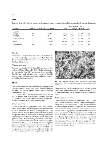
Gilbert syndrome was diagnosed in 21 cases. There were observed<br />
shortening and mal-developed vascular pole microvilli<br />
or loss <strong>of</strong> microvilli, and increase <strong>of</strong> collagen in the Disse<br />
space in various intensity. In all patients an increase <strong>of</strong> the<br />
endoplasmic reticulum pr<strong>of</strong>iles with coexisting dilatation <strong>of</strong><br />
rough endoplasmic reticulum with degranulation and pigment<br />
granules variable in size containing both electron dense material<br />
which alternate with less dense material and lipid droplets<br />
was observed (Fig. 1A). In 10 biopsy samples enlarged or<br />
giant, oval or polymorphic mitochondria with paracrystalline<br />
inclusions (Fig. 1B) were present. In all cases the biliary pole<br />
<strong>of</strong> hepatocytes shown normal caniculi and microvilli, but with<br />
numerous pigment granules around. Lip<strong>of</strong>uscin granules were<br />
Fig. 1 Gilbert syndrome: (A) vascular pole <strong>of</strong> hepatocyte with single microvilli.<br />
Mag. x 40 000, (B) mitochondrium containing paracrystallines, mag. x<br />
22 000<br />
common finding. The remaining group <strong>of</strong> 11 patients showed<br />
uncompleted ultrastructural features <strong>of</strong> Gilbert disease, i.e. lack<br />
<strong>of</strong> giant mitochondria and no paracristalline in mitochondrial<br />
matrix.<br />
Dubin-Johnson syndrome<br />
Dubin-Johnson syndrome was defined in 5 cases. There<br />
were not seen alterations in the vascular pole <strong>of</strong> hepatocytes,<br />
which exhibit normal microvilli. Slightly increased number<br />
<strong>of</strong> smooth endoplasmic reticulum pr<strong>of</strong>iles that formed small<br />
vesicles, normal mitochondria with some variation in size,<br />
well developed Golgi pr<strong>of</strong>iles located around bile canicules<br />
were common features. Dilation <strong>of</strong> bile caniculi with loss <strong>of</strong><br />
microvilli and characteristic pigment granules around were<br />
seen (Fig. 2). Pigment granules were composed mainly <strong>of</strong><br />
granular material, however vesicle-like or lipid components<br />
were also seen. They were polygonal in shape and delineated<br />
by a single membrane. The lack <strong>of</strong> lip<strong>of</strong>uscin in hepatocytes<br />
was common feature.


