On-Off Flushing Reservoirs
On-Off Flushing Reservoirs
On-Off Flushing Reservoirs

Create successful ePaper yourself
Turn your PDF publications into a flip-book with our unique Google optimized e-Paper software.
100674-731-01.qxd 8/25/2004 8:35 AM Page 1<br />
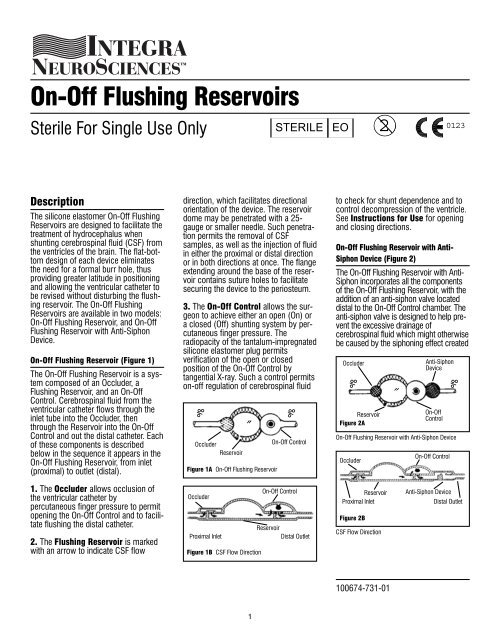
<strong>On</strong>-<strong>Off</strong> <strong>Flushing</strong> <strong>Reservoirs</strong><br />
Sterile For Single Use <strong>On</strong>ly<br />
2<br />
STERILE EO 0123<br />
Description<br />
The silicone elastomer <strong>On</strong>-<strong>Off</strong> <strong>Flushing</strong><br />
<strong>Reservoirs</strong> are designed to facilitate the<br />
treatment of hydrocephalus when<br />
shunting cerebrospinal fluid (CSF) from<br />
the ventricles of the brain. The flat-bottom<br />
design of each device eliminates<br />
the need for a formal burr hole, thus<br />
providing greater latitude in positioning<br />
and allowing the ventricular catheter to<br />
be revised without disturbing the flushing<br />
reservoir. The <strong>On</strong>-<strong>Off</strong> <strong>Flushing</strong><br />
<strong>Reservoirs</strong> are available in two models:<br />
<strong>On</strong>-<strong>Off</strong> <strong>Flushing</strong> Reservoir, and <strong>On</strong>-<strong>Off</strong><br />
<strong>Flushing</strong> Reservoir with Anti-Siphon<br />
Device.<br />
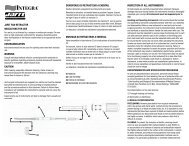
<strong>On</strong>-<strong>Off</strong> <strong>Flushing</strong> Reservoir (Figure 1)<br />
The <strong>On</strong>-<strong>Off</strong> <strong>Flushing</strong> Reservoir is a system<br />
composed of an Occluder, a<br />
<strong>Flushing</strong> Reservoir, and an <strong>On</strong>-<strong>Off</strong><br />
Control. Cerebrospinal fluid from the<br />
ventricular catheter flows through the<br />
inlet tube into the Occluder, then<br />
through the Reservoir into the <strong>On</strong>-<strong>Off</strong><br />
Control and out the distal catheter. Each<br />
of these components is described<br />
below in the sequence it appears in the<br />
<strong>On</strong>-<strong>Off</strong> <strong>Flushing</strong> Reservoir, from inlet<br />
(proximal) to outlet (distal).<br />
1. The Occluder allows occlusion of<br />
the ventricular catheter by<br />
percutaneous finger pressure to permit<br />
opening the <strong>On</strong>-<strong>Off</strong> Control and to facilitate<br />
flushing the distal catheter.<br />
2. The <strong>Flushing</strong> Reservoir is marked<br />
with an arrow to indicate CSF flow<br />
direction, which facilitates directional<br />
orientation of the device. The reservoir<br />
dome may be penetrated with a 25-<br />
gauge or smaller needle. Such penetration<br />
permits the removal of CSF<br />
samples, as well as the injection of fluid<br />
in either the proximal or distal direction<br />
or in both directions at once. The flange<br />
extending around the base of the reservoir<br />
contains suture holes to facilitate<br />
securing the device to the periosteum.<br />
3. The <strong>On</strong>-<strong>Off</strong> Control allows the surgeon<br />
to achieve either an open (<strong>On</strong>) or<br />
a closed (<strong>Off</strong>) shunting system by percutaneous<br />
finger pressure. The<br />
radiopacity of the tantalum-impregnated<br />
silicone elastomer plug permits<br />
verification of the open or closed<br />
position of the <strong>On</strong>-<strong>Off</strong> Control by<br />
tangential X-ray. Such a control permits<br />
on-off regulation of cerebrospinal fluid<br />
‰<br />
Occluder<br />
Reservoir<br />
Figure 1A <strong>On</strong>-<strong>Off</strong> <strong>Flushing</strong> Reservoir<br />
Occluder<br />
Proximal Inlet<br />
˝<br />
Figure 1B CSF Flow Direction<br />
‰<br />
<strong>On</strong>-<strong>Off</strong> Control<br />
<strong>On</strong>-<strong>Off</strong> Control<br />
Reservoir<br />
Distal Outlet<br />
to check for shunt dependence and to<br />
control decompression of the ventricle.<br />
See Instructions for Use for opening<br />
and closing directions.<br />
<strong>On</strong>-<strong>Off</strong> <strong>Flushing</strong> Reservoir with Anti-<br />
Siphon Device (Figure 2)<br />
The <strong>On</strong>-<strong>Off</strong> <strong>Flushing</strong> Reservoir with Anti-<br />
Siphon incorporates all the components<br />
of the <strong>On</strong>-<strong>Off</strong> <strong>Flushing</strong> Reservoir, with the<br />
addition of an anti-siphon valve located<br />
distal to the <strong>On</strong>-<strong>Off</strong> Control chamber. The<br />
anti-siphon valve is designed to help prevent<br />
the excessive drainage of<br />
cerebrospinal fluid which might otherwise<br />
be caused by the siphoning effect created<br />
Occluder<br />
Reservoir<br />
Figure 2A<br />
Occluder<br />
Reservoir<br />
Proximal Inlet<br />
Figure 2B<br />
Anti-Siphon<br />
Device<br />
‰<br />
‰<br />
˝<br />
<strong>On</strong>-<strong>Off</strong> <strong>Flushing</strong> Reservoir with Anti-Siphon Device<br />
CSF Flow Direction<br />
<strong>On</strong>-<strong>Off</strong><br />
Control<br />
<strong>On</strong>-<strong>Off</strong> Control<br />
Anti-Siphon Device<br />
Distal Outlet<br />
100674-731-01<br />
1
100674-731-01.qxd 8/25/2004 8:35 AM Page 2<br />
by the elevation of the ventricular<br />
catheter with respect to the distal catheter<br />
(i.e. when the patient sits, stands or is<br />
held erect. This siphoning effect is<br />
minimized by the anti-siphon valve, which<br />
closes under a negative pressure, yet will<br />
reopen to allow the flow of CSF to resume<br />
before intraventricular pressure becomes<br />
excessive.<br />
Indications<br />
The <strong>On</strong>-<strong>Off</strong> <strong>Flushing</strong> Reservoir, utilized in<br />
the treatment of hydrocephalic patients, is<br />
a component in systems designed to<br />
shunt cerebrospinal fluid from the lateral<br />
ventricles into either the right atrium of the<br />
heart or the peritoneum.<br />
Reservois with an Anti-Siphon Device are<br />
intended to reduce the hazard of negative<br />
intraventricular pressure when the patient<br />
is sitting, semi-recumbent or standing.<br />
Contraindications<br />
Ventriculatrial or ventriculoperitoneal<br />
shunting systems should not be used in<br />
the presence of known or suspected<br />
infections along the course of the shunt<br />
(meningitis, ventriculitis, skin infections,<br />
bacteremia, septicemia or peritonitis). It is<br />
advisable to avoid shunting procedures if<br />
infection is present anywhere in the body.<br />
The ventriculoatrial method of shunting is<br />
contraindicated for hydrocephalic patients<br />
with congenital heart disease or other<br />
anomalies of the cardiopulmonary<br />
system.<br />
Instructions For Use<br />
The introduction of a shunting system<br />
may be accomplished through a variety<br />
of surgical techniques; therefore, the surgeon<br />
is best advised to use the method<br />
which his/her own practice and training<br />
dictate to be best for the patient.<br />
To close the <strong>Flushing</strong> Reservoir, press the<br />
radiopaque, tantalum-impregnated<br />
silicone elastomer plug (attached to the<br />
dome of the <strong>On</strong>-<strong>Off</strong> Control) into the<br />
radiopaque circle of the valve seat.<br />
(Figure 3.)<br />
Re-opening of the system is achieved by<br />
holding the occluder closed with finger<br />
pressure, then pressing the reservoir<br />
dome to open the <strong>On</strong>-<strong>Off</strong> Control.<br />
(Figure 4.) Assurance that the control is<br />
in the desired position can be verified by<br />
tangential X-ray.<br />
To inject fluids into, or check the patency<br />
of, the ventricular catheter, close the <strong>On</strong>-<br />
<strong>Off</strong> Control with percutaneous finger pressure<br />
to prevent flow in the distal direction.<br />
Introduce a 25-gauge or smaller needle<br />
into the reservoir dome only. Never<br />
inject into the Anti-Siphon Device.<br />
Following removal of the needle, pump<br />
reservoir several times to flush the fluids<br />
into the ventricular catheter. Re-open the<br />
<strong>On</strong>-<strong>Off</strong> Control following this procedure.<br />
(Figures 5 and 6.)<br />
To inject fluids into, or check the patency<br />
of the distal catheter, open the <strong>On</strong>-<strong>Off</strong><br />
Control and close the occluder at the inlet<br />
side of the reservoir with percutaneous<br />
finger pressure to prevent flow in the<br />
proximal direction. Following removal of<br />
the needle, pump the reservoir several<br />
times to flush the fluids into the distal<br />
catheter. (Figures 7 and 8.)<br />
How Supplied<br />
The <strong>On</strong>-<strong>Off</strong> <strong>Flushing</strong> <strong>Reservoirs</strong> are<br />
supplied in a sterile, double-wrap,<br />
pyrogen-free packaging system. The<br />
double-wrap system facilitates the<br />
preferred method of sterile product transfer<br />
from the circulating area to the sterile<br />
field.<br />
Do Not Resterilize<br />
This product is for Single Use <strong>On</strong>ly.<br />
Warnings<br />
Hydrocephalic patients with<br />
cerebrospinal fluid drainage systems<br />
must be kept under close observation<br />
for signs and symptoms of increasing<br />
intracranial pressure due to shunt<br />
failure. These signs and symptoms<br />
may vary from patient to patient.<br />
Increasing intracranial pressure is characterized<br />
by headache, vomiting,<br />
irritability, listlessness, drowsiness,<br />
other signs of deterioration of<br />
consciousness, and nuchal rigidity. In<br />
the infant, increased scalp tension at<br />
the anterior fontanelle and congestion<br />
of scalp veins will be noted.<br />
This product has not been tested for<br />
drug compatibility and therefore is not<br />
intended for drug administration.<br />
Integra NeuroSciences makes no claim<br />
for or representation as to the<br />
performance characteristics of this<br />
product if it is used in conjunction with<br />
components of other manufacturers.<br />
Silicone tubing may be easily cut or<br />
torn when instruments are used to<br />
secure it to a connector. The use of<br />
instruments to attach silicone catheters<br />
to connectors should be avoided. When<br />
instruments are used, carefully inspect<br />
the tubing for nicks or other damage<br />
prior to closure.<br />
Precautions<br />
Prior to surgery, prospective patients or<br />
their representatives should be<br />
informed of the possible complications<br />
associated with the use of this product.<br />
The silicone inlet and outlet tubing<br />
should be carefully secured to the connectors<br />
in such a manner as to avoid<br />
cutting or occlusion of the tubing.<br />
Fluid flow through the reservoir should<br />
be verified immediately prior to implantation.<br />
(See Instructions for Use.)<br />
If a hypodermic injection into the<br />
flushing valve is required, use a 25-<br />
gauge or smaller needle and inject<br />
through the reservoir dome only.<br />
The reservoir should be securely<br />
attached to the periosteum to prevent<br />
movement of the device (and<br />
subsequent movement of the<br />
ventricular catheter) during the<br />
operation of the <strong>On</strong>-<strong>Off</strong> Control.<br />
Complications<br />
Complications which may result from<br />
the use of this product include the risks<br />
associated with the medication and<br />
methods utilized in the surgical<br />
procedure, as well as the patient’s<br />
response, reaction or degree of intolerance<br />
to any foreign object implanted in<br />
the body.<br />
The principal complications associated<br />
with cerebrospinal fluid shunting into<br />
the right atrium or peritoneum are shunt<br />
obstruction, functional failure of the<br />
shunt system, infection or intracranial<br />
hypotension.<br />
Shunt obstruction may occur in either<br />
the proximal ventricular catheter or in<br />
the distal, atrial or peritoneal catheters.<br />
Ventricular catheters may be obstructed<br />
2
100674-731-01.qxd 8/25/2004 8:35 AM Page 3<br />
Figure 3<br />
Occluder<br />
Proximal<br />
Inlet<br />
Procedure for Closing <strong>On</strong>-<strong>Off</strong> <strong>Flushing</strong> Reservoir<br />
Figure 4<br />
Proximal<br />
Inlet<br />
Occluder<br />
Procedure for Opening <strong>On</strong>-<strong>Off</strong> Reservoir<br />
Figure 5<br />
Proximal Injection<br />
Figure 6<br />
Proximal <strong>Flushing</strong><br />
Hold Occluder<br />
Closed<br />
Figure 7<br />
Distal Injection<br />
Hold Occluder<br />
Closed<br />
Figure 8<br />
Distal <strong>Flushing</strong><br />
Occluder<br />
Reservoir<br />
<strong>On</strong>-<strong>Off</strong> Control<br />
Reservoir<br />
Reservoir<br />
Inject into reservoir dome only!<br />
Depress Reservoir Dome<br />
Reservoir<br />
Inject into reservoir dome only!<br />
Reservoir<br />
Depress Reservoir Dome<br />
Radiopaque<br />
Ring<br />
Radiopaque<br />
Ring<br />
<strong>On</strong>-<strong>Off</strong> Control<br />
<strong>On</strong>-<strong>Off</strong> Control<br />
in Closed Position<br />
Distal Outlet<br />
Distal Outlet<br />
<strong>On</strong>-<strong>Off</strong> Control<br />
in Closed Position<br />
<strong>On</strong>-<strong>Off</strong> Control<br />
in Open Position<br />
<strong>On</strong>-<strong>Off</strong> Control<br />
in Open Position<br />
by particulate matter such as blood<br />
clots, fibrin or brain fragments. If not<br />
properly located in the lateral ventricle,<br />
the catheter may become embedded in<br />
the ventricular wall or choroid plexus.<br />
Less commonly, the catheter may be<br />
obstructed by excessive reduction of<br />
ventricular size to slit-like proportions.<br />
Cardiac and peritoneal catheters may<br />
also be obstructed by particulate<br />
matter. The intra-atrial segment of the<br />
cardiac catheter may be obstructed by<br />
investment in a thrombus. Emboli from<br />
the latter may seed the pulmonary<br />
circulation sufficiently to result in<br />
pulmonary artery hypertension and cor<br />
pulmonale. Peritoneal catheters may be<br />
obstructed by the omentum or coiled<br />
loops of bowel.<br />
Loss of valve and/or reservoir patency<br />
may result from obstruction of the fluid<br />
pathway by particulate matter such as<br />
blood clots or other biological accumulations.<br />
Functional failure of the shunt system<br />
due to separation of its component<br />
parts can result in serious<br />
complications. Ventricular catheters<br />
may migrate into the lateral ventricles.<br />
Should the cardiac catheter become<br />
detached, it may lodge in the right<br />
atrium or ventricle or, rarely, in the<br />
pulmonary artery. Peritoneal catheters<br />
may migrate completely into the<br />
peritoneal cavity. Volvulus and perforation<br />
of intra-abdominal viscera may<br />
occur or the catheter may be extruded.<br />
Infection is a common and serious<br />
complication of a shunting system and<br />
is most frequently caused by skin contaminants.<br />
Septicemia, which occurs<br />
most frequently in debilitated infants,<br />
can result from infections anywhere in<br />
the body and may develop with few or<br />
no symptoms. It may occur as a result<br />
of a wound infection. The presence of a<br />
foreign body (i.e. the shunting system)<br />
may trigger ventriculitis or a dormant<br />
meningitis. Intracranial infection may<br />
then be disseminated throughout the<br />
body via the distal catheter. Lesions<br />
developing from the breakdown of skin<br />
or tissue over the shunting system may<br />
also serve as foci of serious infections.<br />
In the event of an infection, removal of<br />
the shunt system is indicated in<br />
addition to the appropriate therapy.<br />
Excessive lowering of intracranial pres-<br />
3
100674-731-01.qxd 8/25/2004 8:35 AM Page 4<br />
sure may result in complications,<br />
particularly in the infant. These include<br />
subdural hematomas, markedly sunken<br />
fontanelles, overriding of cranial bones<br />
and the conversion of a communicating<br />
to a noncommunicating hydrocephalus<br />
due to obstruction of the aqueduct of<br />
Sylvius.<br />
Failure of the shunting system may be<br />
evidenced by any or all of the following:<br />
continuing symptoms of increased<br />
intracranial pressure, the subcutaneous<br />
exudation of CSF along the pathway of<br />
the shunt and leakage of fluid through<br />
the surgical wound. These failures<br />
require immediate replacement of the<br />
shunting system or of the affected<br />
component.<br />
Product Information<br />
Disclosure<br />
Integra NeuroSciences has exercised<br />
reasonable care in the choice of materials<br />
and manufacture of this product.<br />
Integra NeuroSciences excludes all<br />
warranties, whether expressed or implied<br />
by operation of law or otherwise,<br />
including, but not limited to any implied<br />
warranties of merchantability or fitness.<br />
Integra NeuroSciences shall not be<br />
liable for any incidental or<br />
consequential loss, damage, or<br />
expense, directly or indirectly arising<br />
from use of this product. Integra<br />
NeuroSciences neither assumes or<br />
authorizes any other person to assume<br />
for it, any other or additional liability or<br />
responsibility in connection with this<br />
device.<br />
Product Order Information<br />
All products can be ordered through<br />
your Integra NeuroSciences Neuro<br />
Specialist or customer service<br />
representative or by contacting :<br />
Integra NeuroSciences<br />
311 Enterprise Drive<br />
Plainsboro, NJ 08536 USA<br />
Telephone: 1-800-654-2873<br />
Outside the US: 1-609-275-0500<br />
Fax: 609-275-5363<br />
or<br />
Integra NeuroSciences<br />
Newbury Road, Andover<br />
Hampshire SP10 4DR England<br />
Tel: +44(0) 1264-345-700<br />
Fax: +44 (0) 1264-332-113<br />
Caution: Federal (USA) law restricts<br />
this device to sale by or on the<br />
order of a physician. Do not use if<br />
the package has been opened or<br />
damaged.<br />
Symbols Used <strong>On</strong> Labeling<br />
2<br />
LOT<br />
STERILE EO<br />
See instructions for use<br />
Expiration date<br />
Do not reuse after opening<br />
Lot number<br />
Sterile unless package is<br />
opened or damaged.<br />
Method of sterilizationethylene<br />
oxide.<br />
0123 Product complies with<br />
requirements of directive<br />
93/42/EEC for medical<br />
devices.<br />
Bibliography<br />
Foltz, Eldon L. and David B. Shurtleff,<br />
“Conversion of Communicating<br />
Hydrocephalus to Stenosis or<br />
Occlusion of the Aqueduct During<br />
Ventricular Shunt,” Journal of<br />
Neurosurgery, 24 (1966), 520-529.<br />
Ignelzi, Ronald J. and Wolff M. Kirsch.<br />
“Follow-up Analysis of Ventriculoperitoneal<br />
and Ventriculoatrial Shunts<br />
for Hydrocephalus,” Journal of<br />
Neurosurgery, 42 (June, 1975),<br />
679-682.<br />
Illingworth, Robin D., Valentine Logue,<br />
Lindsay Symon, et al. “The<br />
Ventriculocaval Shunt in the Treatment<br />
of Adult Hydrocephalus: Results and<br />
Complications in 101 Patients,” Journal<br />
of Neurosurgery, 35 (December, 1971),<br />
681-685.<br />
Little, John R., Albert L. Rhoton, Jr. and<br />
James F. Mellinger. “Comparison of<br />
Ventriculoperitoneal and Ventriculoatrial<br />
Shunts for Hydrocephalus in Children,”<br />
Mayo Clin. Proc., 47 (June, 1972),<br />
396-401.<br />
McCullough, David C, John L. Fox, et<br />
al. “Effects of CSF Shunts on<br />
Intracranial Pressure and CSF<br />
Dynamics,” Cisternography and<br />
Hydrocephalus, edited by John C.<br />
Harbert. Springfield, Illinois 1972.<br />
Milhorat, Thomas H. Hydrocephalus<br />
and the Cerebrospinal Fluid. The<br />
Williams and Wilkins Co. Baltimore,<br />
1972.<br />
Portnoy, Harold D., Rudolf R. Schulte,<br />
John L. Fox, et al. “Anti-Siphon and<br />
Reversible Occlusion Valves for<br />
Shunting in Hydrocephalus and<br />
Preventing Post-Shunt Subdural<br />
Hematomas,” Journal of Neurosurgery,<br />
38 (June, 1973), 729-738.<br />
Portnoy, Harold D. and Paul D.<br />
Croissant. “Combined Drainage of<br />
Ventricular and Subdural Fluid,”<br />
Surgical Neurology, 2 (January, 1974),<br />
41-42.<br />
Pudenz, Robert H. “The Ventriculo-<br />
Atrial Shunt,” Journal of Neurosurgery,<br />
25 (1966), 602-608.<br />
Sugar, Oscar and Orville T. Bailey.<br />
“Subcutaneous Reaction to Silicone in<br />
Ventriculoperitoneal Shunts,” Journal of<br />
Neurosurgery, 41 (September, 1974),<br />
367-371.<br />
4
100674-731-01.qxd 8/25/2004 8:35 AM Page 5<br />
Dimensional Illustrations (All dimensions are nominal)<br />
Catalog Number<br />
Description<br />
NL850-0150*<br />
<strong>On</strong>-<strong>Off</strong> <strong>Flushing</strong> Reservoir<br />
NL850-0155<br />
<strong>On</strong>-<strong>Off</strong> <strong>Flushing</strong> Reservoir with Anti-Siphon Device<br />
<strong>On</strong>-<strong>Off</strong> <strong>Flushing</strong> Reservoir<br />
33.3mm<br />
18.5mm<br />
12mm<br />
7mm<br />
3.9mm<br />
Inlet Tube Size<br />
1.2mm ID, 2.2mm OD<br />
Outlet Tube Size<br />
1.2mm ID, 2.2mm OD<br />
6.2mm<br />
7mm<br />
<strong>On</strong>-<strong>Off</strong> <strong>Flushing</strong> Reservoir with Anti-Siphon Device<br />
42.5mm<br />
18.5mm 12mm 9mm<br />
7mm<br />
6.2mm<br />
3.9mm<br />
Inlet Tube Size<br />
1.2mm ID, 2.2mm OD<br />
Outlet Tube Size<br />
1.2mm ID, 2.2mm OD<br />
3.8mm<br />
7mm<br />
*U.S. Patent No. 3,827,439<br />
3,769,982<br />
5
100674-731-01.qxd 8/25/2004 8:35 AM Page 6<br />
Integra NeuroSciences<br />
311 Enterprise Drive, Plainsboro, NJ 08536 USA © Copyright 2002 Integra NeuroSciences. All rights reserved.<br />
6