Manual: AdEasy Adenoviral Vector System - UCLA Human Genetics
Manual: AdEasy Adenoviral Vector System - UCLA Human Genetics
Manual: AdEasy Adenoviral Vector System - UCLA Human Genetics

Create successful ePaper yourself
Turn your PDF publications into a flip-book with our unique Google optimized e-Paper software.
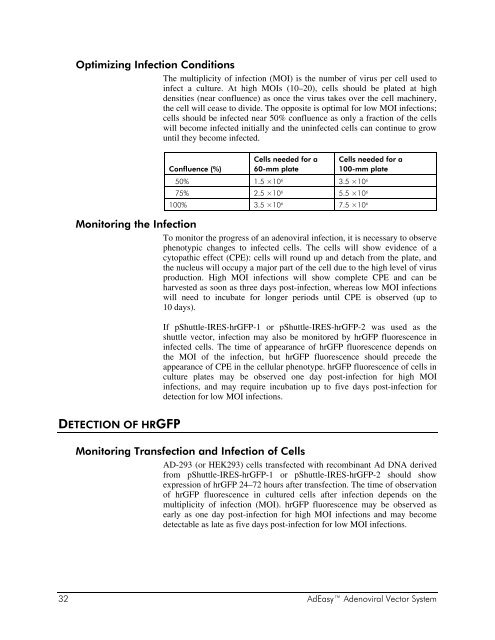
Optimizing Infection Conditions<br />
The multiplicity of infection (MOI) is the number of virus per cell used to<br />
infect a culture. At high MOIs (10–20), cells should be plated at high<br />
densities (near confluence) as once the virus takes over the cell machinery,<br />
the cell will cease to divide. The opposite is optimal for low MOI infections;<br />
cells should be infected near 50% confluence as only a fraction of the cells<br />
will become infected initially and the uninfected cells can continue to grow<br />
until they become infected.<br />
Confluence (%)<br />
Cells needed for a<br />
60-mm plate<br />
Cells needed for a<br />
100-mm plate<br />
50% 1.5 ×10 6 3.5 ×10 6<br />
75% 2.5 ×10 6 5.5 ×10 6<br />
100% 3.5 ×10 6 7.5 ×10 6<br />
Monitoring the Infection<br />
To monitor the progress of an adenoviral infection, it is necessary to observe<br />
phenotypic changes to infected cells. The cells will show evidence of a<br />
cytopathic effect (CPE): cells will round up and detach from the plate, and<br />
the nucleus will occupy a major part of the cell due to the high level of virus<br />
production. High MOI infections will show complete CPE and can be<br />
harvested as soon as three days post-infection, whereas low MOI infections<br />
will need to incubate for longer periods until CPE is observed (up to<br />
10 days).<br />
DETECTION OF HRGFP<br />
If pShuttle-IRES-hrGFP-1 or pShuttle-IRES-hrGFP-2 was used as the<br />
shuttle vector, infection may also be monitored by hrGFP fluorescence in<br />
infected cells. The time of appearance of hrGFP fluorescence depends on<br />
the MOI of the infection, but hrGFP fluorescence should precede the<br />
appearance of CPE in the cellular phenotype. hrGFP fluorescence of cells in<br />
culture plates may be observed one day post-infection for high MOI<br />
infections, and may require incubation up to five days post-infection for<br />
detection for low MOI infections.<br />
Monitoring Transfection and Infection of Cells<br />
AD-293 (or HEK293) cells transfected with recombinant Ad DNA derived<br />
from pShuttle-IRES-hrGFP-1 or pShuttle-IRES-hrGFP-2 should show<br />
expression of hrGFP 24–72 hours after transfection. The time of observation<br />
of hrGFP fluorescence in cultured cells after infection depends on the<br />
multiplicity of infection (MOI). hrGFP fluorescence may be observed as<br />
early as one day post-infection for high MOI infections and may become<br />
detectable as late as five days post-infection for low MOI infections.<br />
32 <strong>AdEasy</strong> <strong>Adenoviral</strong> <strong>Vector</strong> <strong>System</strong>