Orthobiologics in Shoulder Surgery - ShoulderDoc.co.uk
Orthobiologics in Shoulder Surgery - ShoulderDoc.co.uk
Orthobiologics in Shoulder Surgery - ShoulderDoc.co.uk

You also want an ePaper? Increase the reach of your titles
YUMPU automatically turns print PDFs into web optimized ePapers that Google loves.
<strong>Orthobiologics</strong><br />
<strong>in</strong><br />
<strong>Shoulder</strong> <strong>Surgery</strong><br />
Carl J. Basamania, MD, FACS<br />
The PolyCl<strong>in</strong>ic and Swedish<br />
Orthopaedic Institute<br />
Seattle, Wash<strong>in</strong>gton
“The future a<strong>in</strong>’t what it used to be”<br />
“You've got to be very careful if<br />
you don't know where you're go<strong>in</strong>g,<br />
because you might not get there.”<br />
Yogi Berra
What’s so special about<br />
shoulder surgery<br />
• Often work<strong>in</strong>g with chronic, degenerative<br />
<strong>co</strong>nditions<br />
• Rotator cuff tears are very <strong>co</strong>mmon<br />
– If you live long enough, you probably will have<br />
a cuff tear<br />
• Re-tear rates after traditional surgery is<br />
much higher than previously appreciated<br />
– 40-95% retear rate!<br />
• Annual f<strong>in</strong>ancial burden of $3 billion on the<br />
US e<strong>co</strong>nomy
My Introduction to <strong>Orthobiologics</strong><br />
• What is it<br />
– “highly <strong>in</strong>ductive,<br />
scaffold<strong>in</strong>g material”<br />
» Steve Arnockzy<br />
• How does it work<br />
– “I haven’t the slightest<br />
idea”<br />
» Steve Arnockzy
Realities of the “Orthopaedic<br />
Marketplace”<br />
• Most surgeons know noth<strong>in</strong>g about<br />
“orthobiologics”<br />
• Many surgeons know noth<strong>in</strong>g about soft<br />
tissue heal<strong>in</strong>g<br />
• Many surgeons are desperate for anyth<strong>in</strong>g<br />
to give them better results<br />
• Some surgeons will try anyth<strong>in</strong>g to get a<br />
“<strong>co</strong>mpetitive edge” over other surgeons
Biologics <strong>in</strong> <strong>Shoulder</strong> <strong>Surgery</strong><br />
• Soft tissue substitutes<br />
– Biologic<br />
– Structural<br />
• Hard tissue substitutes<br />
– Graft extenders<br />
– Graft substitutes<br />
• Heal<strong>in</strong>g “enhancers”
<strong>Orthobiologics</strong> <strong>in</strong> Orthopaedic <strong>Surgery</strong><br />
• Alternatives:<br />
– Bone Substitutes<br />
• Tricalcium phosphate<br />
• Amorphous Calcium Phosphate<br />
• Dicalcium Phosphate Dihydrate Precursors<br />
• Dem<strong>in</strong>eralized bone matrix<br />
–Soft tissue scaffolds<br />
• ECM grafts<br />
– Cell based heal<strong>in</strong>g response<br />
• Platelet Rich Plasma<br />
– Factor based heal<strong>in</strong>g
Bone Graft Substitutes<br />
Class Description Examples<br />
Allograft based<br />
Factor based<br />
Cell based<br />
Ceramic based<br />
Polymer based<br />
Allograft bone used alone or <strong>in</strong><br />
<strong>co</strong>mb<strong>in</strong>ation with other<br />
materials<br />
Natural and re<strong>co</strong>mb<strong>in</strong>ant<br />
growth factors used alone or <strong>in</strong><br />
<strong>co</strong>mb<strong>in</strong>ation with other<br />
materials<br />
Cells used to generate new<br />
tissue alone or seeded onto a<br />
support matrix<br />
Includes calcium phosphate,<br />
calcium sulfate, and bioglass<br />
used alone or <strong>in</strong> <strong>co</strong>mb<strong>in</strong>ation<br />
Both degradable and<br />
nondegradable polymers used<br />
alone and <strong>in</strong> <strong>co</strong>mb<strong>in</strong>ation with<br />
other materials<br />
Allogro, Othroblast, Opteform,<br />
Grafton<br />
TGF-beta, PDGF, FGF, BMP<br />
Mesenchymal stem cells,<br />
Cellect<br />
Osteograf, Norian SRS,<br />
ProOsteon, Osteoset, Conduit<br />
Cortoss, OPLA, Immix
Bone Graft Substitutes<br />
• 1998 - >300K bone grafts, 1999 ~500K<br />
• Total <strong>co</strong>st ~ $2.5 billion/year<br />
• However:<br />
– Relatively high <strong>co</strong>mplication rate ~20-30%<br />
– Donor site pa<strong>in</strong><br />
– Increased operative times<br />
– Increased operative <strong>co</strong>sts (supplies, OR time,<br />
etc)<br />
– Limited supply/availability
Bone Morphogenetic Prote<strong>in</strong><br />
• Also known as BMP<br />
– Synthetics: INFUSE ® and OP-1 ®<br />
• Signal<strong>in</strong>g prote<strong>in</strong> that tells mesenchymal<br />
stem cells what to be<strong>co</strong>me (i.e. bone,<br />
muscle or <strong>co</strong>nnective tissue)<br />
• 30 known human BMPs<br />
• “Key” that fits <strong>in</strong>to one keyhole on the<br />
surface of the MSC
Problems with BMP<br />
• Needs MSC’s<br />
• Expensive<br />
• Non-reimbursable<br />
• Questions of us<strong>in</strong>g<br />
factor <strong>in</strong> far greater<br />
than normal quantities
Problems with Factor Based<br />
Heal<strong>in</strong>g<br />
• Not only are the factors important<br />
– Relationship of the <strong>co</strong>mb<strong>in</strong>ation of factors<br />
– Quantity of factors<br />
– 3 dimensional presentation of factors<br />
– Host status
Platelet Rich Plasma & Fibr<strong>in</strong> Glue<br />
• Most important wound heal<strong>in</strong>g tissue<br />
factors are platelet derived<br />
– PDGF – platelet derived growth factor<br />
– VEGF – vascular/endothelial growth factor<br />
– TGFβ – transform<strong>in</strong>g growth factor<br />
– IFG-I – <strong>in</strong>sul<strong>in</strong> like growth factor<br />
• Can we enhance heal<strong>in</strong>g by application of<br />
a platelet <strong>co</strong>ncentrate at the time of<br />
surgery<br />
• Various systems available to <strong>co</strong>ncentrate<br />
autologous plate
Background<br />
• Multiple studies have shown enhanced<br />
wound heal<strong>in</strong>g, improved hemostasis and<br />
improved bone formation with application<br />
of PRP and PPP<br />
• However, other studies have shown no<br />
benefit and even decreased bone<br />
formation with PRP
PRP Causes Migration of MSCs<br />
250<br />
Cell number/chamber<br />
200<br />
150<br />
100<br />
50<br />
0<br />
SF 10% PPP 10% PRP<br />
Chemotactic Test Solution
PRP Causes Proliferation of MSCs<br />
900<br />
Cell Number X 1000<br />
800<br />
700<br />
600<br />
500<br />
400<br />
300<br />
200<br />
*<br />
*<br />
*<br />
*<br />
100<br />
0<br />
Serum Free<br />
Medium<br />
Growth<br />
Medium<br />
Peripheral<br />
Blood<br />
PPP<br />
0.625x 1.25x 2.5x 5x<br />
Fold Increase of Platelet Releasate<br />
*P
PRP and Implant Coat<strong>in</strong>g<br />
• Most studies are <strong>in</strong> dental, ENT and<br />
maxillofacial areas<br />
• Fontana, et al (2004) found greater<br />
volume of peri-implant bone on implants<br />
treated with PRP
• Gardner MJ, Demetrakopoulos D, Klepchick PR, et al.<br />
The efficacy of autologous platelet gel <strong>in</strong> pa<strong>in</strong> <strong>co</strong>ntrol and<br />
blood loss <strong>in</strong> total knee arthroplasty. An analysis of the<br />
haemoglob<strong>in</strong>, nar<strong>co</strong>tic requirement and range of motion.<br />
Int Orthop. 2007;31:309–313.<br />
• Siebrecht MA, De Rooij PP, Arm DM, et al. Platelet<br />
<strong>co</strong>ncentrate <strong>in</strong>creases bone <strong>in</strong>growth <strong>in</strong>to porous<br />
hydroxyapatite. Orthopedics. 2002;25:169–172.
PRP <strong>in</strong> My Practice
Observations<br />
• No longer need dra<strong>in</strong> post-op<br />
• Less swell<strong>in</strong>g and ecchymosis<br />
• Better/earlier range of motion and function<br />
• Incisions “mature” sooner<br />
• *Patients have been us<strong>in</strong>g significantly<br />
less post-op nar<strong>co</strong>tics
Humeral Nonunion 68yo female heavy smoker 1<br />
year s/p failed ORIF<br />
3 mos s/p Revision with PRP and TCP
Humeral Osteotomy Nonunion <strong>in</strong> 28 yo<br />
male now 1 month s/p revision with PRP<br />
and TCP
Revision shoulder arthroplasty<br />
PRP + TCP
PRP plus DBM/Cancellous Bone
72 yo home O 2 dependent, EF 25%<br />
3 weeks s/p revsion ORIF c/ PRP, 15cc IC Graft
Soft Tissue Scaffolds<br />
• CuffPatch (Arthrotek)<br />
– Porc<strong>in</strong>e SIS<br />
• Graftjacket (Wright<br />
Medical)<br />
– Human dermal tissue<br />
• Graftjacket Xpress<br />
(Wright Medical)<br />
– Flowable Soft Tissue<br />
Scaffold<br />
– micronized human<br />
dermal tissue<br />
• AlloPatch (MTF)<br />
– Human fascia lata,<br />
• Perma<strong>co</strong>l (Tissue<br />
Science Lab/Zimmer)<br />
– Porc<strong>in</strong>e dermal tissue<br />
• Restore (DePuy Ortho)<br />
– Porc<strong>in</strong>e SIS<br />
• TissueMend (Stryker<br />
Howmedica)<br />
– Bov<strong>in</strong>e fetal dermal<br />
tissue<br />
• OrthADAPT (Pegasus<br />
Biologics)<br />
– Equ<strong>in</strong>e Pericardium
Uses of the Restore® Graft<br />
• *FDA approved for:<br />
– Rotator cuff repair<br />
• 1 and 2 tendon repairs<br />
• NOT TO BE USED WITH MORE THAN MILD<br />
FATTY DEGENERATION<br />
– Re<strong>in</strong>forcement of soft tissue where<br />
weakness exists<br />
• Replacement of torn/ruptured ligaments<br />
• Replacement of torn/ruptured tendons
• 6 years post-op<br />
revision left rotator<br />
cuff repair<br />
• Patient rates left<br />
shoulder as<br />
“normal”<br />
• MRI at 1, 3 and 6<br />
year f/u show<br />
<strong>in</strong>tact left cuff<br />
• Patient has reruptured<br />
right RC<br />
repair done<br />
without Restore<br />
– Revised 10/2004<br />
with Restore
6 year postop MRI & CT
5 weeks postop Right revision
Lateral Ret<strong>in</strong>acular Defect <strong>in</strong> 36yo w/HIV –<br />
failed 2 prior attempts at primary repair<br />
Pre-op<br />
6 mos post-op
Re<strong>in</strong>forcement of Triceps Rupture<br />
“Overlay Technique”
Levator Scapulae Avulsion Repair
Post-acromioplasty deltoid avulsion
Subscapularis Re<strong>in</strong>forcement<br />
The “Ta<strong>co</strong>” Technique
Subscapularis<br />
re<strong>in</strong>forcement
Pectoralis Rupture
History<br />
• Cook and Arnoczky:<br />
– SIS appears to <strong>in</strong>duce regeneration of<br />
meniscal-like tissue <strong>in</strong> large, avascular<br />
meniscal defects <strong>in</strong> dogs<br />
– Control dogs had significantly more articular<br />
cartilage damage than grafted dogs<br />
– Tissue Eng 2001 Jun;7(3):321-34
History<br />
• Fox and Arnoczky:<br />
– SIS disks underwent cellular and extracellular<br />
matrix modification result<strong>in</strong>g <strong>in</strong> fibrocartilagelike<br />
tissue when placed free <strong>in</strong> dog jo<strong>in</strong>ts<br />
– Suggested that SIS scaffolds can support cell<br />
attachment and <strong>in</strong>growth <strong>in</strong> a diarthroadial<br />
jo<strong>in</strong>t<br />
– Tissue Eng 2004 Jan;10(1-2):129-37
Glenoid Resurfac<strong>in</strong>g Arthroplasty<br />
Not FDA approved for this application
Glenoid Resurfac<strong>in</strong>g Arthroplasty<br />
Not FDA approved for this application
Dog Glenoid Resurfac<strong>in</strong>g Study
Dog Glenoid Resurfac<strong>in</strong>g Results<br />
• There was significantly less surface<br />
erosion (12% VS. 39%, p>0.05%) <strong>in</strong> the<br />
group with SIS resurfac<strong>in</strong>g by 6 months<br />
• Good remodel<strong>in</strong>g on histopathology<br />
assessment was also seen <strong>in</strong> the SIS<br />
group at both 3 and 6 months.<br />
• There were no differences <strong>in</strong> lameness at<br />
3 and 6 months and no difference <strong>in</strong><br />
surface erosions at 3 months.
3 months<br />
Control<br />
SIS
6 months<br />
Control (2x)<br />
SIS (10x)
Necropsy photos obta<strong>in</strong>ed at 6 months<br />
Control<br />
SIS
Where Are We Go<strong>in</strong>g With Soft<br />
Tissue Grafts
Concerns:<br />
• Only 1 <strong>co</strong>ntrolled study<br />
– Questionable patient selection<br />
• Difficult to prove effectiveness <strong>in</strong> humans<br />
– Especially difficult <strong>in</strong> rotator cuff tears<br />
• No good studies show<strong>in</strong>g long-term effectiveness <strong>in</strong><br />
cuff repairs<br />
– What is a success<br />
• No pa<strong>in</strong><br />
• Good function<br />
• Intact cuff on<br />
re-imag<strong>in</strong>g
ECM’s <strong>in</strong> Rotator Cuff <strong>Surgery</strong><br />
• 2-year follow-up of 12 patients<br />
• Arthros<strong>co</strong>pic repair of massive chronic rotator cuff tears<br />
• Restore SIS as an augmentation device.<br />
• MRI scans showed significant thicken<strong>in</strong>g of the cuff tendon<br />
with the <strong>in</strong><strong>co</strong>rporation of the SIS graft <strong>in</strong> 11 patients<br />
• Cl<strong>in</strong>ical failure was observed with<strong>in</strong> 12 weeks with <strong>co</strong>mplete<br />
resorption of the graft <strong>in</strong> 1 patient<br />
• No evidence of local or systemic rejection or <strong>in</strong>fection <strong>in</strong> any<br />
patient<br />
• Mean postoperative UCLA s<strong>co</strong>re was 19.9 (out of 35)<br />
– Preoperative 9.9 (P.01)<br />
• Functional improvement <strong>in</strong> all patients based on Simple<br />
<strong>Shoulder</strong> Test<br />
• Metcalf, Savioe, Kellum. Oper Tech Orthop, 2002;12:204-8.
2 year Canadian Multicenter Study<br />
• Chronic one and two tendon tears<br />
• With<strong>in</strong> FDA clearance for use<br />
– No more than mild fatty <strong>in</strong>filtration<br />
– No significant retraction<br />
• One year data:<br />
– Restore patch decreased re-tear rate by at<br />
least 25%<br />
– No significant rejection or hyper-immune<br />
responses
ECM’s <strong>in</strong> Rotator Cuff <strong>Surgery</strong><br />
• Sclamberg et al evaluated the use of<br />
Restore as an augmentation device <strong>in</strong> a<br />
<strong>co</strong>hort of 11 patients undergo<strong>in</strong>g open<br />
repair of large or massive rotator cuff tears<br />
• At 6 months postoperatively, MRI scans<br />
revealed that 10 of 11 Restore augmented<br />
repairs had failed<br />
Sclamberg SG, Tibone JE, Itamura JM, Kasraeian S. Six-month<br />
magnetic resonance imag<strong>in</strong>g follow-up of large and massive<br />
rotator cuff repairs re<strong>in</strong>forced with porc<strong>in</strong>e small <strong>in</strong>test<strong>in</strong>al submu<strong>co</strong>sa.<br />
J <strong>Shoulder</strong> Elbow Surg 2004;13:538-41.
Use of Restore Orthobiologic Implant for<br />
Agumentation of Chronic Two Tendon<br />
Rotator Cuff Tears: A Controlled<br />
Randomized Study<br />
Joseph P. Iannotti MD, PhD, Jim Cic<strong>co</strong>ne CRNA,<br />
Michael Codsi MD,Young Kwon MD, PhD,<br />
Kathe Derw<strong>in</strong> PhD, John Brems MD<br />
JBJS, 2006
Results<br />
• Prospective, randomized, <strong>co</strong>ntrolled trial of 30<br />
patients with repairable, large 2-tendon cuff<br />
tears<br />
• The unaugmented group was 7% more likely to<br />
heal than the Restore augmented group<br />
• The postoperative PENN s<strong>co</strong>res <strong>in</strong> the<br />
unaugmented group (91 po<strong>in</strong>ts) were also<br />
significantly higher than those <strong>in</strong> the augmented<br />
group (83 po<strong>in</strong>ts) (P .007)
“Larger Sized Tear”<br />
Moderate to Severe Atrophy:<br />
Group II
Problem with these studies<br />
• Restore patch was used <strong>in</strong> cl<strong>in</strong>ical sett<strong>in</strong>g<br />
<strong>co</strong>ntra<strong>in</strong>dicated by the FDA clearance<br />
– Massive cuff tears with fatty degeneration and<br />
retraction<br />
• Is it reasonable to use a graft material <strong>in</strong> a<br />
non-<strong>in</strong>dicated sett<strong>in</strong>g and then question it’s<br />
value
Cellular Hysteria<br />
• Cellular elements<br />
• Zheng MH, Chen J, Kirilak Y, Willers C, Xu J, Wood D. Porc<strong>in</strong>e<br />
small <strong>in</strong>test<strong>in</strong>e submu<strong>co</strong>sa (SIS) is not an acellular <strong>co</strong>llagenous<br />
matrix and <strong>co</strong>nta<strong>in</strong>s porc<strong>in</strong>e DNA: possible implications <strong>in</strong> human<br />
implantation. J Biomed Mater Res B Appl Biomater 2005;73:61-7.<br />
• Is this functional DNA<br />
• What are the chances of cross – species DNA disease<br />
transmission versus species specific
What I Have Learned:<br />
• There is a great deal is misunderstand<strong>in</strong>g<br />
about the heal<strong>in</strong>g of rotator cuff<br />
– What is the goal of surgery<br />
• A functional muscle unit<br />
• There is great deal of misunderstand<strong>in</strong>g<br />
about ECMs<br />
• There has been a great deal of “sp<strong>in</strong>” by<br />
the manufacturers
“…there is no such th<strong>in</strong>g as an<br />
irreparable cuff…”
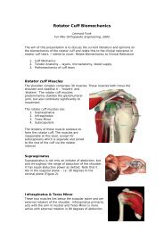
Native nerve<br />
orientation<br />
Suprascapular nerve<br />
Scapular notch<br />
First motor branch to<br />
suprasp<strong>in</strong>atus<br />
An Anatomical Study of the Effects on the Suprascapular Nerve Se<strong>co</strong>ndary to Retraction of the Suprasp<strong>in</strong>atus Muscle after a Rotator Cuff Tear,<br />
JSES, 2003
3cm retraction<br />
Suprascapular nerve<br />
Scapular notch<br />
First motor branch to<br />
suprasp<strong>in</strong>atus<br />
An Anatomical Study of the Effects on the Suprascapular Nerve Se<strong>co</strong>ndary to Retraction of the Suprasp<strong>in</strong>atus Muscle after a Rotator Cuff<br />
Tear,<br />
JSES, 2003
Biology of a Cuff Tear<br />
• Cytok<strong>in</strong>e-mRNAs <strong>in</strong> the shoulder synovium<br />
expressed more significantly <strong>in</strong> full-thickness<br />
tears<br />
• Significant <strong>in</strong>crease <strong>in</strong> <strong>co</strong>llagenase-3 (MMP-<br />
13) mRNA levels<br />
• Decrease <strong>in</strong> stromelys<strong>in</strong>-1 (MMP-3) mRNA<br />
levels<br />
• Decrease <strong>in</strong> tissue <strong>in</strong>hibitor of<br />
metalloprote<strong>in</strong>ase-2, -3, and -4 mRNA levels<br />
• Increase <strong>in</strong> the active form of <strong>co</strong>llagenase-3<br />
(MMP-13) <strong>in</strong> rotator cuff tendon tears.
Can We Change the Biology<br />
• In theory, <strong>in</strong>hibition of <strong>co</strong>llagenases and<br />
cytok<strong>in</strong>es should promote heal<strong>in</strong>g<br />
• If so, why do nonsteriodal anti<strong>in</strong>flammatory<br />
drugs decrease heal<strong>in</strong>g
What’s The Po<strong>in</strong>t of No Return<br />
• There are numerous adaptive and<br />
“maladaptive” responses to a cuff tear<br />
• These <strong>in</strong>clude biological, mechanical and<br />
neurological changes<br />
• Some of these make any attempt repair<br />
futile<br />
• Repair of these tears need to <strong>co</strong>nsider not<br />
only the mechanics of the repair but also<br />
the biology of the repair
Thank You
Comparative study of Soft Tissue<br />
Scaffolds<br />
• Badylak studied 5 different extracellular<br />
soft tissue scaffolds and <strong>co</strong>mpared them<br />
with an autologous tissue graft<br />
– GraftJacket – Wright Medical<br />
– Restore – DePuy<br />
– TissueMend – TEI Biosciences<br />
– Perma<strong>co</strong>l – Tissue Sciences<br />
– CuffPatch – Arthrotek<br />
• Critical abdom<strong>in</strong>al wall defect model
Results – Autologous Graft<br />
• Replaced with poorly<br />
organized <strong>co</strong>llagen<br />
<strong>co</strong>nsistent with scar<br />
A = 1 week<br />
C = 16 weeks
Results – Restore Graft<br />
• Rapidly <strong>in</strong>flitratred<br />
with dense<br />
mononuclear cells<br />
• Followed by<br />
progressive<br />
organization <strong>in</strong>to a<br />
mixture of skeletal<br />
muscle and organized<br />
<strong>co</strong>nnective tissue<br />
A = 1 week<br />
C = 16 weeks
Results – CuffPatch<br />
• Prolonged <strong>in</strong>filtration<br />
by mononuclear cells<br />
AND foreign body<br />
giant cells<br />
• Followed by partial<br />
scaffold remodel<strong>in</strong>g<br />
<strong>in</strong>to organized<br />
<strong>co</strong>nnective tissue<br />
A = 1 week<br />
C = 16 weeks
Results – GraftJacket<br />
• Intense cellular<br />
<strong>in</strong>filtrate<br />
• Eventual replacement<br />
by partially organized,<br />
dense <strong>co</strong>nnective<br />
tissue<br />
A = 1 week<br />
C = 16 weeks
Results – TissueMend<br />
• Little cellular<br />
<strong>in</strong>filtration<br />
• Eventually<br />
surrounded by fibrous<br />
<strong>co</strong>nnective tissue and<br />
adipose tissue<br />
A = 1 week<br />
C = 16 weeks
Results – Perma<strong>co</strong>l<br />
• Rema<strong>in</strong>ed <strong>in</strong>tact with<br />
virtually no cellular<br />
<strong>in</strong>filtration<br />
• Eventually enveloped<br />
by a dense fibrous<br />
capsule<br />
A = 1 week<br />
C = 16 weeks
Conculsion<br />
• Host response to different soft tissue<br />
scaffolds will vary based on their<br />
<strong>co</strong>mposition and process<strong>in</strong>g<br />
• Stronger with less <strong>in</strong>flammation is not<br />
necessarily better