2011 Annual Report - Center for Integrated Nanotechnologies - Los ...
2011 Annual Report - Center for Integrated Nanotechnologies - Los ...
2011 Annual Report - Center for Integrated Nanotechnologies - Los ...

Create successful ePaper yourself
Turn your PDF publications into a flip-book with our unique Google optimized e-Paper software.
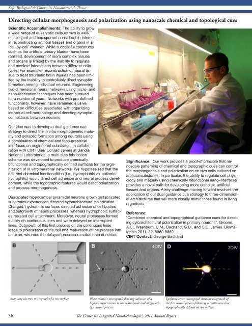
Soft, Biological & Composite Nanomaterials Thrust<br />
Directing cellular morphogenesis and polarization using nanoscale chemical and topological cues<br />
Scientific Accomplishments: The ability to grow<br />
a wide range of eukaryotic cells ex vivo is wellestablished<br />
and has spurred considerable interest<br />
in reconstructing artificial tissues and organs in a<br />
“cell-by-cell” manner. While successful constructs<br />
such as the artificial urinary bladder have been<br />
realized, development of more complex tissues<br />
and organs is limited by the inability to regulate<br />
and mediate interactions between different cells<br />
types. For example, reconstruction of neural tissue<br />
to treat traumatic brain injuries has been limited<br />
by the inability to controllably direct synaptic<br />
<strong>for</strong>mation among individual neurons. Engineering<br />
two-dimensional neural networks using micro- and<br />
nano-fabrication techniques has been pursued<br />
<strong>for</strong> a number of years. Networks with pre-defined<br />
functionality, however, have remained elusive<br />
based on difficulties associated with organizing<br />
individual cell morphology and directing synaptic<br />
connections between neurons.<br />
Our idea was to develop a dual guidance cue<br />
strategy to direct the in vitro morphogenetic maturity<br />
and synaptic <strong>for</strong>mation among neurons using<br />
a combination of chemical and topo-graphical<br />
interfaces on engineered substrates. In collaboration<br />
with CINT User Conrad James at Sandia<br />
National Laboratories, a multi-step fabrication<br />
scheme was developed to produce chemically<br />
bifunctional and topographically defined surfaces <strong>for</strong> the organization<br />
of in vitro neuronal networks. We hypothesized that the<br />
different chemical functionalities (i.e., hydrophobic vs. cationic/<br />
hydrophilic) would direct cell adhesion and neural process development,<br />
while the topographic features would direct polarization<br />
and process morphogenesis.<br />
Dissociated hippocampal pyramidal neurons grown on fabricated<br />
substrates experienced directed cytoarchitectural polarization.<br />
Charged, hydrophilic surfaces directed adhesion of cell bodies<br />
and outgrowth of neural processes, whereas hydrophobic surfaces<br />
resisted cell attachment. Moreover, neural processes <strong>for</strong>med<br />
quickly on continuous lines and were delayed on interrupted<br />
lines. Outgrowth of this first process on the continuous lines<br />
leads to polarization of the cell and maturation of the process into<br />
an axon, whereas the delayed processes mature into dendrites.<br />
Significance: Our work provides a proof-of principle that nanoscale<br />
patterning of chemical and topographic cues can control<br />
the morphogenesis and polarization on ex vivo cells cultured on<br />
artificial substrates. In particular, the ability to regulate cell physiology<br />
and maturity using chemically bifunctional nano-interfaces<br />
provides a novel path <strong>for</strong> developing more complex, artificial<br />
tissues and organs. A key challenge moving <strong>for</strong>ward involves the<br />
application of our dual guidance cue strategy to three-dimensional<br />
architectures that will more closely mimic those found in living<br />
organisms.<br />
Reference:<br />
“Combined chemical and topographical guidance cues <strong>for</strong> directing<br />
cytoarchitectural polarization in primary neurons”, Greene,<br />
A.C., Washburn, C.M., Bachand, G.D., and C.D. James. Biomaterials<br />
<strong>2011</strong>, 32, 8860-8869.<br />
CINT Contact: George Bachand<br />
Scanning electron micrograph of a test surface.<br />
Phase contrast micrograph showing adhesion of a<br />
hippocampal neuron to the centralnode and outgrowth<br />
of a neural process.<br />
Epifluorescence micrograph showing outgrowth of<br />
the first neural process following a continuous line<br />
topographically defined on the surface.<br />
36<br />
36The <strong>Center</strong> <strong>for</strong> <strong>Integrated</strong> <strong>Nanotechnologies</strong> | <strong>2011</strong> <strong>Annual</strong> <strong>Report</strong>