PathScan® ELISA Products and Cellular Analysis Tools - Lab-JOT
PathScan® ELISA Products and Cellular Analysis Tools - Lab-JOT
PathScan® ELISA Products and Cellular Analysis Tools - Lab-JOT

You also want an ePaper? Increase the reach of your titles
YUMPU automatically turns print PDFs into web optimized ePapers that Google loves.
PathScan ®<br />
<strong>ELISA</strong> <strong>Products</strong><br />
<strong>and</strong> <strong>Cellular</strong> <strong>Analysis</strong> <strong>Tools</strong><br />
www.cellsignal.com<br />
UNPARALLELED PRODUCT QUALITY, VALIDATION, AND TECHNICAL SUPPORT
PathScan ® S<strong>and</strong>wich <strong>ELISA</strong> Kits<br />
Cell Signaling Technology (CST) has applied<br />
its antibody expertise to identify antibody<br />
pairs with optimal activity in solid phase<br />
s<strong>and</strong>wich enzyme-linked immunosorbent<br />
assays (<strong>ELISA</strong>). These assays enable the<br />
detection of low<br />
amounts of target<br />
protein from cell<br />
lysates. Over 170<br />
PathScan ® S<strong>and</strong>wich<br />
<strong>ELISA</strong> kits are available<br />
for signaling proteins<br />
that serve as assay endpoints in<br />
drug discovery screening campaigns.<br />
Kits <strong>and</strong> pairs are developed,<br />
produced, <strong>and</strong> validated<br />
in-house, ensuring the highest<br />
quality.<br />
» PathScan ® S<strong>and</strong>wich <strong>ELISA</strong> Kits (both Colormetric<br />
<strong>and</strong> Chemiluminescent) contain all necessary<br />
components for detection of endogenous levels of<br />
key signaling molecules. Matched phospho <strong>and</strong> total<br />
protein <strong>ELISA</strong> kits are available for many targets.<br />
» PathScan ® Multi-Target <strong>ELISA</strong> Kits examine several<br />
important <strong>and</strong> well-characterized signaling events in<br />
a single assay.<br />
» PathScan ® S<strong>and</strong>wich <strong>ELISA</strong> Antibody Pairs provide<br />
scientists with an economical alternative to our<br />
complete <strong>ELISA</strong> kits.<br />
» PathScan ® <strong>ELISA</strong> Control Cell Extracts provide the<br />
appropriate positive <strong>and</strong> negative controls, <strong>and</strong> also<br />
allow the st<strong>and</strong>ardization of signal obtained from<br />
different plates.<br />
» Custom <strong>ELISA</strong> products allow researchers the option<br />
of different detection methods <strong>and</strong> plate formats.<br />
Convenient bulk packaging is available upon request.<br />
XP Monoclonal Antibodies<br />
XP Monoclonal Antibodies are a line of high<br />
quality rabbit monoclonal antibodies exclusively<br />
available from Cell Signaling Technology (CST).<br />
Any product labeled with XP has been carefully<br />
selected based on superior performance in all<br />
approved applications.<br />
XP Monoclonal Antibodies are generated using XMT , a proprietary<br />
monoclonal technology developed at Cell Signaling Technology. The<br />
technology provides access to a broad range of antibody-producing<br />
B cells unattainable with traditional monoclonal technologies,<br />
allowing more comprehensive screening <strong>and</strong> the identification of XP <br />
monoclonal antibodies with:<br />
eXceptional specificity<br />
As with all CST antibodies, the antibody is specific to your target<br />
of interest, saving you valuable time <strong>and</strong> resources.<br />
+ eXceptional sensitivity<br />
The antibody will provide a stronger signal for your target protein in<br />
cells <strong>and</strong> tissues, allowing you to monitor expression of low levels<br />
of endogenous proteins, saving you valuable materials.<br />
+ eXceptional stability <strong>and</strong> reproducibility<br />
XMT combined with our stringent quality control ensures maximum<br />
lot-to-lot consistency <strong>and</strong> the most reproducible results.<br />
EGF Receptor (D38B1) XP Rabbit mAb #4267:<br />
Confocal IF analysis of A549 cells, untreated (top) or<br />
treated with human epidermal growth factor (bottom),<br />
using #4267 (green). Blue = DRAQ5 ® #4084 (fluorescent<br />
DNA dye).<br />
= eXceptional Performance <br />
XMT coupled with our extensive antibody validation <strong>and</strong> stringent<br />
quality control delivers XP monoclonal antibodies with eXceptional<br />
Performance in the widest range of applications.
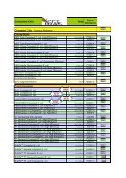
PathScan ® S<strong>and</strong>wich <strong>ELISA</strong> <strong>Products</strong><br />
Colormetric<br />
S<strong>and</strong>wich <strong>ELISA</strong> Kits<br />
Chemiluminescent<br />
S<strong>and</strong>wich <strong>ELISA</strong> Kits<br />
Antibody<br />
Pairs<br />
<strong>ELISA</strong> Control<br />
Cell Extract<br />
Target<br />
Multi-Target Kits<br />
Phospho-4E-BP1 (Thr37/Thr46) #7216 #7854<br />
4E-BP1 #7179<br />
Phospho-Acetyl CoA Carboxylase (Ser79) #7986<br />
β-Actin #7880 #7881<br />
Phospho-AMPKα (Thr172) #7959 #7955<br />
Phospho-Akt (Thr308) #7252 #7135 #7144 Cell Growth #7239 #7989<br />
Phospho-Akt1 (Ser473) #7160 #7134 #7143<br />
Cell Growth #7239<br />
Signaling Nodes #7272<br />
#7988<br />
Akt1 #7170 #7132 #7142<br />
Cell Growth #7239<br />
Signaling Nodes #7272<br />
#7989<br />
Phospho-Akt2 (Ser474) #7048<br />
Phospho-Akt2 (Ser474) (mouse preferred) #7932<br />
Akt2 #7046<br />
Phospho-Akt3 (Ser472) (mouse preferred) #7942<br />
Akt3 (mouse preferred) #7934<br />
Phospho-ALK (Tyr1586) #7159<br />
Phospho-ALK (Tyr1604) #7324 #7020<br />
ALK #7322 #7084<br />
Phospho-ATF-2 (Thr71) #7185 #7989<br />
Phospho-Aurora A (Thr288) #7114 #7115<br />
Aurora A #7116 #7117<br />
Axl (pan p-Tyr) #7042<br />
Axl #7040<br />
Phospho-Bad (Ser112) #7182 #7842 Apoptosis #7105 #7989<br />
Bad #7162 #7840 Apoptosis #7105 #7988<br />
β-Catenin #7308 #7309<br />
E-Cadherin #7886 #7887<br />
Cleaved Caspase-3 (Asp175) #7190 Apoptosis #7105<br />
Phospho-cdc2 (Tyr15) #7176 #7838<br />
Phospho-Chk1 (Ser317) #7870 #7989<br />
Chk1 #7872 #7873<br />
Phosho-Chk2 (Thr68) #7037<br />
Chk2 #7045 #7090<br />
Phospho-c-Jun (Ser63) #7145 #7027 #7141<br />
c-Jun #7150 #7028 #7314<br />
Phospho-DDR1 (pan p-Tyr) #7863<br />
DDR1 #7845<br />
Phospho-EGFR (Tyr845) #7189<br />
Phospho-EGFR (Tyr1068) #7240<br />
Phospho-EGFR (Tyr1173) #7187<br />
EGFR #7250<br />
Phospho-eIF2α (Ser51) #7948 #7988<br />
eIF2α #7952 #7988<br />
Phospho-eIF4E (Ser209) #7938<br />
eIF4E #7940<br />
Phospho-eNOS (Ser1177) #7980<br />
Phospho-Erk1 (Thr202/Tyr204) #7315 #7278<br />
Phospho-Erk1/2 (Thr202/Tyr204) #7177 #7246<br />
Cell Growth #7239<br />
MAP Kinase #7274<br />
Erk 1/2 #7050<br />
Phospho-Flt3 (pan p-Tyr) #7761<br />
Phospho-Flt3 (Tyr591) #7206<br />
Flt3 #7202<br />
GFP #7878 #7879<br />
Phospho-HER2 (pan p-Tyr) #7968<br />
Phospho-HER2 (Tyr1221/1222) #7148 #7817<br />
HER2 #7310<br />
Phospho-HER3 (pan p-Tyr) #7890<br />
HER3 #7888<br />
Acetyl-Histone H2A #7233
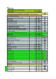
PathScan ® S<strong>and</strong>wich <strong>ELISA</strong> products continued<br />
Colormetric<br />
S<strong>and</strong>wich <strong>ELISA</strong> Kits<br />
Chemiluminescent<br />
S<strong>and</strong>wich <strong>ELISA</strong> Kits<br />
Antibody<br />
Pairs<br />
Target<br />
Multi-Target Kits<br />
Acetyl-Histone H2B #7178<br />
Acetyl-Histone H2B (Lys5) #7218<br />
Acetyl-Histone H2B (Lys20) #7222<br />
Acetyl-Histone H3 #7232 #7209<br />
Acetylated Histone H3 (Lys9) #7121<br />
Acetylated Histone H3 (Lys18) #7122<br />
Mono-Methyl Histone H3 (Lys4) #7123<br />
Di-Methyl-Histone H3 (Lys4) #7124<br />
Tri-Methyl Histone H3 (Lys4) #7125<br />
Pan-Methyl Histone H3 (Lys9) #7864<br />
Di-Methyl Histone H3 (Lys9) #7862<br />
Tri-Methyl Histone H3 (Lys27) #7866<br />
Di-Methyl Histone H3 (Lys36) #7868<br />
Phospho-Histone H3 (Ser10) #7155 #7207<br />
Histone H3 #7253<br />
Acetyl-Histone H4 #7238<br />
Acetyl-Histone H4 (Lys8) #7224<br />
Acetyl-Histone H4 (Lys12) #7228<br />
Phospho-HSP27 (Ser78) #7290<br />
Phospho-HSP27 (Ser82) #7152<br />
HSP27 #7295<br />
Phospho-IGF1 Receptor (Tyr1131) #7302 #7820<br />
Phospho-IκBα (Ser32) #7355 #7343 Inflammation #7276<br />
IκBα #7360 #7831<br />
Phospho-IKKα (Ser176/180) #7073<br />
Total-IKKα #7078<br />
Phospho-IKKβ (Ser177/181) #7080<br />
iNOS #7097<br />
Phospho-Insulin Receptor B (Tyr1146) #7254 #7827<br />
Phospho-Insulin Receptor B (Tyr1150/1151) #7258 #7828<br />
Phospho-Insulin Receptor B (Tyr1345) #7326 #7823<br />
Phospho-IRS-1 (pan p-Tyr) #7133 #7347<br />
Phospho-IRS-1 (Ser302) #7283 #7284<br />
Phospho-IRS-1 (Ser307) #7287 #7288<br />
Phospho-IRS-1 (Ser612) #7332<br />
IRS-1 #7328<br />
Phospho-IRS-2 (pan p-Tyr) #7860 #7861<br />
IRS-2 #7884 #7885<br />
Phospho-c-Kit (pan p-Tyr) #7231 #7294<br />
Phospho-c-Kit (Tyr719) #7298 #7299<br />
c-Kit #7197<br />
Phospho-LAT (Tyr191) #7936 #7937<br />
Phospho-Lck (Tyr505) #7941 #7993<br />
<strong>ELISA</strong> Control<br />
Cell Extract<br />
Phospho-MEK1 (Ser217/221) #7175 #7029 #7211<br />
Signaling Nodes #7272<br />
MAP Kinase #7274<br />
#7988<br />
MEK1 #7165 #7030 #7215 MAP Kinase #7274<br />
Phospho-Met (pan p-Tyr) #7333 #7334<br />
Phospho-Met (Tyr1003) #7241<br />
Phospho-Met (Tyr1234/1235) #7227 #7229<br />
Phospho-Met (Tyr1349) #7896<br />
Met #7242<br />
Phospho-NF-κB p65 (Ser536) #7173 #7834<br />
Inflammation #7276<br />
Signaling Nodes #7272<br />
NF-κB p65 #7174 #7836 Inflammation #7276 #7988<br />
p21 WAF1/CIP1 #7167 #7856<br />
Phospho-p38 (Thr180/Tyr182) #7946<br />
MAP Kinase #7274<br />
Inflammation #7276<br />
Signaling Nodes #7272<br />
Acetyl-p53 #7236 #7848<br />
Phospho-p53 (Ser15) #7365 #7846 Apoptosis #7105<br />
p53 #7370 #7844 Apoptosis #7105
Colormetric<br />
S<strong>and</strong>wich <strong>ELISA</strong> Kits<br />
Chemiluminescent<br />
S<strong>and</strong>wich <strong>ELISA</strong> Kits<br />
Antibody<br />
Pairs<br />
<strong>ELISA</strong> Control<br />
Cell Extract<br />
Target<br />
Multi-Target Kits<br />
Phospho-p70 S6 Kinase (Thr389) #7063 #7053<br />
p70 S6 Kinase #7038 #7039<br />
Phospho-p90 Rsk1 (Ser380) #7965<br />
p90 Rsk1 #7966<br />
Cleaved PARP (Asp214) #7262 #7858 Apoptosis #7105<br />
Phospho-PDGFR α/β (pan p-Tyr) #7235 #7307<br />
Phospho-PDGFR α (Tyr849) #7296 #7317<br />
PDGFR α #7318 #7264<br />
Phospho-PDGFR β (Tyr751) #7345 #7826<br />
Phospho-PTEN (Ser380) #7285<br />
PTEN #7882 #7883<br />
Phospho-Ret (pan p-Tyr) #7034<br />
Ret #7032<br />
Phospho-Ros (pan p-Tyr) #7093<br />
Phospho-S6 Ribosomal Protein (Ser235/236) #7205 #7201 Cell Growth #7239 #7988<br />
S6 Ribosomal Protein #7225 #7203 Cell Growth #7239 #7988<br />
Phospho-SAPK/JNK1/2/3 (Thr183/Tyr185) #7325 #7849 #7217<br />
MAP Kinase #7274<br />
Inflammation #7276<br />
#7989<br />
Phospho-Src (Tyr416) #7953 #7963<br />
Src #7984 #7992<br />
SAPK/JNK1/2/3 #7330 #7869 #7219 MAP Kinase #7274 #7989<br />
Phospho-Smad2 (Ser465/467) #7348<br />
Smad2 #7244<br />
Phospho-Stat1 (Tyr701) #7234<br />
Phospho-Stat3 (Tyr705) #7300 #7149 #7146<br />
Signaling Nodes #7272<br />
Inflammation #7276<br />
Phospho-Stat3 (Ser727) #7995<br />
Stat3 #7305<br />
Phospho-Stat5 (Tyr694) #7113 #7281<br />
Survivin #7169<br />
Phospho-Syk (pan p-Tyr) #7928 #7929<br />
Phospho-Syk (Tyr525/526) #7970<br />
Phospho-TrkA (Tyr490) #7210<br />
Phospho-TrkA (Tyr674/675) #7212<br />
TrkA #7208<br />
Phospho-TrkB (pan p-Tyr) #7108<br />
Phospho-TrkB (Tyr516) #7111<br />
Phospho-TrkB (Tyr706/707) #7118<br />
TrkB #7106<br />
α-Tubulin #7944 #7945 #7989<br />
Acetyl-Tubulin #7204<br />
Phospho-VEGFR-2 (Tyr1175) #7335 #7842<br />
VEGFR-2 #7340 #7825<br />
Phospho-Zap-70 (Tyr319) #7171 #7852<br />
Zap-70 #7172 #7850<br />
Companion <strong>Products</strong><br />
» BSA #9998<br />
» Cell Lysis Buffer (10X) #9803<br />
» Phosphate Buffered Saline (PBS-20X) #9808<br />
» Phosphate Buffered Saline with Tween 20 (PBST-20X) #9809<br />
» STOP Solution #7002<br />
» TMB Substrate #7004
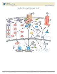
PathScan ® RTK Signaling Antibody<br />
Array Kits<br />
These kits offer the user the opportunity to monitor<br />
a multitude of targets simultaneously with minimal<br />
sample requirements. The PathScan ® RTK Signaling<br />
Antibody Array Kits are slide-based antibody arrays<br />
founded on the s<strong>and</strong>wich immunoassay principle.<br />
These kits include all necessary reagents for either<br />
fluorescent or chemiluminescent detection.<br />
4 4 9 9 14 14 19 19 24 24 29 29 34 34<br />
1 1 5 5 10 10 15 15 20 20 25 25 30 30 35 35<br />
2 2 6 6 11 11 16 16 21 21 26 26 31 31 36 36 39<br />
3 3 7 7 12 12 17 17 22 22 27 27 32 32 37 37 39<br />
8 8 13 13 18 18 23 23 28 28 33 33 38 38<br />
Positive Control<br />
Negative Control<br />
Each kit contains two 8-pad slides, allowing the user to test up to<br />
16 samples. Each pad is spotted in duplicate with 39 targetspecific<br />
capture antibodies, biotinylated protein (positive control),<br />
<strong>and</strong> nonspecific IgG (negative control). Following incubation with<br />
sample lysate, a biotinylated secondary antibody cocktail recognizes<br />
captured protein targets. Sample readout can be performed by<br />
either chemiluminescent or fluorescent detection.<br />
» Arrays are produced <strong>and</strong> optimized in-house,<br />
incorporating the highest quality antibodies<br />
<strong>and</strong> ensuring results you can trust.<br />
» Arrays allow the analysis of phosphorylation<br />
levels of 39 proteins per assay, saving valuable<br />
time <strong>and</strong> reagents.<br />
» Arrays are designed to detect RTKs <strong>and</strong> key<br />
intracellular signaling molecules, allowing the<br />
most comprehensive readout of downstream<br />
signaling events.<br />
» Technical support is provided by the same<br />
scientists who developed <strong>and</strong> produce the<br />
product, allowing us to provide a thorough,<br />
fast, <strong>and</strong> accurate response.<br />
» The option of chemiluminescent readout<br />
allows convenient <strong>and</strong> easy detection by<br />
conventional chemiluminescent film without<br />
specialized instrumentation.<br />
PathScan ® Antibody Array Kits currently offered<br />
#7982 PathScan ® RTK Signaling Antibody Array Kit<br />
(Chemiluminescent Readout)<br />
#7949 PathScan ® RTK Signaling Antibody Array Kit<br />
(Fluorescent Readout)<br />
Fluorescence Intensity<br />
16000<br />
14000<br />
12000<br />
10000<br />
8000<br />
6000<br />
4000<br />
2000<br />
0<br />
EGFR<br />
HER2<br />
No Lysate<br />
Karpas-299<br />
K-562<br />
HER3<br />
FGFR1<br />
FGFR3<br />
FGFR4<br />
INSR<br />
IGF-IR<br />
TrkA<br />
TrkB<br />
Met<br />
Ron<br />
*All targets are phospho-specific with pan-tyrosine recognition<br />
unless a specific phophorylation site is designated.<br />
Screening of Karpas-299 <strong>and</strong> K-562 cell lines<br />
using the PathScan ® RTK Signaling Antibody<br />
Array Kits reveals various phosphorylated<br />
RTKs <strong>and</strong> signaling nodes. The fluorescent<br />
readout (middle panel) <strong>and</strong> the corresponding<br />
quantification (upper panel) were obtained<br />
using PathScan ® RTK Signaling Antibody<br />
Array Kit (Fluorescent Readout) #7949. The<br />
chemiluminescent readout (lower panel) was<br />
obtained using PathScan ® RTK Signaling<br />
Antibody Array Kit (Chemiluminescent Readout)<br />
#7982 <strong>and</strong> chemiluminescent film.<br />
Ret<br />
ALK<br />
PDGFR<br />
c-Kit<br />
FLT3<br />
M-CSFR<br />
EphA1<br />
EphA2<br />
EphA3<br />
EphB1<br />
EphB3<br />
EphB4<br />
Tyro3<br />
Axl<br />
Tie2<br />
VEGFR2<br />
Akt (Thr308)<br />
p44/42 (Thr202/Tyr204)<br />
Akt (Ser473)<br />
No Lysate Karpas-299<br />
K-562<br />
ALK (pan-Tyr)<br />
ALK (pan-Tyr)<br />
Akt (Thr308)<br />
Akt (Thr308)<br />
Stat3<br />
(Tyr705)<br />
Stat3<br />
(Tyr705)<br />
S6 (Ser235/236)<br />
c-Abl<br />
IRS-1<br />
Zap-70<br />
Src<br />
Lck<br />
Stat1 (Tyr701)<br />
Stat3 (Tyr705)<br />
c-Abl (pan-Tyr)<br />
S6 Ribosomal Protein (Ser235/236)<br />
c-Abl (pan-Tyr)<br />
S6 Ribosomal Protein (Ser235/236)
<strong>Cellular</strong> <strong>Analysis</strong> <strong>Tools</strong><br />
BrdU Cell Proliferation Assay Kit<br />
The BrdU Cell Proliferation Assay Kit #6813 from<br />
Cell Signaling Technology (CST) is a plate-based<br />
immunoassay that provides a straight forward<br />
means of assaying fundamental cellular activity.<br />
This CST kit offers an accurate, sensitive, <strong>and</strong><br />
direct readout of cell division unattainable with<br />
viability dyes.<br />
Advantages of the CST BrdU<br />
Cell Proliferation Assay Kit include:<br />
» Ability to interface with microplate<br />
environment, allowing higher throughput.<br />
» Elimination of the need for microscopy,<br />
yielding results without specialized equipment.<br />
» Elimination of the need for radioactive<br />
isotope labeling, providing a safer <strong>and</strong><br />
simpler protocol.<br />
4<br />
HRP<br />
TMB<br />
BrdU<br />
Add BrdU (5-bromo-2’-deoxyuridine)<br />
to the culture media of proliferating cells<br />
BrdU is a pyrimidine analog <strong>and</strong> is<br />
incorporated into newly synthesized<br />
DNA in place of thymidine<br />
Cells are fixed <strong>and</strong> DNA is exposed;<br />
BrdU mouse mAb dectects BrdU<br />
incorporated into DNA<br />
Anti-mouse secondary antibody<br />
conjugated to HRP is added<br />
TMB substrate is added <strong>and</strong> turns<br />
color in the presence of HRP (strength<br />
of color is directly proportional to<br />
amount of BrdU incorporated)<br />
Measure absorbance at 450 nm<br />
Absorbance<br />
450nm<br />
3<br />
2<br />
1<br />
0<br />
0.001 0.01 0.1 1 10 100<br />
hEGF (ng/ml)<br />
< Treatment of MCF7 10A cells with Human Epidermal Growth Factor (hEGF) #8916 increases<br />
cell proliferation as detected by BrdU Cell Proliferation Assay Kit #6813. MCF7 10A cells were<br />
seeded at 1x10 4 cells/well in a 96-well plate <strong>and</strong> incubated overnight. Cells were then starved<br />
in serum free medium overnight. hEGF was added to the plate <strong>and</strong> cells were incubated for 24<br />
hours. Finally, 10 μM BrdU was added to the plate <strong>and</strong> cells were incubated for 4 hours.<br />
Cyclic AMP <strong>and</strong> GMP Assay Kits<br />
CST now offers new Cyclic AMP <strong>and</strong> Cyclic GMP Assay Kits to<br />
measure the activation of many G protein coupled receptors<br />
(GPCRs). Both kits are immunoassays based on competitive<br />
binding.<br />
In the Cyclic AMP XP Assay Kit #4339, cAMP in the sample<br />
of interest competes with a fixed amount of cAMP-HRP<br />
conjugate provided in the kit for the binding to a cAMP XP<br />
rabbit monoclonal antibody that is pre-coated on the assay<br />
plate. Because of the competitive nature of this assay, the<br />
magnitude of the absorbance is inversely proportional to the<br />
quantity of cAMP in the sample.<br />
CST’s highest quality XP monoclonal antibodies employed in<br />
the assay ensure the greatest possible sensitivity <strong>and</strong> specificity.<br />
Enzymatic Immunoassays<br />
#4339 Cyclic AMP XP Assay Kit<br />
#4360 Cyclic GMP XP Assay Kit<br />
Other <strong>Cellular</strong> <strong>Analysis</strong> <strong>Tools</strong><br />
#9860 Senescence β-Galactosidase<br />
Staining Kit<br />
Absorbance 450 nm<br />
3.0<br />
2.5<br />
2.0<br />
1.5<br />
1.0<br />
0.5<br />
0.0<br />
-3 -2 -1 0<br />
Log [Isoproterenol] (µM)<br />
Treatment of 293 cells with isoproterenol increases<br />
the cAMP concentration as detected by Cyclic AMP<br />
XP Assay Kit #4339. 293 cells were seeded at<br />
3*104 cells/well in a 96-well plate <strong>and</strong> incubated<br />
overnight. Cells were pretreated with 0.5 mM IBMX<br />
for 30 minutes prior to isoproterenol treatment (3<br />
minutes) <strong>and</strong> lysed with 1X Cell Lysis Buffer #9803.<br />
The absorbance values (left) <strong>and</strong> percentage of activity<br />
(right) are shown above. The percentage of activity<br />
is calculated as follows: % activity=100X[(A-Abasal )/<br />
(Amax-Abasal )], where A is the absorbance of the sample,<br />
Amax is the absorbance at maximum stimulation (i.e.,<br />
high isoproterenol concentration), <strong>and</strong> Abasal is the<br />
absorbance at basal level (no isoproterenol).<br />
1<br />
% Activity<br />
110<br />
90<br />
70<br />
50<br />
30<br />
10<br />
-10<br />
-2 -1<br />
Log [Isoprot
USA Headquarters<br />
Cell Signaling Technology<br />
Technical Support: (toll-free) 1-877-678-8324<br />
Tel: 978-867-2300 / Fax: 978-867-2400<br />
E-mail: info@cellsignal.com / www.cellsignal.com<br />
In the U.S. for Bulk Volumes or Pricing Information,<br />
contact our sales team: sales@cellsignal.com<br />
International Subsidiaries<br />
Cell Signaling Technology (China) Limited<br />
Technical Support: (toll-free) 4006 GreatQ (473287)<br />
Tel: (86) 21-5835-6288 / Fax: (86) 21-5835-6116<br />
E-mail: info@cst-c.com.cn / www.cst-c.com.cn<br />
Cell Signaling Technology Japan, K.K.<br />
Tel: 03 (5652) 0213 / Fax: 03 (3249) 1170<br />
E-mail: info@cstj.co.jp / www.cstj.co.jp<br />
Cell Signaling Technology Europe<br />
Tel: +31 (0)71 568 1060 / Fax: +31 (0)71 568 1065<br />
E-mail: info@cellsignal.eu / www.cellsignal.eu<br />
ANTIBODIES AND RELATED REAGENTS<br />
FOR SIGNAL TRANSDUCTION RESEARCH<br />
Printed in the USA on recycled paper using soy inks <strong>and</strong> processed chlorine free.<br />
© 11/2010 Cell Signaling Technology, Inc.<br />
Cell Signaling Technology ® , XP, XMT, eXceptional Performance, CST, <strong>and</strong> PathScan ® are trademarks of Cell Signaling Technology, Inc. Selected rabbit monoclonal antibodies are produced under license (granting<br />
certain rights including those under U. S. Patents No. 5,675,063 <strong>and</strong> in some instances 7,429,487) from Epitomics, Inc. .<br />
All content of this Brochure <strong>and</strong> Technical Reference is protected by U.S. <strong>and</strong> foreign intellectual property laws. You may not copy, modify, upload, download, post, transmit, republish or distribute any of the content without<br />
our prior written permission except for your own personal <strong>and</strong> non-commercial purposes. Except as provided in the preceding sentence, nothing contained in this Brochure <strong>and</strong> Technical Reference shall be construed as<br />
granting a license or other rights under any patent, trademark, copyright or other intellectual property of Cell Signaling Technology or any third party. Unauthorized use of any Cell Signaling Technology trademark, service<br />
mark or logo may be a violation of federal <strong>and</strong> state trademark laws.