2011 Apop.pdf - Lab-JOT
2011 Apop.pdf - Lab-JOT
2011 Apop.pdf - Lab-JOT

Create successful ePaper yourself
Turn your PDF publications into a flip-book with our unique Google optimized e-Paper software.
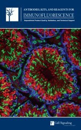
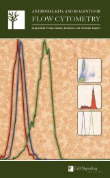
XP Monoclonal Antibodies<br />
for <strong>Apop</strong>tosis and Autophagy<br />
XP Monoclonal Antibodies are a line of high quality<br />
rabbit monoclonal antibodies exclusively available<br />
from Cell Signaling Technology (CST). Any product<br />
labeled with XP has been carefully selected based on<br />
superior performance in all approved applications.<br />
XP Monoclonal Antibodies are generated using<br />
XMT ® technology, a proprietary monoclonal method<br />
developed at CST. This technology provides access to a<br />
broad range of antibody-producing B cells unattainable<br />
with traditional monoclonal technologies, allowing more<br />
comprehensive screening and the identification of XP<br />
monoclonal antibodies with:<br />
eXceptional specificity<br />
As with all CST antibodies, the antibody is specific to your<br />
target of interest, saving you valuable time and resources.<br />
+ eXceptional sensitivity<br />
The antibody will provide a stronger signal for your target protein<br />
in cells and tissues, allowing you to monitor expression of low<br />
levels of endogenous proteins, saving you valuable materials.<br />
+ eXceptional stability and reproducibility<br />
XMT technology combined with our stringent quality control ensures<br />
maximum lot-to-lot consistency and the most reproducible results.<br />
= eXceptional Performance <br />
XMT technology coupled with our extensive antibody validation and<br />
stringent quality control delivers XP monoclonal antibodies with<br />
eXceptional Performance in the widest range of applications.<br />
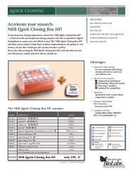
LC3A (D50G8) XP Rabbit mAb #4599<br />
is an example of an antibody with superior performance in a<br />
wide range of tested applications.<br />
Events<br />
A<br />
LC3A<br />
C<br />
LC3A (D50G8) XP Rabbit mAb #4599: Flow cytometric analysis of chloroquine-treated<br />
HeLa cells (A) using #4599 (blue) compared to Rabbit (DA1E) mAb IgG XP Isotype Control<br />
#3900 (red). IHC analysis of paraffin-embedded human glioblastoma multiforme (B) using<br />
#4599. Confocal IF analysis of HeLa cells, untreated (C) or chloroquine-treated (D), using #4599<br />
(green). Actin filaments were labeled with DY-554 phalloidin (red). Blue pseudocolor = DRAQ5 ®<br />
#4084 (fluorescent DNA dye).<br />
B<br />
D<br />
Antibodies and Kits for the Study of<br />
<strong>Apop</strong>tosis<br />
and Autophagy<br />
Cell Signaling Technology provides the highest possible quality<br />
activation-state and total protein antibodies available for the study of<br />
signaling pathways central to cell survival and programmed cell death.<br />
CST antibodies have been extensively validated by our in-house<br />
scientists in applications including western blotting, immunofluorescence,<br />
flow cytometry, immunohistochemistry, chromatin immunoprecipitation<br />
(ChIP), and ELISA. CST phosphorylation-specific<br />
antibodies are the most highly cited and serve as core reagents in<br />
multiple drug discovery platforms. Comprehensive and up-to-date<br />
information can be found at our website.<br />
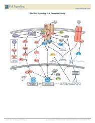
The Bcl-2 family of proteins is composed of pro- and anti-apoptotic members that homo and heterodimerize<br />
with one another. Together, these serve as a rheostat mechanism to regulate the onset of apoptosis at the<br />
mitochondrion. In this image, the balance is in favor of the pro-apoptotic Bad proteins (red) that outnumber<br />
the anti-apoptotic Bcl-xL proteins (green). As a result, the mitochondrial pore composed of VDAC in the<br />
inner membrane and ANT in the outer membrane (both in brown) is shown releasing small molecules into<br />
the cytosol during the permeability transition. Cytochrome c (blue) below the outer mitochondrial membrane<br />
is poised to spill into the cytosol and trigger apoptosis once the outer mitochondrial membrane ruptures.<br />
Table of Contents<br />
4 Antibody Sampler Kits<br />
5 Autophagy<br />
6 Caspase Signaling<br />
8 Cleaved Substrates<br />
9 Antibody Validation<br />
10 Bcl-2 Family Members<br />
12 PathScan ® ELISA Kits and Antibody Pairs<br />
13 <strong>Apop</strong>tosis Inhibitor Proteins / Mitochondrial Proteins<br />
14 TNFR Family<br />
15 Adaptor Proteins<br />
16 NF-kB<br />
18 p53 / Other Transcriptional Regulators<br />
Other Signaling Proteins /<br />
19 Granzymes and Other Proteases<br />
20 Signaling Pathways<br />
Visit our website for more experimental details, additional information,<br />
and a complete list of available XP monoclonal antibodies.<br />
© 2/<strong>2011</strong> Cell Signaling Technology, Inc.<br />
eXceptional Performance , Cell Signaling Technology ® , CST , PathScan ® , SignalSilence ® , XMT ® and XP are registered trademarks or trademarks of Cell Signaling Technology, Inc.<br />
Selected rabbit monoclonal antibodies are produced under license (granting certain rights including those under U. S. Patents No. 5,675,063 and in some instances 7,429,487) from Environmental commitment: eco.cellsignal.com<br />
Epitomics, Inc. Alexa Fluor ® and MitoTracker ® are registered trademark of Molecular Probes, Inc. / DRAQ5 ® is a registered trademark of Biostatus Limited. / Acumen ® is a registered<br />
trademark of TTP <strong>Lab</strong>tech.<br />
PathScan ® <strong>Apop</strong>tosis and Proliferation MultiPlex IF Kit: Some kit components are provided under an agreement between Life Technologies Corporation and Cell Signaling Technology, Inc., and the manufacture, use, sale or import of antibody conjugate in this product is subject to one or more US patents and<br />
corresponding non-US equivalents, owned or controlled by Life Technologies Corporation or its affiliates. The purchase of this product conveys to the buyer the non-transferable right to use the purchased amount of the product and components of the product only in research conducted by the buyer (whether<br />
the buyer is an academic or for-profit entity), for immunocytochemistry, high content screening (HCS) analysis, or flow cytometry applications. The sale of this product or its components (1) in manufacturing; (2) to provide a service, information, or data to an unaffiliated third party for payment; (3) for therapeutic,<br />
diagnostic or prophylactic purposes; (4) resale, whether or not such product or its components are resold for use in research; or for any other commercial purpose. For information on purchasing a license to this product for purposes other than research, contact Life Technologies Corporation, Cellular<br />
Analysis Business Unit, Busi ness Development, 29851 Willow Creek Road, Eugene, OR 97402, Tel: (541) 465-8300. Fax: (541) 335-0354.<br />
All content of this Brochure and Technical Reference is protected by U.S. and foreign intellectual property laws. You may not copy, modify, upload, download, post, transmit, republish or distribute any of the content without our prior written permission except for your own personal and non-commercial<br />
purposes. Except as provided in the preceding sentence, nothing contained in this Brochure and Technical Reference shall be construed as granting a license or other rights under any patent, trademark, copyright or other intellectual property of Cell Signaling Technology or any third party. Unauthorized use of<br />
any Cell Signaling Technology trademark, service mark or logo may be a violation of federal and state trademark laws.