

Phytopathogenic Dothideomycetes - CBS - KNAW
Phytopathogenic Dothideomycetes - CBS - KNAW
Phytopathogenic Dothideomycetes - CBS - KNAW

You also want an ePaper? Increase the reach of your titles
YUMPU automatically turns print PDFs into web optimized ePapers that Google loves.
Phylogenetic lineages in Pseudocercospora<br />
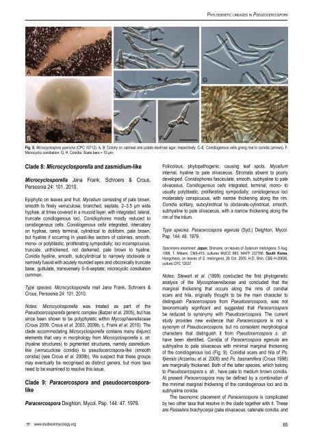
Fig. 8. Microcyclospora quercina (CPC 10712). A, B. Colony on oatmeal and potato-dextrose agar, respectively. C–E. Conidiogenous cells giving rise to conidia (arrows). F.<br />
Microcyclic conidiation. G, H. Conidia. Scale bars = 10 μm.<br />
Clade 8: Microcyclosporella and zasmidium-like<br />
Microcyclosporella Jana Frank, Schroers & Crous,<br />
Persoonia 24: 101. 2010.<br />
Epiphytic on leaves and fruit. Mycelium consisting of pale brown,<br />
smooth to finely verruculose, branched, septate, 2–3.5 μm wide<br />
hyphae, at times covered in a mucoid layer, with integrated, lateral,<br />
truncate conidiogenous loci. Conidiophores mostly reduced to<br />
conidiogenous cells. Conidiogenous cells integrated, intercalary<br />
on hyphae, rarely terminal, cylindrical to doliiform, pale brown,<br />
but hyaline if occurring in yeast-like sectors of colonies, smooth,<br />
mono- or polyblastic, proliferating sympodially; loci inconspicuous,<br />
truncate, unthickened, not darkened, pale brown to hyaline.<br />
Conidia hyaline, smooth, subcylindrical to narrowly obclavate or<br />
narrowly fusoid with acutely rounded apex and obconically truncate<br />
base, guttulate, transversely 0–6-septate; microcyclic conidiation<br />
common.<br />
Type species: Microcyclosporella mali Jana Frank, Schroers &<br />
Crous, Persoonia 24: 101. 2010.<br />
Notes: Microcyclosporella was treated as part of the<br />
Pseudocercosporella generic complex (Batzer et al. 2005), but has<br />
since been shown to be polyphyletic within Mycosphaerellaceae<br />
(Crous 2009, Crous et al. 2003, 2009b, c, Frank et al. 2010). The<br />
clade accommodating Microcyclosporella contains many disjunct<br />
elements that vary in morphology from Microcyclosporella s. str.<br />
(hyaline structures) to pigmented structures, namely zasmidiumlike<br />
(verrucuclose conidia) to pseudocercospora-like (smooth<br />
conidia) (see Crous et al. 2009b). We suspect that these groups<br />
may eventually be recognised as distinct genera, but more taxa<br />
need to be examined to resolve this issue.<br />
Clade 9: Paracercospora and pseudocercosporalike<br />
Paracercospora Deighton, Mycol. Pap. 144: 47. 1979.<br />
Foliicolous, phytopathogenic, causing leaf spots. Mycelium<br />
internal, hyaline to pale olivaceous. Stromata absent to poorly<br />
developed. Conidiophores fasciculate, smooth, subhyaline to pale<br />
olivaceous. Conidiogenous cells integrated, terminal, mono- to<br />
usually polyblastic, proliferating sympodially; conidiogenous loci<br />
moderately conspicuous, with narrow thickening along the rim.<br />
Conidia solitary, subcylindrical to obclavate-cylindrical, smooth,<br />
subhyaline to pale olivaceous, with a narrow thickening along the<br />
rim of the hilum.<br />
Type species: Paracercospora egenula (Syd.) Deighton, Mycol.<br />
Pap. 144: 48. 1979.<br />
Specimens examined: Japan, Shimane, on leaves of Solanum melongena, 5 Aug.<br />
1998, T. Mikami, CNS-415, cultures MUCC 883, MAFF 237766. South Korea,<br />
Hongcheon, on leaves of S. melongena, 26 Oct. 2005, H.D. Shin, <strong>CBS</strong> H-20836,<br />
culture CPC 12537.<br />
Notes: Stewart et al. (1999) conducted the first phylogenetic<br />
analysis of the Mycosphaerellaceae and concluded that the<br />
marginal thickening that occurs along the rims of conidial<br />
scars and hila, originally thought to be the main character to<br />
distinguish Paracercospora from Pseudocercospora, was not<br />
taxonomically significant and suggested that Paracercospora<br />
be reduced to synonymy with Pseudocercospora. The current<br />
study provides new evidence that Paracercospora is not a<br />
synonym of Pseudocercospora, but no consistent morphological<br />
characters that distinguish it from Pseudocercospora s. str.<br />
have been identified. Conidia of Paracercospora egenula are<br />
subhyaline to pale olivaceous with minimal marginal thickening<br />
of the conidiogenous loci (Fig. 9). Conidial scars and hila of Ps.<br />
fijiensis (Arzanlou et al. 2008) and Ps. basiramifera (Crous 1998)<br />
are marginally thickened. Both of the latter species, which belong<br />
to Pseudocercospora s. str., have pale to medium brown conidia.<br />
At present Paracercospora may be defined by a combination of<br />
the minimal marginal thickening of the conidiogenous loci and its<br />
subhyaline conidia.<br />
The taxonomic placement of Paracercospora is complicated<br />
by two other taxa that resolve in the clade together with it. These<br />
are Passalora brachycarpa (pale olivaceous, catenate conidia, and<br />
www.studiesinmycology.org<br />
65


