Experimental Study of Bone Response to Hydroxyapatite CoatingImplants: BIC and Removal Torque TestSurface Property Evaluation According to the High Crystallinity of the<strong>TS</strong>III HA ImplantTae-Gwan Eom, Gyeo-Rok Jeon, Chang-Mo Jeong, Young-Kyun Kim, Su-Gwan Kim, In-Hee Cho, Yong-Seok Cho, Ji-Su OhSubmitted to Oral Surg Oral Med Oral Pathol Oral Radiol Endod 2011June-Cheol Hwang, Hong-Young Choi, Tae-Gwan EomScientific Poster, <strong>Osstem</strong> World Meeting 2011<strong>TS</strong> <strong>System</strong> Pre-Clinical StudyObjective:The objective of this study was to evaluate the earlyosseointegration of hydroxyapatite (HA) coated implantversus resorbable blast media (RBM) and sand-blasted withalumina and acid etched (SA) surface tapered implants.Study design:Twelve adult male miniature pigs (Medi Kinetics Micropigs,Medi Kinetics Co., Ltd., Korea) were used in this study. Theremoval torque of implants placed in the tibia of miniaturepigs was measured. For implants placed in the mandible,histomorphometric evaluation was performed for theevaluation of the bone-implant contact (BIC) ratio.Results:After 4, 8, and 12 weeks, removal torque values wereincreased. Among the 3 groups, the HA coated group showedthe highest value (p < .05). When the HA surface, RBM, andSA surface group were compared at each time point, the HAgroup showed statistically significantly high removal torquevalue (RTV) values (p < .05). At 2 weeks, in comparison withRBM, SA showed an 11 % increase, and HA showed a 42 %increase; nonetheless, they were not statistically significant. At4 weeks, the BIC ratio of HA was significantly higher than thatof SA (p < .05). Nonetheless, RBM and SA were notsignificantly different (p > .05). At 8 weeks, the BIC of HA wasshown to be significantly higher than RBM or SA (p < .05).Nonetheless, RBM and SA were not significantly different (p >.05).Fig. 1. Removal torque value graph.Fig. 2. BIC ratio.Conclusions:The early osseointegration of HA coated implants was found tobe excellent, and HA coated implants will be available in poorquality bone.Objective:To evaluate the characteristics of the HA (Hydroxyapatite)surfacedue to the high crystal growth of the <strong>TS</strong>III HA implant.Study design:The <strong>TS</strong>III HA implant (∅4.0 x 10 mm) by <strong>Osstem</strong> and <strong>TS</strong>VHA implant (∅4.1 x 10 mm) by Zimmer were used toevaluate solubility. For the solution, 1M Tris buffer with pH7.4 was used and solubility was evaluated. For themeasurement of the eluted calcium, Arsenazo III andMalachite Green were used to produce the color reactionwith calcium. Absorbance was measured with BeckmanCoulter Spectrophotometer to quantify the reaction.Table 1. Crystallinity of conventional and <strong>TS</strong>III HA[Ca 2+ , ppm]ConventionalHA<strong>TS</strong>III HACrystalplaneFWHM(deg.)CrystalliteSize (A)(002) 0.1490 1181(211) 0.1515 1155(002) 0.1387 1389(211) 0.1340 1530HACrystallinity68.2%98%Results:Based on the result of the evaluation of HA solubility withcalcium ion in relation to HA crystallinity, after 4 days in thesolution, the high crystalline <strong>TS</strong>III HA implant showed similardissolution results compared to the <strong>TS</strong>V HA implant. Theelution of calcium ion gradually decreased with accordancewith the increase of HA crystallinity over the number of daysand tests.Conclusions:The highly crystallized <strong>TS</strong>III HA implant has a stable surfacecoating layer and exhibits safe dissolution characteristics. Thus,it is evaluated as an excellent product that secures long-termsafety.Fig. 2. The surface observation according to the calcium ion dissolutioncharacteristic of HAa) Conventional HA-(24hr)b) <strong>TS</strong>III HA-(24hr)c) Conventional HA-(28day)d) <strong>TS</strong>III HA-(28day).<strong>TS</strong> <strong>System</strong> Pre-Clinical StudyFig. 1. The calcium ion dissolution characteristic of the HA implant.1819
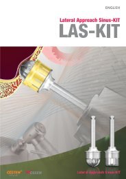
Architectural Features of the <strong>TS</strong>III HA Implant<strong>TS</strong> <strong>System</strong> ReferencesClinical StudyJune-Cheol Hwang, Hong-Young Choi, Tae-Gwan EomScientific Poster, <strong>Osstem</strong> World Meeting 20111. Choon-Mo Yang. Immediate Implant Placement and Immediate Loading with <strong>Osstem</strong> <strong>TS</strong>III Implant <strong>System</strong> and Chair-side Provisional Restoration inMandibular Anterior Partial Edentulism. Scientific Poster, <strong>Osstem</strong> World Meeting 2011.2. Hyun-Ki Cho. Immediate placement of dental implant - <strong>TS</strong>III SA Implant clinical result. Scientific Poster, <strong>Osstem</strong> World Meeting 2011.<strong>TS</strong> <strong>System</strong> Pre-Clinical StudyObjective:The highly crystallized <strong>TS</strong>III HA implant was uniquely treatedafter the hydroxyapatite (HA) coating process.In this study, the structural characteristics of the HA-coated<strong>TS</strong>III HA implant were evaluated.Study design:The structural characteristics of two products: the <strong>Osstem</strong><strong>TS</strong>III HA implant (∅4.0 x 10mm) and the Zimmer <strong>TS</strong>V HAimplant (∅4.1 x 11mm) were compared and examinedbased on product design, surface image, degree ofcrystallinity, stability of HA coating layer after implantationin pig bone, tensile bonding strength, and shear bondingstrength.Results:1) Surface morphology observation:In Fig. 1, the typical HA surface with a plasma spray isshown through the SEM image of a <strong>TS</strong>III and <strong>TS</strong>V implantsurface.<strong>TS</strong>III HA<strong>TS</strong>V HAFig. 1. SEM morphology observation of the implanta) <strong>TS</strong>III HA, b)<strong>TS</strong>V HA.3) Stability of HA coating layer after implantationThe SEM observation result of the HA coating layer wasaltogether excellent after implanting a <strong>TS</strong>III HA and <strong>TS</strong>V HAimplant with the implanted torque of a 35 Ncm in the pigbone.Fig. 3. HA coating surface as observed with SEM after implanting it with theimplanted torque of a 35Ncm in the pig bone. a)<strong>TS</strong>III HA, b)<strong>TS</strong>V HA.4) Tensile and shear strength evaluation[Mpa][Mpa]3. Hak Oh. Immediate Loading of <strong>TS</strong>III HA, <strong>TS</strong>III SA Implant system. Scientific Poster, <strong>Osstem</strong> World Meeting 2011.4. Young-Kyun Kim, Ji-Hyun Bae. Subjective satisfaction of clinician and Short-termClinical Evaluation of <strong>Osstem</strong> <strong>TS</strong>III SA Implant. J Korean Cilnical Implant2010;30(7):430-43.Pre-Clinical StudyBiology1. Hong-Young Choi, Su-Kyung Kim, Jae-Jun Park, Tae-Gwan Eom. Enhancement of in Vitro Osteogenesis on Sandblasted and Acid Etched Surfaces withRough Micro-topography. Scientific Poster, <strong>Osstem</strong> World Meeting 2011.2. Hong-Young Choi, Jae-June Park, Su-Kyung Kim, Tae-Gwan Eom. The Effects of Surface Roughness on the Sandblasted with Large Grit Alumina andAcid Etched Surface Treatment: In Vitro Evaluation. Scientific Poster, <strong>Osstem</strong> World Meeting 2011.3. Hong-Young Choi, Jae-June Park, In-Hee Cho, Tae-Gwan Eom. The Effects of Surface Roughness on the Sandblasted with Large Grit Alumina and AcidEtched Surface Treatment: In Vivo Evaluation. Scientific Poster, <strong>Osstem</strong> World Meeting 2011.4. In-Hee Cho, Hong-Young Choi, Woo-Jung Kim, Tae-Gwan Eom. Biomechanical and Histomorphometrical Evaluation of Bone-Implant Integration at SandBlasting with Alumina and Acid Etching (SA) Surface. Scientific Poster, 19th Annual Scientific Congress of EAO 2010.5. Tae-Gwan Eom, Gyeo-Rok Jeon, Chang-Mo Jeong, Young-Kyun Kim, Su-Gwan Kim, In-Hee Cho, Yong-Seok Cho, Ji-Su Oh. Experimental Study of BoneResponse to Hydroxyapatite Coating Implants: BIC and Removal Torque Test. Submitted to Oral Surg Oral Med Oral Pathol Oral Radiol Endod 2011.6. Tae-Gwan Eom, Young-Kyun Kim, Jong-Hwa Kim, In-Hee Cho, Chang-Mo Jeong, Yong-Seok Cho. Experimental Study about the Bony Healing ofHydroxyapatite Coating Implants. J Korean Assoc Oral Maxillofac Surg 2011;37(4):295-300.7. Min-Woo Kim, Tae-Gwan Eom. Surface Property Evaluation According to the High Crystallinity of the <strong>TS</strong>III HA Implant. Scientific Poster, <strong>Osstem</strong> WorldMeeting 2011.8. June-Cheol Hwang, Hong-Young Choi, Tae-Gwan Eom. Architectural Feature of <strong>TS</strong>III HA Implant. Scientific Poster, <strong>Osstem</strong> World Meeting2011<strong>TS</strong> <strong>System</strong> References2) Degree of crystallinityCrystallinity 68.2%Amorphous CaPPre-Clinical StudyBiomechanics1. Tae-Gwan Eom, Seung-Woo Suh, Myung-Duk Kim, Young-Kyun Kim, Seok-Gyu Kim. Influence of Implant Length and Insertion Depth on Stress Analysis:a Finite Element Study. Scientific Poster, Academy of KSME 2011.2. Tae-Gwan Eom, In-Ho Kim, Insertion Torque Sensitivity of <strong>TS</strong>IV SA Fixture in Artificial Bone Model. Scientific Poster, <strong>Osstem</strong> World Meeting 2011.IntensityCrystallinity 98%Method- Tensile TEST : ASTM F1147-05 / - Shear TEST : ASTM F1044-052-theta(degree)Conclusions:The <strong>TS</strong>III HA implant displayed excellent initial stability andplacement convenience in terms of design in the structuralanalysis. The quality and performance of this HA coatingproduct are considered equal to the products of renownedmanufacturers overseas.Fig. 2. The comparison of the X-ray diffraction pattern of HA specialprocessing. Before (a) and after (b).2021