
Create successful ePaper yourself
Turn your PDF publications into a flip-book with our unique Google optimized e-Paper software.
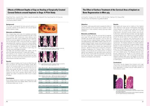
Effects of Different Depths of Gap on Healing of Surgically CreatedCoronal Defects around Implants in Dogs: A Pilot StudyThe Effect of Surface Treatment of the Cervical Area of Implant onBone Regeneration in Mini-pigHong-Cheol Yoon, Jung-Yoo Choi, Ui-Won Jung, Eun-Kyung Bae, Seong-Ho Choi, Kyoo-Sung Cho, Ho-Yong Lee,Chong-Kwan Kim, June-Sung ShimJ Periodontol 2008;79: 355-61Jin-Yong Cho, Young-Jun Kim, Min-Gi Yu, Min-Suk Kook, Hee-Kyun Oh, Hong-Ju ParkJ Korean Assoc Oral Maxillofac Surg 2008;34:285-92US <strong>System</strong> Pre-Clinical StudyBackground:This study investigated the bone growth pattern insurgically created coronal defects with various depthsaround implants in dogs.Materials and Methods:Four mongrel dogs were used. All mandibular premolarswere extracted under general anesthesia and left to healfor 2 months. After ostectomy, bony defects wereprepared in test sites, using a stepped drill with a diameterof 6.3 mm and two depths: 2.5 mm (test sites 1 [T1]) and5.0 mm (test sites 2 [T2]). In the control sites, the implantswere placed after ostectomy without any coronal defects.T1, T2, and control sites were prepared in the right and leftsides of the mandible. Six implants, 3.3 mm in diameterand 10 mm in length, were placed in each dog; theimplants were submerged completely.Two dogs were sacrificed 8 weeks after surgery, and theother two dogs were sacrificed 12 weeks after surgery.The stability of all implants was measured with aresonance frequency analyzer after placement and aftersacrifice. All sites were block-dissected for groundsectioning and histologic examination.Results:After 12 weeks of healing, only T2 were not filled fully withbone. At week 8, the mean bone-to-implant contact (BIC)was 47.7% for control, 43.6% for T1, and 22.2% for T2. Atweek 12, the control BIC was 56.7% and the 2.5 mmdefect had a greater BIC (58.8%). However, in the 0.5 mmdefect, the BIC was 35.1%. At insertion, stability wasreduced at sites with a greater defect depth. Similarstability was noted in all specimens after 8 and 12 weeksof healing.Conclusions:Bone healing between an implant and marginal bone wascompromised at sites with a deeper defect when the widthof the bone defect was 1.5 mm.Fig. 1. Clinical photograph of control, T1, and T2.A B CFig. 2. Longitudinal sections after 8 weeks of healing in control (A),T1 (B), and T2 (C) (original magnification×10).A B CFig. 3. Longitudinal sections after 12 weeks of healing in control (A),T1 (B), and T2 (C) (original magnification×10).Table 1. BIC (%; mean ± SD) in the coronal 5 mm of the implantControl Ti (2.5 mm) T2 (5.0 mm)8 weeks 47.7 ± 14.7 43.6 ± 19.0 22.2 ± 14.712 weeks 56.7 ± 17.0 58.8 ± 12.0 35.1 ± 8.0Table 2. Distance (mm; mean ± SD) from the implant margin to themost coronal level of contact between bone and implantControl Ti (2.5 mm) T2 (5.0 mm)8 weeks 0.75 ± 0.26 1.20 ± 0.59 1.98 ± 1.4512 weeks 0.59 ± 0.36 0.36 ± 0.40 2.52 ± 1.06Objective:The present study was performed to evaluate the effect ofsurface treatment of the cervical area of implant on boneregeneration in fresh extraction socket following implantinstallation.Materials and Methods:The four minipigs, 18 months old and 30 kg weighted,were used. Four premolars of the left side of both themandible and maxilla were extracted. ∅3.3 mm and 11.5mm long US II plus implants (<strong>Osstem</strong> Implant, Korea) withresorbable blasting media (RBM) treated surface and US IIimplants (<strong>Osstem</strong> Implant, Korea) with machined surface atthe top and RBM surface at lower portion were installed inthe socket. Stability of the implant was measured withOsstell TM (Model 6 Resonance Frequency Analyser:Integration Diagnostics Ltd., Sweden). After 2 months ofhealing, the procedures and measurement of implantstability were repeated in the right side by same method ofleft side. At four months after first experiment, the animalswere sacrificed after measurement of stability of allimplants, and biopsies were obtained.Results:Well healed soft tissue and no mobility of the implantswere observed in both groups. Histologically satisfactoryosseointegration of implants was observed with RBMsurface, and no foreign body reaction as well asinflammatory infiltration around implant were found.Furthermore, substantial bone formation and high degreeof osseointegration were exhibited at the marginal defectsaround the cervical area of US II plus implants. However,healing of US II implants was characterized by theincomplete bone substitution and the presence of theconnective tissue zone between the implant and newlyformed bone. The distance between the implant platform(P) and the most coronal level of bone-to-implant contact(B) after 2 months of healing was 2.66 ± 0.11 mm at US IIimplants group and 1.80 ± 0.13 mm at US II plus implantgroup. The P-B distance after 4 months of healing was 2.29± 0.13 mm at US II implants group and 1.25 ± 0.10 mm atUS II plus implants group. The difference between bothgroups regarding the length of P-B distance wasstatistically significant (p < .05). Concerning the resonancefrequency analysis (RFA) value, the stability of US II plusimplants group showed relatively higher RFA value than USII implants group.Conclusions:The current results suggest that implants with roughsurface at the cervical area have an advantage in process ofbone regeneration on defect around implant placed in afresh extraction socket.US <strong>System</strong> Pre-Clinical StudyTable 3. Implant stability quotient values (mean ± SD)Control Ti (2.5 mm) T2 (5.0 mm)Insertion 72.8 ± 5.2 65.0 ± 9.2 55.3 ± 9.08 weeks 79.7 ± 9.2 79.3 ± 2.5 78.1 ± 9.612 weeks 74.8 ± 9.0 78.0 ± 6.3 72.0 ± 6.6Fig. 1. The ground sections illustrate the result of healing.A, The defect adjacent to coronal portion of US II plus implants is filledwith newly formed bone.B, The defect adjacent to coronal portion of US II implants is separatedfrom the implant surface by a connective tissue.5657