
Create successful ePaper yourself
Turn your PDF publications into a flip-book with our unique Google optimized e-Paper software.
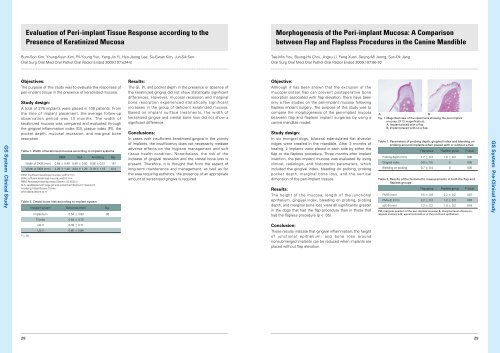
Evaluation of Peri-implant Tissue Response according to thePresence of Keratinized MucosaMorphogenesis of the Peri-implant Mucosa: A Comparisonbetween Flap and Flapless Procedures in the Canine MandibleBum-Soo Kim, Young-Kyun Kim, Pil-Young Yun, Yang-Jin Yi, Hyo-Jeong Lee, Su-Gwan Kim, Jun-Sik SonOral Surg Oral Med Oral Pathol Oral Radiol Endod 2009;107:e24-8Tae-Min You, Byung-Ho Choi, Jingxu Li, Feng Xuan, Seung-Mi Jeong, Sun-Ok JangOral Surg Oral Med Oral Pathol Oral Radiol Endod 2009;107:66-70Objectives:The purpose of this study was to evaluate the responses ofperi-implant tissue in the presence of keratinized mucosa.Results:The GI, PI, and pocket depth in the presence or absence ofthe keratinized gingiva did not show statistically significantdifferences. However, mucosal recession and marginalbone resorption experienced statistically significantincreases in the group of deficient keratinized mucosa.Based on implant surface treatments, the width ofkeratinized gingiva and crestal bone loss did not show asignificant difference.Objective:GS <strong>System</strong> Clinical StudyStudy design:A total of 276 implants were placed in 100 patients. Fromthe time of implant placement, the average follow-upobservation period was 13 months. The width ofkeratinized mucosa was compared and evaluated throughthe gingival inflammation index (GI), plaque index (PI), thepocket depth, mucosal recession, and marginal boneresorption.Table 1. Width of keratinized mucosa according to implant systemsRBM SLA Anodizing Sig.Width of DKM (mm) 0.64 ± 0.49 0.40 ± 0.50 0.56 ± 0.51 .157Width of SKM (mm) 3.26 ± 1.40 3.04 ± 1.29 3.19 ± 1.18 .614DKM, Insufficient keratinized mucosa, width 2 mmSKM, sufficient keratinized mucosa, width 2 mmRBM, Resorbable blasting media (<strong>Osstem</strong> US II/GS II)SLA, sandblasted with large grit and acid-etched (Dentium Implantium)Anodizing, Nobel Biocare TiUniteother abbreviations as inTable 2. Crestal bone loss according to implant systemImpalant systemBone loss (mm)Sig.Implantium 0.54 ± 0.83 .36TiUnite 0.44 ± 0.72GS II 0.39 ± 0.71US II 0.60 ± 0.84Conclusions:In cases with insufficient keratinized gingiva in the vicinityof implants, the insufficiency does not necessarily mediateadverse effects on the hygiene management and softtissue health condition. Nonetheless, the risk of theincrease of gingival recession and the crestal bone loss ispresent. Therefore, it is thought that from the aspect oflong-term maintenance and management, as well as forthe area requiring esthetics, the presence of an appropriateamount of keratinized gingiva is required.Although it has been shown that the exclusion of themucoperiosteal flap can prevent postoperative boneresorption associated with flap elevation, there have beenonly a few studies on the peri-implant mucosa followingflapless implant surgery. The purpose of this study was tocompare the morphogenesis of the peri-implant mucosabetween flap and flapless implant surgeries by using acanine mandible model.Study design:In six mongrel dogs, bilateral edentulated flat alveolarridges were created in the mandible. After 3 months ofhealing, 2 implants were placed in each side by either theflap or the flapless procedure. Three months after implantinsertion, the peri-implant mucosa was evaluated by usingclinical, radiologic, and histometric parameters, whichincluded the gingival index, bleeding on probing, probingpocket depth, marginal bone loss, and the verticaldimension of the peri-implant tissues.Results:The height of the mucosa, length of the junctionalepithelium, gingival index, bleeding on probing, probingdepth, and marginal bone loss were all significantly greaterin the dogs that had the flap procedure than in those thathad the flapless procedure (p < .05).ABFig. 1. Magnified view of the specimens showing the peri-implantmucosa. (X 12 magnification).A, Implant placed with a flap.B, Implant placed without a flap.Table 1. Parameters of probing depth, gingival index and bleeding onprobing around implants when placed with or without a flapFlap groupFlapless groupP valueProbing depth (mm) 1.7 ± 0.3 1.0 ± 0.3 .006Gingival index 0.9 ± 0.5 0 .005Bleeding on probing 0.7 ± 0.4 0 .005Table 2. Results of the histometric measurements in both the flap andflapless groupsFlap groupFlapless groupP valuePM-B (mm) 3.5 ± 0.8 2.2 ± 0.2 .007PM-aJE (mm) 2.2 ± 0.3 1.2 ± 0.3 .003aJE-B (mm) 1.3 ± 0.2 1.0 ± 0.2 .018PM, marginal position of the peri-implant mucosa; B, marginal level of bone-toimplantcontact; aJE, apical termination of the junctional epithelium.GS <strong>System</strong> Pre-Clinical StudyConclusion:These results indicate that gingival inflammation, the heightof junctional epithelium, and bone loss aroundnonsubmerged implants can be reduced when implants areplaced without flap elevation.P > .052829