
You also want an ePaper? Increase the reach of your titles
YUMPU automatically turns print PDFs into web optimized ePapers that Google loves.
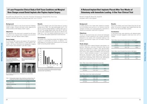
A 1-year Prospective Clinical Study of Soft Tissue Conditions and MarginalBone Changes around Dental Implants after Flapless Implant SurgeryA Relaxed Implant Bed: Implants Placed After Two Weeks ofOsteotomy with Immediate Loading- A One Year Clinical TrialSeung-Mi Jeong, Byung-Ho Choi, Ji-Hun Kim, Feng Xuan, Du-Hyeong Lee, Dong-Yub Mo, Chun-Ui LeeOral Surg Oral Med Oral Pathol Oral Radiol Endod. 2011 Jan;111(1):41-6Bansal DJ, Kedige DS, Bansal DA, Anand DSAccepted in 2010 J Oral ImplantolBackground:Results:None of the implants were lost during follow-up, giving asuccess rate of 100%. The mean probing depth was 2.1 mm(SD 0.7), and the average bleeding on probing index was 0.1(SD 0.3). The average gingival index score was 0.1 (SD 0.3),and the mean marginal bone loss was 0.3 mm (SD 0.4 mm;range 0.0-1.1 mm). Ten implants exhibited bone loss of >1.0mm, whereas 125 implants experienced no bone loss at all.Background:A waiting period of two weeks after osteotomy increasesthe surrounding tissue activity to its maximum level ascollagen formation and neoangiogenesis represents arelaxed and acceptable implant bed configuration.Results:None of the implants were found mobile during the one yearperiod. The amount of average mean marginal bone loss was0.4 mm over the one year follow up period.GS <strong>System</strong> Clinical StudyDespite several reports on the clinical outcomes of flaplessimplant surgery, limited information exists regarding theclinical conditions after flapless implant surgeryObjectives:The objective of this study was to evaluate the soft tissueconditions and marginal bone changes around dentalimplants 1 year after flapless implant surgery.Study design:For the study, 432 implants were placed in 241 patients byusing a flapless 1-stage procedure. In these patients, periimplantsoft tissue conditions and radiographic marginalbone changes were evaluated 1 year after surgery.Fig. 1. Clinical features afterpunching the soft tissue at theproposed implant sites with a 3-mmsoft tissue punch.Fig. 2. Clinical features after healingabutments were connected to thefixtures.Conclusions:The results of this study demonstrate that flapless implantsurgery is a predictable procedure. In addition, it isadvantageous for preserving crestal bone and mucosalhealth surrounding dental implants.Objectives:The aim of the present study was a clinical and radiologicalevaluation of early osteotomy with implant placementdelayed for two weeks with immediate loading in theanterior and premolar region with minimally invasiveapproach.Study design:A total of seven GS II implants (<strong>Osstem</strong>) were placed in sixpatients. Osteotomy was done followed by flap closurewithout the placement of implant. After approximatelywaiting for a period of two weeks, implant placement wasdone which were loaded immediately with provisionalcrown in implant protected occlusion. It was replaced bydefinitive restoration after 6-8 weeks which wasconsidered as baseline. Implant stability and marginal bonelevels were assessed with clinical and radiologicalparameters at baseline, 6th and 12th month intervals.Conclusions:In the present study, early osteotomy with delayed implantplacement showed negligible crestal bone loss with nomobility.Table 3. Mean values of marginal bone levels on mesial aspectAssessment TimeMean ± SD% Change frombaselineBaseline 0.36 ± 0.54 -6th Month 0.37 ± 0.36 13.88%12th Month 0.36 ± 0.41 -13.88%Table 4. Mean values of marginal bone levels on distal aspectAssessment TimeMean ± SD% Change frombaselineBaseline 0.54 ± 0.50 -6th Month 0.50 ± 0.41 5.55%12th Month 0.53 ± 0.42 1.85%GS <strong>System</strong> Clinical StudyTable 1. Mean values of width of keratinized mucosa indexAssessment TimeMean ± SD% Change frombaselineBaseline 2.00 ± 0.63 -Fig. 4. Number of implants that exhibited varying amounts ofbone loss during the healing period from the time of implantplacement to the 1-year follow-up.6th Month 2.17 ± 0.41 -8.5%12th Month 2.33 ± 0.52 -16.5%Fig. 3. Periapical radiograph taken immediately (A) and 1 year (B) afterimplant placement.Table 2. Mean values of peri-implant probing depthAssessment TimeMean ± SD% Change frombaselineBaseline 2.38 ± 0.54 -6th Month 2.29 ± 0.33 3.36%12th Month 2.08 ± 0.34 12.18%Table 1. Probing depth, gingival index, bleeding on probing index, andcrestal bone loss when implants were placed without a flapIndex1 yearProbing depth (mm) 2.1 ± 0.7Bleeding on probing index 0.1 ± 0.3Gingival index 0.1 ± 0.3Crestal bone loss 0.3 ± 0.42425