Diapositive 1 - de l'Université libre de Bruxelles
Diapositive 1 - de l'Université libre de Bruxelles
Diapositive 1 - de l'Université libre de Bruxelles

You also want an ePaper? Increase the reach of your titles
YUMPU automatically turns print PDFs into web optimized ePapers that Google loves.
UNIVERSITÉ LIBRE DE BRUXELLES (ULB)Faculté <strong>de</strong>s SciencesDépartement <strong>de</strong> Biologie <strong>de</strong>s OrganismesLaboratoire <strong>de</strong> Biologie marineINSTITUT ROYAL DES SCIENCES NATURELLES DE BELGIQUE (IRSNB)Département <strong>de</strong>s InvertébrésIncorporation du magnésium dans les squelettes calcitiques<strong>de</strong>s échino<strong>de</strong>rmes et <strong>de</strong>s éponges hypercalcifiéesJulie HERMANSThèse présentée en vue <strong>de</strong> l'obtention du titre <strong>de</strong>Docteur en SciencesJUILLET 2010Promoteurs <strong>de</strong> thèseDr Philippe Dubois (ULB)Dr Philippe Willenz (IRSNB)
UNIVERSITÉ LIBRE DE BRUXELLES (ULB)UNIVERSITÉ D’EUROPEFaculté <strong>de</strong>s SciencesDépartement <strong>de</strong> Biologie <strong>de</strong>s OrganismesLaboratoire <strong>de</strong> Biologie marineINSTITUT ROYAL DES SCIENCES NATURELLES DE BELGIQUE (IRSNB)Département <strong>de</strong>s InvertébrésIncorporation du magnésium dans les squelettes calcitiques<strong>de</strong>s échino<strong>de</strong>rmes et <strong>de</strong>s éponges hypercalcifiéesJulie HERMANSThèse présentée en vue <strong>de</strong> l'obtention du titre <strong>de</strong>Docteur en SciencesPromoteurs <strong>de</strong> thèseDr Philippe Dubois (ULB)Dr Philippe Willenz (IRSNB)Comité <strong>de</strong> lectureDr Christiane Lancelot (ULB)Dr Guy Josens (ULB)Dr Frank Dehairs (ULB-VUB)Dr Bruno David (Université <strong>de</strong> Bourgogne, France)Dr Luc André (Musée Royal <strong>de</strong> l’Afrique Centrale-ULB) JUILLET 2010
RemerciementsJ’aimerais tout d’abord remercier le Pr Jackie Van Goethem <strong>de</strong> m’avoir accueillie au sein<strong>de</strong> son département <strong>de</strong> l’Institut royal <strong>de</strong>s sciences naturelles <strong>de</strong> Belgique et <strong>de</strong> m’avoirsoutenue dans l’obtention du contrat sans lequel cette thèse n’aurait pas été possible. Jelui suis reconnaissante, ainsi qu’à son successeur à la tête du département, le Pr ThierryBackeljau, pour la confiance qu’ils m’ont témoignée.Je remercie vivement le Pr Michel Jangoux qui, par <strong>de</strong>ux fois, m’a ouvert les portes <strong>de</strong>son laboratoire <strong>de</strong> Biologie marine à l’ULB. Je crois pouvoir dire que je m’y sensactuellement comme un poisson dans l’eau.Mes remerciements les plus sincères vont à mon promoteur Philippe Dubois. Mercid’avoir été la personne sur laquelle j’ai toujours pu compter durant ces années <strong>de</strong> thèse.Merci pour ton investissement <strong>de</strong> tous les jours, ton soutien et ton optimisme. Ta passion<strong>de</strong> la recherche et tes encouragements dans les jours difficiles ont été une pièce maîtresse<strong>de</strong> l’aboutissement <strong>de</strong> cette thèse. Merci aussi pour ta patience face à mes difficultésrécurrentes avec les statistiques et les colles <strong>de</strong> chimie ;-). Enfin, merci pour ton humouret ton côté humain. Saches que ce fut un réel plaisir <strong>de</strong> travailler avec toi.Je remercie aussi mon co-promoteur Philippe Willenz. Tu m’auras enseigné la rigueur etle sens du détail. J’aurai beaucoup appris sur les relations humaines en travaillant à tescôtés. Je n’oublierai pas les bons moments passés en mission, <strong>de</strong> Marseille à Rho<strong>de</strong>s, niton humour qui aura souvent détendu l’atmosphère.Je tiens à remercier l’équipe du service <strong>de</strong> Minéralogie et Pétrographie du Musée Royal<strong>de</strong> l’Afrique Centrale, qui m’a fait un excellent accueil tout au long <strong>de</strong> cette thèse.J’exprime toute ma reconnaissance au Pr Luc André pour ses précieux conseils et pournous avoir toujours ouvert les portes <strong>de</strong> son laboratoire. Un merci tout particulier àLaurence Monin et Nourdine Dakhani pour leur ai<strong>de</strong> lors <strong>de</strong>s (nombreuses) analysesd’ICP-AES <strong>de</strong> cette thèse et pour leur sympathie. Je suis reconnaissante à Jacques Navezpour les analyses en HR-ICP-MS et pour son ai<strong>de</strong> dans le traitement <strong>de</strong>s résultats. Merci àFrédéric Planchon pour les discussions riches en magnésium que nous avons eues… toutsimplement fortifiantes !Je remercie aussi toute l’équipe <strong>de</strong> la VUB, que j’ai eu l’occasion <strong>de</strong> côtoyer dans le cadredu projet CALMARS, notamment les Pr Frank Dehairs et Eddy Keppens, ainsi queRémy Mas, Michael Korntheuer et Emilie Chevalier pour leurs explications sur lesanalyses IRMS.Je tiens à remercier le Pr Jean Vacelet. Nos échanges d’idées au cours <strong>de</strong> la rédaction <strong>de</strong>l’article sur la croissance <strong>de</strong> P. massiliana ont été extrêmement intéressants.Je remercie le Pr Yannicke Dauphin pour sa collaboration pour les analyses XANES,réalisées au synchrotron <strong>de</strong> Grenoble.Un tout grand merci au Pr Philippe Compère pour ses conseils sur les extractions <strong>de</strong>matrices organiques et le prêt à long terme <strong>de</strong> son dispositif d’extraction <strong>de</strong> protéines.Je remercie le Pr Alain Bernard pour les analyses XRD qu’il a effectuées à ma <strong>de</strong>man<strong>de</strong>.
Je tiens à remercier Jean et Véro du club <strong>de</strong> plongée « Au-<strong>de</strong>là plongée » pour le bonaccueil que notre équipe y a toujours reçu. Merci aussi à André <strong>de</strong> « Waterhoppers » pourson ai<strong>de</strong> lors <strong>de</strong>s plongées à Rho<strong>de</strong>s.Merci à Helmut Zibrowius <strong>de</strong> m’avoir aidé à localiser les grottes à Petrobiona <strong>de</strong> l’île <strong>de</strong>Rho<strong>de</strong>s. Sa précision et son excellente mémoire nous ont été d’un précieux secours.Un tout grand merci à toute la joyeuse équipe du laboratoire. Merci à Chantal, Viviane,David, ainsi que Edith et Saloua, toujours hyper efficaces pour les comman<strong>de</strong>s d’urgenced’un produit. Merci aussi à Thierry Dupont pour ses conseils d’aquariophile sans lesquelsje n’aurai pu monter mon propre Nausicaa et pour son ai<strong>de</strong> dans cette entreprise, ainsi quepour les 1001 petites réparations <strong>de</strong> sauvetage qu’il a faites. Enormes mercis à Ana(« Lunch time ! ») pour sa bonne humeur <strong>de</strong> tous les jours et ses qualités <strong>de</strong> chercheusebibliographique invétérée, Claire (« Patriiiiiiiick the starfish !!! ») pour sa sympathie etses conseils toujours avisés, Colin (« moi j’aime pas… ») pour un intérim <strong>de</strong> plongeurquand j’en ai eu besoin et une mission express ron<strong>de</strong>ment menée à Marseille, Mathieupour son ai<strong>de</strong> et sa sympathie, Gauthier pour ses conversations sans fin et ses petitsrappels <strong>de</strong> bio toujours très didactiques, et Bruno pour sa bonne humeur lors <strong>de</strong> sesincursions dans notre bureau. Merci aussi à la ribambelle <strong>de</strong> mémorants qui se sontsuccédé pour mettre un peu d’ambiance dans ce grand laboratoire (« Faites votrevaisselle! »).Je tiens à adresser un merci tout spécial à mes compagnes <strong>de</strong> bureau successives,Catherine Borremans et Stéphanie Bonnet. Votre présence et nos longues conversations(parfois intellectuelles !) ont contribué à faire <strong>de</strong> cette thèse un bon souvenir dans mamémoire… Je n’oublie pas non plus le palmier gonflable qui a orné notre bureau pendanttoutes ces années, égaillant la pièce <strong>de</strong> ses couleurs vives.Un tendre merci à Philippe Pernet pour son ai<strong>de</strong> précieuse et toujours efficace, que ce soitau cours <strong>de</strong>s missions, pour les analyses AAS, les interminables séances <strong>de</strong> découpage <strong>de</strong>piquants, les régénérations <strong>de</strong> résines, et j’en passe. Merci pour ta présence, tadisponibilité et ton oreille attentive. Merci du fond du cœur pour ton soutien sans faillelors <strong>de</strong> la stressante pério<strong>de</strong> <strong>de</strong>s <strong>de</strong>rniers mois <strong>de</strong> thèse. Une chose est sûre, tu m’es<strong>de</strong>venu indispensable.Je remercie aussi la petite équipe <strong>de</strong> l’institut : Melany pour sa sympathie et Laetitia pourson ai<strong>de</strong> toujours souriante... Un tout grand merci à Yves Barette, pour son ai<strong>de</strong> rapi<strong>de</strong> etefficace face aux problèmes <strong>de</strong> PC et un sauvetage <strong>de</strong> disque dur que je n’ai pas oublié.Merci aussi à Pol Gosselin sur lequel, comme à son habitu<strong>de</strong>, j’ai toujours pu compterpour un conseil sur les préférences alimentaires <strong>de</strong>s oursins ou une mission express à Lucsur-Mer.Merci aussi à tous les membres du laboratoire <strong>de</strong> biologie marine <strong>de</strong> Mons, qui m’onttoujours fait bon accueil, avec une spéciale dédicace à Fifi, Elise et Aline. Merci aussi àPaul Postiau pour sa gentillesse et sa disponibilité.Merci à la mafia bibliographique portugaise (Johana, Elizabete, Màrio) pour lesnombreux pdf que j’ai pu obtenir grâce à leur complicité.
Ce travail a été rendu possible par la collaboration <strong>de</strong> l’Institut <strong>de</strong>s Sciences naturelles etdu laboratoire <strong>de</strong> biologie marine <strong>de</strong> l’Université Libre <strong>de</strong> <strong>Bruxelles</strong>. Il a pu être réaliségrâce à l’appui financier d’un programme « Action 2 » financé par la PolitiqueScientifique fédérale belge, et à celui <strong>de</strong> la Fondation David et Alice Van Buuren.Enfin, je remercie ma famille et tous mes amis, qui m’ont toujours soutenue et sanslesquels je n’aurai pu <strong>de</strong>venir celle que je suis aujourd’hui. Vous êtes dans mon cœur et yresterez, quelle que soit la distance.
RÉSUMÉDe nombreux organismes marins précipitent <strong>de</strong>s squelettes en calcite magnésienne.Depuis près d’un siècle, il est connu que les concentrations en magnésium <strong>de</strong> cessquelettes sont influencées par les conditions environnementales, telle la température,régnant au moment <strong>de</strong> leur dépôt. Dans le contexte actuel <strong>de</strong> changement climatique, cettepropriété a promu l’usage <strong>de</strong> plusieurs taxons en tant qu’archive naturelle <strong>de</strong>s conditionsenvironnementales du passé. Cependant, les squelettes d’espèces sympatriques, voired’individus <strong>de</strong> la même espèce, peuvent présenter <strong>de</strong>s concentrations en magnésium trèsdifférentes, attestant <strong>de</strong> l’influence <strong>de</strong> facteurs biologiques sur la détermination <strong>de</strong> laconcentration squelettique en cet élément. Une parfaite compréhension <strong>de</strong>s mécanismesd’incorporation du magnésium dans les squelettes est donc requise pour vali<strong>de</strong>r l’usage <strong>de</strong>ce paléotraceur. De plus, la solubilité <strong>de</strong>s calcites augmentant avec leur concentration enmagnésium, l’incorporation <strong>de</strong> cet élément conditionne en partie la stabilité <strong>de</strong>s squelettescalcitiques dans un océan en cours d’acidification.Le présent travail contribue à l’étu<strong>de</strong> <strong>de</strong>s différents facteurs, tant environnementaux quephysiologiques et minéralogiques, susceptibles d’affecter l’incorporation du magnésiumdans les squelettes en calcite <strong>de</strong> trois taxons présentant <strong>de</strong>s concentrations en cet élémentparticulièrement élevées, une éponge hypercalcifiée, Petrobiona massiliana, et <strong>de</strong>uxéchino<strong>de</strong>rmes, Paracentrotus lividus et Asterias rubens.Dans une première partie, les effets <strong>de</strong> plusieurs facteurs environnementaux ont étéétudiés, en milieu naturel dans le cas <strong>de</strong> l’éponge, étant donné son incapacité à survivre enaquarium, et en conditions contrôlées d’aquarium dans le cas <strong>de</strong>s <strong>de</strong>ux échino<strong>de</strong>rmes.Une influence environnementale prépondérante <strong>de</strong> la température sur la concentration enmagnésium squelettique a été mise en évi<strong>de</strong>nce dans les 3 modèles biologiques étudiés.Une fois les facteurs génétiques (espèce) et structurels (élément squelettique) fixés, unerelation positive liant la température à la concentration en magnésium squelettique a étécaractérisée en milieu naturel chez l’éponge hypercalcifiée P. massiliana et en conditionscontrôlées chez l’oursin P. lividus. Chez ce <strong>de</strong>rnier, cette relation, non linéaire, sestabilise aux plus hautes températures envisagées, probablement suite à la saturation d’unprocessus biologique intervenant dans l’incorporation <strong>de</strong> cet élément. La salinité, un autrefacteur environnemental majeur en milieu marin, influence elle aussi positivement laconcentration en magnésium dans le squelette <strong>de</strong> l’étoile <strong>de</strong> mer A. rubens. A nouveau, ilest proposé que cette influence <strong>de</strong> l’environnement soit modulée par un processusbiologique: chez les échino<strong>de</strong>rmes, la concentration en magnésium, contrairement à celledu calcium, n’est pas régulée dans le liqui<strong>de</strong> coelomique. Elle est donc directementinfluencée par la salinité, et affecte probablement la concentration en cet élément dans lesquelette formé. La diffusion <strong>de</strong>puis l’eau <strong>de</strong> mer jusqu’au site <strong>de</strong> calcification parl’intermédiaire <strong>de</strong>s flui<strong>de</strong>s internes a en effet été suggérée sur base du fait que le rapportMg/Ca <strong>de</strong> l’eau <strong>de</strong> mer influence celui <strong>de</strong>s squelettes calcairesUne fois l’influence, directe ou indirecte, <strong>de</strong>s facteurs environnementaux exclue, 44% <strong>de</strong>la variabilité du rapport Mg/Ca du squelette <strong>de</strong>s échino<strong>de</strong>rmes restent à expliquer. Lesexpériences <strong>de</strong> croissance d’échino<strong>de</strong>rmes réalisées en conditions contrôlées indiquentque ce rapport est indépendant <strong>de</strong> la vitesse <strong>de</strong> croissance dans ce groupe, contrairementaux hypothèses émises dans la littérature.
Dans la secon<strong>de</strong> partie, la modulation <strong>de</strong>s facteurs minéralogiques par les facteursbiologiques a été investiguée. Pour ce faire, d’une part, les interactions entre rapportMg/Ca en solution et matrice organique <strong>de</strong> minéralisation ont été étudiées dans un modèlein vitro. D’autre part, les relations entre soufre et magnésium dans le squelette ont étédécryptées.Le rapport Mg/Ca <strong>de</strong> la solution <strong>de</strong> précipitation a une influence prépondérante sur laconcentration en magnésium du carbonate <strong>de</strong> calcium précipité in vitro, attestant <strong>de</strong>l’importance <strong>de</strong> la régulation <strong>de</strong> la composition du flui<strong>de</strong> <strong>de</strong> calcification et <strong>de</strong>smécanismes <strong>de</strong> transport la contrôlant. Deux mécanismes biologiques complémentairespermettent <strong>de</strong> favoriser l’incorporation, dans les calcites biogéniques, <strong>de</strong> quantités <strong>de</strong>magnésium largement supérieures à celles observées dans les calcites inorganiques, et ce,malgré la forte hydratation <strong>de</strong> ce cation : l’intervention d’agents chélateurs du magnésiumet le passage par une phase <strong>de</strong> carbonate <strong>de</strong> calcium amorphe (CCA). Les molécules <strong>de</strong> lamatrice organique <strong>de</strong> minéralisation jouent entre autres le rôle <strong>de</strong> chélateur dumagnésium, réduisant son état d’hydratation et facilitant ainsi son incorporation dans leminéral. Un rôle similaire a été suggéré pour les sulfates en solution, au vu <strong>de</strong> lacorrélation observée dans ce travail entre les rapports Mg/Ca et S/Ca dans la phaseminérale <strong>de</strong>s calcites biogéniques étudiées. La matrice organique affecte elle aussi laconcentration en magnésium dans le cristal, probablement via la stabilisation <strong>de</strong> la phase<strong>de</strong> CCA nécessaire à l’incorporation <strong>de</strong> concentrations élevées <strong>de</strong> cet élément: ainsi, lesmacromolécules <strong>de</strong> la matrice organique du test d’oursin induisent in vitro la formation <strong>de</strong>calcites plus riches en magnésium que celles formées en présence <strong>de</strong> matrice <strong>de</strong> piquant,un résultat concordant avec le fait que, in vivo, le test contient <strong>de</strong>s concentrations enmagnésium plus élevées que les piquants.Cette thèse <strong>de</strong> doctorat a donc soulevé l’importance <strong>de</strong>s effets biologiques dans ladétermination du rapport Mg/Ca dans les calcites biogéniques. Les résultats obtenusmontrent que le décryptage <strong>de</strong>s mécanismes impliqués dans l’incorporation dumagnésium se doit <strong>de</strong> considérer la phase amorphe transitoire qui précè<strong>de</strong> lacristallisation. Des effets environnementaux affectent eux aussi la concentrationsquelettique en magnésium, mais nos résultats suggèrent qu’ils agissent au travers d’unemodulation <strong>de</strong>s effets biologiques, et non par une influence thermodynamique directe.Cette hypothèse, si elle est confirmée, impose la plus gran<strong>de</strong> pru<strong>de</strong>nce lors <strong>de</strong> l’utilisation<strong>de</strong>s squelettes en calcite en tant que paléotraceurs.
SUMMARYThe magnesium concentration in calcite skeletons produced by marine invertebrates isknown to be <strong>de</strong>pen<strong>de</strong>nt on several environmental parameters, including temperature,salinity and seawater Mg/Ca ratio. This property prompted the use of this concentration asa proxy of the consi<strong>de</strong>red parameters. However, skeletal magnesium contents insympatric species and even in individuals of the same species may be rather different.These inter and intra-individual variabilities indicate that biological factors also affectmagnesium incorporation into biogenic calcites. Magnesium incorporation mechanismsare still unknown in calcifying invertebrates, a fact that questions the validity of thiselement as a paleoproxy. Moreover, higher magnesium contents increase calcite solubilityand could therefore worsen the case of calcifying organisms facing ocean acidificationlinked to global change.The present thesis is a contribution to the study of the environmental, biological andmineralogical factors affecting magnesium incorporation into the calcitic skeletons of 3taxa, i.e. one hypercalcified sponge, Petrobiona massiliana, and two echino<strong>de</strong>rms,Paracentrotus lividus and Asterias rubens.The first part of this work was <strong>de</strong>dicated to the study of several environmental factorsaffecting the magnesium concentration in the calcite skeleton of the 3 studied organisms.Consequently to its low survival in aquarium, the sponge was studied using fieldspecimens collected along an environmental gradient. Echino<strong>de</strong>rms were grown incontrolled conditions in aquarium. Once the genetic (species) and structural (skeletalelement) factors were fixed, skeletal magnesium concentration was positively related totemperature in the 3 studied species. The Mg/Ca ratio of the test of aquarium-grownP. lividus increased with temperature until a plateau which was probably due to thesaturation of a biological process involved in magnesium incorporation. A positive effectof salinity, an other major environmental parameter, on skeletal Mg/Ca was <strong>de</strong>monstratedin aquarium-grown A. rubens. This influence can also be linked to a biological process:contrary to magnesium, calcium concentration is controlled in the coelomic fluid, fromwhich ions probably diffuse through the living tissues to the calcification site. Thus, theobserved positive relation can be explained by the fact that a salinity increase raises thecoelomic Mg/Ca ratio, which, according to previous studies, affected the Mg/Ca ratio ofthe precipitated skeleton.In addition to the reported environmental influences, 44% of the skeletal Mg/Ca ratiovariation remained unexplained in echino<strong>de</strong>rms. The absence of growth rate effect onmagnesium incorporation into the echino<strong>de</strong>rm skeleton was <strong>de</strong>monstrated in aquariumexperiments, contrary to previous literature statements. Other biological factors musttherefore affect the incorporation of this element.In the second part of this work, the modulation of mineralogical factors by biologicalfactors was investigated. The interaction between Mg/Ca ratio in the precipitationsolution and organic matrix was studied in an in vitro precipitation experiment. Inaddition, the relation between skeletal Mg/Ca and S/Ca ratios was investigated.A major influence of the precipitation solution Mg/Ca ratio on the magnesiumconcentration of in vitro precipitated minerals was evi<strong>de</strong>nced, highlighting the importanceof transport mechanisms which <strong>de</strong>termine the composition of the calcifying solution. The
higher magnesium concentrations presented in some biogenic calcites in comparison toinorganic calcites can be attributed to the action of chelating molecules and to thetransition trough an amorphous phase. The strong ten<strong>de</strong>ncy of magnesium towardshydration can be overcome by the involvement of molecules that can function asmagnesium chelators and, therefore, favour the formation of calcite with a highmagnesium content. Organic matrix macromolecules have been suggested to proceed asmagnesium chelators, reducing the hydration of this ion and facilitating its incorporationinto calcite. A similar function was suggested for sulphates that were measured in theechino<strong>de</strong>rm skeleton. This would explain the positive correlation between skeletal Mg/Caand S/Ca ratios observed in the studied species. Organic matrix macromolecules alsoincreased the magnesium concentration of minerals precipitated in vitro, probablystabilizing the transient phase of amorphous calcium carbonate, which can incorporatehigh quantities of magnesium in its structure. The enhancement of magnesiumincorporation was more pronounced with the organic matrix extracted from the test of seaurchin than with that extracted from their spines. This result was in agreement with the invivo skeletal Mg/Ca ratios in P. lividus skeleton that were higher in the test than in thespines.This study <strong>de</strong>monstrated the importance of the biological effects in the <strong>de</strong>termination ofMg/Ca ratios in biogenic calcites. According to the suggested hypotheses, theun<strong>de</strong>rstanding of mechanisms involved in magnesium incorporation should take thetransient amorphous phase into account. Magnesium concentration in biogenic calcite wasalso affected by environmental parameters, but these influences could proceed throughthe indirect modulation of biological rather than a direct thermodynamic control. Thishypothesis, if proved correct, would have <strong>de</strong>ep implications for the use of magnesium incalcite skeletons as a paleoproxy.
TABLE DES MATIÈRESIntroduction générale...............................................................................................................11. Les biominéraux et l’environnement....................................................................................12. Les calcites magnésiennes....................................................................................................32.1. L’ion magnésium...........................................................................................................32.2. La magnésium dans les calcites inorganiques et biogéniques.......................................62.2.1. Facteurs contrôlant la concentration en magnésium dans les calcitesinorganiques……………………………………………………………………....82.2.1.1.Composition <strong>de</strong> la solution <strong>de</strong> précipitation……………………………….82.2.1.2.Température………………………………………………………………..92.2.2. Facteurs contrôlant la concentration en magnésium dans les calcitesbiogéniques…………………………………………………………...…………102.2.2.1.Température………………………………………………………………102.2.2.2.Rapport Mg/Ca <strong>de</strong> l’eau <strong>de</strong> mer…………………………………………..122.2.2.3.Autres facteurs environnementaux……………………………….......…...122.2.2.4.Effets vitaux et contrôle biologique............................................................143. Modèles biologiques étudiés..............................................................................................193.1. Le squelette postmétamorphique <strong>de</strong>s échino<strong>de</strong>rmes....................................................193.1.1. Structure et morphogenèse................................................................................193.1.2. Le magnésium dans le squelette <strong>de</strong>s échino<strong>de</strong>rmes..........................................223.2. Les squelettes <strong>de</strong>s éponges calcaires............................................................................233.2.1. Les spicules calcaires........................................................................................243.2.2. Les squelettes massifs.......................................................................................26Buts du travail.........................................................................................................................29PREMIÈRE PARTIE : Facteurs environnementaux et physiologiques affectantl’incorporation du magnésium…………………………………………………........31Chapitre 1. Growth rate and chemical features of the massive calcium carbonate……….35skeleton of Petrobiona massiliana (Baeriida, Calcaronea, Calcispongiae).Journal of the Marine Biological Association of the United Kingdom, June2010, 90:749-754; doi: 10.1017/S0025315409991081Chapitre 2. Temperature, salinity and growth rate <strong>de</strong>pen<strong>de</strong>nces of Mg/Ca and………….47Sr/Ca ratios of the skeleton of the sea urchin Paracentrotus lividus(Lamarck): an experimental approach. Marine Biology, June 2010, 157(6): 1293-1300; doi: 10.1007/s00227-010-1409-5Chapitre 3. Salinity effects on the Mg/Ca and Sr/Ca in starfish skeletons and……………61the echino<strong>de</strong>rm relevance for paleoenvironmental reconstructions. Geology,April 2009, 4: 351-354; doi : 10.1130/G25411A.1;2
DEUXIÈME PARTIE: Processus minéralogiques affectant l’incorporation dumagnésium………………………………………………………………………………….75Chapitre 4. An intriguing relationship between Mg/Ca and S/Ca skeletal ratios………….77in biogenic calcites: is sulphur linked to magnesium incorporation?(Submitted in Chemical Geology)Chapitre 5. Relative influences of solution composition and presence of………………….93intracrystalline proteins on magnesium incorporation in calciumcarbonate minerals: an in vitro precipitation study (In preparation)Discussion générale et conclusions.......................................................................................1091. Facteurs biologiques et environnementaux...........................................................1092. Le rapport Mg/Ca en tant qu’enregistreur <strong>de</strong>s conditions environnementales......1163. Perspectives...........................................................................................................118Références bibliographiques................................................................................................121
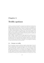
Introduction généraleINTRODUCTION GÉNÉRALE1. LES BIOMINÉRAUX ET L’ENVIRONNEMENTLe terme biominéral désigne un minéral dont la précipitation dépend <strong>de</strong> l’activité d’unorganisme vivant. Un minéral est classiquement défini comme un composé inorganiqueen phase soli<strong>de</strong> <strong>de</strong> composition chimique donnée (Steen 1971). La formation <strong>de</strong>biominéraux est un processus largement répandu dans le mon<strong>de</strong> vivant, recensé dans plus<strong>de</strong> 55 phylums différents (Lowenstam & Weiner 1989). Ces structures biogéniquesremplissent <strong>de</strong>s fonctions biologiques diverses et variées, comprenant notamment lesfonctions squelettiques <strong>de</strong> maintien <strong>de</strong> la forme statique, <strong>de</strong> transmission <strong>de</strong>s mouvementset <strong>de</strong> protection, mais aussi les fonctions <strong>de</strong> préhension et <strong>de</strong> mastication <strong>de</strong> la nourriture.Les biominéraux interviennent également dans la réception et la transmission sonores,l’orientation (dans un champ gravitationnel ou magnétique) et ont une gran<strong>de</strong> importancedans les processus <strong>de</strong> détoxication et <strong>de</strong> stockage d’ions à fonction métabolique.On connaît actuellement plus <strong>de</strong> 64 biominéraux <strong>de</strong> nature différente (Weiner & Dove2003). La calcification est un cas particulier <strong>de</strong> biominéralisation où le cation principal estle calcium. Elle concerne environ la moitié <strong>de</strong>s biominéraux recensés à ce jour, lecarbonate <strong>de</strong> calcium constituant le squelette <strong>de</strong> nombreux invertébrés marins. Dans lanature, le carbonate <strong>de</strong> calcium (CaCO 3 ) existe sous différentes formes (qui diffèrent parla disposition spatiale <strong>de</strong> leurs atomes): une forme amorphe (sans arrangement particulier<strong>de</strong>s atomes) et 5 polymorphes, à savoir l’aragonite (système cristallin orthorhombique), lacalcite (système rhomboédrique), la vatérite (système hexagonal) et les carbonates <strong>de</strong>calcium mono-hydratés (système rhomboédrique) et hexa-hydratés (systèmemonoclinique) (Lippman 1973, Dahl & Buchardt 2006). En conditions inorganiques, lacalcite et l’aragonite sont stables à température et pression normales. Par contre, lavatérite et les formes amorphes et hydratées du carbonate <strong>de</strong> calcium sont instables dansces mêmes conditions (Raz et al 2000, Neumann & Epple 2007). La calcite et l’aragonitesont <strong>de</strong> loin les principales formes <strong>de</strong> carbonate <strong>de</strong> calcium précipitées par les organismesvivants et constituent, entre autres, le squelette <strong>de</strong>s coraux, <strong>de</strong> certaines éponges, <strong>de</strong>sforaminifères, <strong>de</strong>s coccolithophores, <strong>de</strong>s mollusques, <strong>de</strong>s brachiopo<strong>de</strong>s, <strong>de</strong>s bryozoaires et<strong>de</strong>s échino<strong>de</strong>rmes. Le carbonate <strong>de</strong> calcium amorphe (CCA) constitue une phase1
Introduction généraletransitoire ou permanente du squelette <strong>de</strong> nombreux invertébrés, où il est stabilisé par <strong>de</strong>smolécules biologiques (Raz et al 2002). On recense également quelques rares exemples<strong>de</strong> biominéralisation <strong>de</strong> vatérite et <strong>de</strong> carbonate <strong>de</strong> calcium mono-hydraté, dont lesspicules <strong>de</strong> certaines ascidies (Lowenstam & Abbott 1975, Addadi et al 2003).Les biominéraux peuvent être classés selon le niveau <strong>de</strong> contrôle exercé lors <strong>de</strong> leurprécipitation : on distingue les biominéralisations biologiquement induites <strong>de</strong>sbiominéralisations biologiquement contrôlées (Mann 2001). Dans le premier cas, lebiominéral est précipité sous un contrôle biologique minimal, par simple réaction <strong>de</strong>métabolites produits par l’organisme avec <strong>de</strong>s ions du milieu extérieur. Le minéral ainsiformé présente <strong>de</strong>s propriétés similaires à celles <strong>de</strong> son homologue inorganique. Dans lecas <strong>de</strong> la biominéralisation strictement contrôlée par l’organisme, largement répandue ausein du mon<strong>de</strong> vivant, un contrôle strict <strong>de</strong> la formation du minéral est assuré par unefraction souvent quantitativement faible <strong>de</strong> molécules organiques. Le cristal ainsi forméprésente <strong>de</strong>s propriétés particulières par rapport à son équivalent inorganique. Le site <strong>de</strong>calcification est en général isolé <strong>de</strong> l’environnement extérieur, ce qui permet un contrôlelocal strict <strong>de</strong> la composition et <strong>de</strong> la sursaturation du microenvironnement à partir duquelle minéral est formé (Simkiss & Wilbur 1989). La présence <strong>de</strong> cette fraction organiquefait <strong>de</strong>s biominéraux <strong>de</strong>s matériaux composites, enjambant la frontière stricte opposantmon<strong>de</strong>s vivant et minéral <strong>de</strong>s premières classifications <strong>de</strong>s objets naturels (Aristote -343dans Barthélemy Saint Hilaire 1887).La composition <strong>de</strong>s biominéraux est en relation directe avec l’environnement. D’une part,comme nous le verrons, la composition <strong>de</strong>s biominéraux est influencée par les conditions<strong>de</strong> l’environnement (température, concentration <strong>de</strong>s ions dans l’eau pour les organismesaquatiques, etc…). Les biominéraux sont donc susceptibles d’enregistrer certainescaractéristiques environnementales prévalant lors <strong>de</strong> leur formation. De ce fait, plusieursbiominéraux sont utilisés comme paléoenregistreurs permettant la reconstruction <strong>de</strong>sconditions environnementales passées. D’autre part, la composition <strong>de</strong>s biominérauxinfluence leur stabilité et donc la résilience <strong>de</strong>s organismes qui les synthétisent face auxchangements environnementaux. La composition <strong>de</strong>s biominéraux joue donc un rôleimportant dans la problématique <strong>de</strong>s changements environnementaux, ce qui m’a amenéeà y consacrer ma thèse <strong>de</strong> doctorat. Je me suis intéressée plus particulièrement à l’ionmagnésium dans les calcites, dont la concentration est d’une part, contrôlée parl’environnement et d’autre part influence la solubilité du minéral formé.2
Introduction générale2. LES CALCITES MAGNÉSIENNESDe nombreux organismes marins produisent <strong>de</strong>s squelettes <strong>de</strong> carbonate <strong>de</strong> calcium,majoritairement sous la forme d’aragonite ou <strong>de</strong> calcite. Cette <strong>de</strong>rnière cristallise dans lesystème rhomboédrique, et est caractérisée par un axe c perpendiculaire à <strong>de</strong>ux axes asitués à 120° l’un <strong>de</strong> l’autre (Lippman 1973, Figure 1).Figure 1 : Structure <strong>de</strong> la calcite (d’après Young et al 1999).Cette polymorphe du carbonate <strong>de</strong> calcium peut incorporer <strong>de</strong>s concentrations nonnégligeables d’ions magnésium dans sa maille. Une calcite est définie comme hautementmagnésienne, lorsque sa concentration en magnésium est supérieure à 4 mol% <strong>de</strong> MgCO 3(Lippman 1973) et faiblement magnésienne, lorsque cette concentration est inférieure à4 mol%. Les aragonites sont généralement plus pauvres en magnésium que les calcites.C’est notamment le cas dans les carbonates biogéniques (Clarke & Wheeler 1922), où lesformes aragonitiques présentent rarement <strong>de</strong>s concentrations en magnésium supérieures à1 mol% <strong>de</strong> MgCO 3 , alors que les formes calcitiques contiennent souvent plus <strong>de</strong> 20 mol%<strong>de</strong> MgCO 3 , et rarement moins <strong>de</strong> 1 mol% (Chave 1954, Dodd 1967, Figure 2). Lesorganismes auxquels est consacrée ma thèse <strong>de</strong> doctorat produisent un squeletteprincipalement constitué <strong>de</strong> calcite magnésienne. Je me focaliserai par conséquent sur ceminéral. Dans le présent chapitre, je décrirai l’état actuel <strong>de</strong>s connaissances concernantles propriétés chimiques <strong>de</strong> l’ion magnésium et la formation <strong>de</strong>s calcites magnésiennes,3
Introduction généraleFigure 2 : Contenu en magnésium <strong>de</strong>s aragonites (A) et calcites (B) <strong>de</strong>s squelettes <strong>de</strong> différents taxa(d’après Dodd 1967).en conditions inorganiques et organiques, ainsi que l’ensemble <strong>de</strong>s facteurs susceptiblesd’influencer leur synthèse.2.1 L’ION MAGNÉSIUML’ion Mg 2+ est le cation divalent le plus abondant dans les cellules vivantes. C’est unélément essentiel au niveau biologique : il intervient dans la formation <strong>de</strong>s tissus dusquelette, la chimie <strong>de</strong>s flui<strong>de</strong>s biologiques, la régulation <strong>de</strong>s fonctions cellulaires ettissulaires, la régulation d’enzymes, la photosynthèse, … (Vidolin et al 2007, Bentov &Erez 2006). L’ion Mg 2+se distingue <strong>de</strong>s autres cations retrouvés dans les systèmesbiologiques par sa haute <strong>de</strong>nsité <strong>de</strong> charge 1 et sa chimie en solution tout à fait particulière(Smith & Maguire 1998, Wolf & Cittadini 2003).Tableau 1 : Propriétés <strong>de</strong>s cations communs dans les systèmes biologiques (d’après Maguire &Cowan 2002).IonRayonionique(Å)Rayonhydraté(Å)Rapport<strong>de</strong>srayonsVolumeionique(Å 3 )Volumehydraté(Å 3 )Rapport<strong>de</strong>svolumesNombre<strong>de</strong>coordinationTauxd’échangeaqueux (s -1 )Na + 0,95 2,75 2,9 3,6 88,3 24,5 6 8×10 8K + 1,38 2,32 1,7 11,0 52,5 4,8 6-8 10 9Ca 2+ 0,99 2,95 3,0 4,1 108 26,3 6-8 3×10 8Mg 2+ 0,65 4,76 7,3 1,2 453 394 6 10 51 La <strong>de</strong>nsité <strong>de</strong> charge d’un ion correspond à sa charge divisée par son rayon. Elle permet <strong>de</strong> prédire lecomportement d’un élément en solution. La solubilité <strong>de</strong>s cations en solution aqueuse dépend <strong>de</strong>l’attraction électrostatique <strong>de</strong>s molécules d’eau sur cet ion, elle-même fonction <strong>de</strong> la <strong>de</strong>nsité <strong>de</strong> charges <strong>de</strong>l’ion (Railsback 2006).4
Introduction généraleEn effet, parmi les cations abondants dans les systèmes biologiques, l’ion Mg 2+ possè<strong>de</strong> leplus petit rayon ionique et la plus gran<strong>de</strong> <strong>de</strong>nsité <strong>de</strong> charge (Tableau 1). L’ion Mg 2+ ensolution aqueuse a tendance à organiser les molécules d’eau autour <strong>de</strong> lui : il forme ainsiavec l’eau un complexe structuré en une sphère d’hydratation interne <strong>de</strong> 6 moléculesd’eau liées par <strong>de</strong>s liens électrostatiques (Wolf & Cittadini 2003, Günther 2006), entouréepar une sphère externe <strong>de</strong> 12 molécules d’eau supplémentaires (Markham et al 2002,Figure 3). Si le rayon ionique du Mg 2+ est plus petit que celui <strong>de</strong>s ions Ca 2+ , Na + et K + , ilpossè<strong>de</strong> le plus grand rayon hydraté. Ainsi, l’ion Mg 2+ hydraté a un volume 400 foissupérieur à celui <strong>de</strong> l’ion Mg 2+ non hydraté. Par comparaison, le facteur d’accroissement<strong>de</strong> volume du rayon hydraté n’est que <strong>de</strong> 25 pour les ions Ca 2+ et Na + , et <strong>de</strong> 4 pour l’ionK + (Maguire & Cowan 2002). De plus, le taux d’échange <strong>de</strong>s molécules d’eau <strong>de</strong> lasphère d’hydratation est considérablement plus faible dans le cas du magnésium que danscelui <strong>de</strong>s autres cations biologiques (Maguire & Cowan 2002, Wolf & Cittadini 2003). Enconditions biologiques, l’ion Mg 2+ est donc fortement hydraté et possè<strong>de</strong> une enthalpie <strong>de</strong>déshydratation élevée (-1922kJ/mol contre -1592 kJ/mol pour le Ca 2+ , Wolf & Cittadini2003).Figure 3 : Modèle d’arrangement <strong>de</strong>s sphères d’hydratation primaires et secondaires autour d’uncation Mg 2+ (Markham et al 2002).Il résulte <strong>de</strong> cette chimie unique que les interactions biologiques du magnésium sontdifférentes <strong>de</strong> celles <strong>de</strong>s autres cations. De nombreuses interactions biologiques ont lieuau travers <strong>de</strong> la sphère d’hydratation du Mg 2+ , plutôt que directement avec le cation Mg 2+ .L’eau <strong>de</strong> mer contient <strong>de</strong> fortes concentrations en magnésium (1294 mg/kg pour une eau<strong>de</strong> 35 psu, Libes 1992) (Tableau 2). Dans l’eau <strong>de</strong> mer, le magnésium hydraté est5
Introduction généralemajoritairement présent sous forme <strong>libre</strong> (87 %) ou forme une paire ionique avec <strong>de</strong>ssulfates (11 %) (Garrels & Thompson 1962, Kester & Pytkowicz 1969). Lesbiogéochimistes expriment les concentrations en magnésium sous forme d’un rapportmolaire avec le calcium, indépendant <strong>de</strong> la salinité. Les temps <strong>de</strong> rési<strong>de</strong>nce du magnésiumet du calcium dans l’eau <strong>de</strong> mer étant relativement longs (respectivement <strong>de</strong> 13 et 1millions d’années, Lea et al 1999), le rapport Mg/Ca <strong>de</strong>s eaux océaniques est relativementconstant sur une échelle <strong>de</strong> temps <strong>de</strong> l’ordre du million d’années : la valeur actuelle en est<strong>de</strong> 5,2 mol/mol, mais elle a varié au cours <strong>de</strong>s <strong>de</strong>rniers 540 millions d’années, atteignant<strong>de</strong>s minima <strong>de</strong> 1 mol/mol (Stanley 2006, Ries 2009). La variation du rapport Mg/Ca <strong>de</strong>seaux océaniques serait notamment liée aux variations <strong>de</strong>s taux <strong>de</strong> production <strong>de</strong> la croûteocéanique, qui déterminent en partie les concentrations en Ca 2+ , K + , Mg 2+ , SO 2- 4 dansl’eau <strong>de</strong> mer et seraient responsables <strong>de</strong> ces variations (Hardie 1996).Tableau 2 : Composition <strong>de</strong> l’eau <strong>de</strong> mer (Libes 1992).2.2 LE MAGNÉSIUM DANS LES CALCITES INORGANIQUES ETBIOGÉNIQUESLes cristaux <strong>de</strong> carbonate <strong>de</strong> calcium peuvent incorporer <strong>de</strong>s ions divalents autres que lecalcium dans leur maille cristalline, par substitution du calcium, en solution soli<strong>de</strong>. Ceprocessus est sélectif et dépend <strong>de</strong> la taille, la charge et la polarisation <strong>de</strong> l’ion (Simkiss &Wilbur 1989). C’est notamment le cas du magnésium incorporé dans la calcite, dont ilconstitue un élément trace, mineur ou majeur 2 selon son abondance dans le cristal.D’autres cations divalents, tels que le strontium, le fer, le manganèse, le zinc, le plomb et2 Un élément est considéré comme présent à l’état <strong>de</strong> traces lorsque sa concentration est inférieure au µg/g(ou ppm), tandis qu’il est dit majeur lorsqu’il est présent à <strong>de</strong>s concentrations supérieures au mg/g, etmineur lorsqu’il est présent à <strong>de</strong>s concentrations intermédiaires.6
Introduction généralele baryum, peuvent aussi être substitués au calcium dans la calcite (Weber 1969,Auernheimer & Chinchon 1997, Ree<strong>de</strong>r et al 1999). La substitution en solution soli<strong>de</strong>induit une déformation <strong>de</strong> la maille cristalline, dont la modification <strong>de</strong>s paramètres estproportionnelle au taux <strong>de</strong> substitution et détectable en diffraction <strong>de</strong>s rayons X(Goldschmidt et al 1955, Goldsmidth & Graf 1958). De ce fait, elle influence la stabilité<strong>de</strong> la calcite formée : à partir d’une concentration <strong>de</strong> 8 à 12 mol% <strong>de</strong> MgCO 3 (la valeurexacte étant encore indéterminée à ce jour), les calcites magnésiennes sont plus solublesque les aragonites, et voient leur solubilité augmenter avec leur concentration enmagnésium (An<strong>de</strong>rsson et al 2008, Figure 4). L’inclusion d’autres ions influence aussicette solubilité.De plus, il a récemment été démontré que les groupements carbonates peuvent êtresubstitués par <strong>de</strong>s sulfates dans la calcite, provoquant un agrandissement <strong>de</strong>s paramètres<strong>de</strong> la maille (Kontrec et al 2004).Figure 4 : Solubilité <strong>de</strong> la calcite dans l’eau <strong>de</strong> mer en fonction <strong>de</strong> sa concentration en magnésium ;les symboles pleins représentent les calcites biogéniques, tandis que les symboles vi<strong>de</strong>s représententles calcites synthétiques (d’après Morse et al 2006).La substitution du calcium par le magnésium est régie par <strong>de</strong>s lois physico-chimiquesdans les calcites inorganiques, mais on observe dans les calcites organiques <strong>de</strong>s déviationspar rapport à ces prédictions purement physico-chimiques, regroupées sous le termed’effets vitaux par les biogéochimistes. Les principaux facteurs affectant la précipitation<strong>de</strong>s calcites inorganiques et biogéniques et le taux <strong>de</strong> substitution du magnésium dans cesminéraux sont développés dans la section suivante.7
Introduction générale2.2.1 Facteurs contrôlant la concentration en magnésium dans les calcitesinorganiques2.2.1.1 Composition <strong>de</strong> la solution <strong>de</strong> précipitationEn conditions inorganiques, les lois thermodynamiques et cinétiques gouvernent lesréactions <strong>de</strong> précipitation en solution. A température et pression normales, la mise enprésence <strong>de</strong> calcium et <strong>de</strong> bicarbonate en solution sursaturée résulte en la précipitation <strong>de</strong>calcite. Parmi les formes stables du carbonate <strong>de</strong> calcium à température et pressionambiantes, cette polymorphe est la moins soluble, suivie par l’aragonite ou par la calcitemagnésienne selon son contenu en magnésium (An<strong>de</strong>rsson et al 2008). La présenced’autres ions dans la solution <strong>de</strong> précipitation, comme c’est systématiquement le cas enmilieu naturel, influence les propriétés du minéral formé (Morse & Mackenzie 1990). Parexemple, la présence <strong>de</strong> magnésium dans cette solution exerce une forte influence sur lapolymorphe <strong>de</strong> carbonate <strong>de</strong> calcium précipitée: en présence <strong>de</strong> fortes concentrations <strong>de</strong>ce cation (comme c’est le cas dans l’eau <strong>de</strong> mer), la formation d’aragonite est favoriséepar rapport à celle <strong>de</strong> la calcite (Kitano 1962, Kitano & Hood 1962, Lippman 1973, Razet al 2000). En effet, l’ion magnésium entouré <strong>de</strong> sa sphère d’hydratation s’adsorbe sur lasurface du nucléus <strong>de</strong> calcite en croissance (Lippman 1973). Son incorporation dans lamaille <strong>de</strong> la calcite requiert une énergie <strong>de</strong> déshydratation élevée et crée une barrièreénergétique inhibant la croissance ultérieure <strong>de</strong> la calcite (Raz et al 2000, Loste et al2003). Par contre, suite à <strong>de</strong>s contraintes stériques, l’aragonite n’incorpore pas l’ionmagnésium hydraté dans sa structure orthorhombique (sa structure est trop <strong>de</strong>nse pourincorporer la large sphère d’hydratation <strong>de</strong> cet ion, Addadi & Weiner 1992). Ellecristallise sans barrière cinétique en solution sursaturée. En présence <strong>de</strong> magnésium, lesystème en équi<strong>libre</strong> résulte donc en une précipitation nette d’aragonite.Des calcites magnésiennes peuvent toutefois être précipitées dans <strong>de</strong>s solutions au rapportMg/Ca élevé (supérieur à 4 :1), en augmentant la sursaturation <strong>de</strong> la solution ce quiaccélère la cinétique <strong>de</strong> réaction (Loste et al 2003). Lors <strong>de</strong> ces précipitationsinorganiques in vitro, la présence d’une phase transitoire <strong>de</strong> CCA a été mise en évi<strong>de</strong>nce.Cette phase est instable et tend à se transformer en une forme cristalline plus stable parexpulsion <strong>de</strong> l’eau (Raz et al 2000). La concentration en magnésium <strong>de</strong> cette phasetransitoire amorphe est principalement déterminée par le rapport Mg/Ca <strong>de</strong> la solution <strong>de</strong>8
Introduction généraleprécipitation, et détermine celui <strong>de</strong> la phase cristalline subséquente (Loste et al 2003).Cette conclusion est en désaccord avec celle d’Oomori et al (1987), selon lesquels lerapport Mg/Ca <strong>de</strong> la solution <strong>de</strong> précipitation n’aurait qu’une faible influence sur lerapport Mg/Ca <strong>de</strong> la calcite formée, principalement déterminé par la température.Selon Meldrum & Hy<strong>de</strong> (2001), le type <strong>de</strong> polymorphe formé dépendrait aussi <strong>de</strong> lavitesse <strong>de</strong> croissance du minéral, elle-même dépendante <strong>de</strong> plusieurs autrescaractéristiques <strong>de</strong> la solution <strong>de</strong> précipitation, telles que les conditions <strong>de</strong> saturation et laconcentration en carbonates (Morse & Mackenzie 1990, Lopez et al 2009). Cettehypothèse permet d’expliquer la variabilité <strong>de</strong> la concentration en magnésium <strong>de</strong>s calciteshautement magnésiennes formées dans une solution riche en magnésium.2.2.1.2 TempératureLes lois <strong>de</strong> la thermodynamique prédisent que la substitution du calcium par lemagnésium dans la formation <strong>de</strong> la calcite dépend <strong>de</strong> la température. Toute réaction àlaquelle est associé un changement d’enthalpie (∆H 0 ) serait sensible à la température,comme l’exprime l’équation <strong>de</strong> Van t’Hoff : d ln K/ d (1/T) = - ∆H/Roù T= température en °KR=constante <strong>de</strong>s gazK= constante d’équi<strong>libre</strong>Les réactions avec la plus gran<strong>de</strong> valeur <strong>de</strong> ∆H 0 auront la plus gran<strong>de</strong> dépendance vis-àvis<strong>de</strong> la température.La substitution isomorphique du calcium par le magnésium dans la calcite est caractériséepar un ∆H 0 <strong>de</strong> 21 kJ/mol (Lea 2003). C’est donc une réaction endothermique favoriséeaux températures élevées. L’équation <strong>de</strong> Van t’Hoff permet <strong>de</strong> prédire une augmentationexponentielle du rapport Mg/Ca en fonction <strong>de</strong> la température (Lea et al 1999, Lea 2003),et <strong>de</strong> la chiffrer à 3 % par °C entre 0 et 30 °C. Des expériences in vitro sur <strong>de</strong>s calcitesinorganiques confirment que la concentration en magnésium incorporé dépend <strong>de</strong> latempérature (Katz 1973, Oomori et al 1987).9
Introduction générale2.2.2 Facteurs contrôlant la concentration en magnésium dans les calcitesbiogéniquesLes concentrations en magnésium <strong>de</strong>s calcites biogéniques peuvent largement surpassercelles <strong>de</strong>s calcites <strong>de</strong> synthèse et <strong>de</strong>s « ciments inorganiques» <strong>de</strong> calcite magnésienneprécipités à partir <strong>de</strong> l’eau <strong>de</strong> mer (Mucci & Morse 1983 dans Cheng et al 2007). Desconcentrations en magnésium élevées ont par exemple été rapportées dans les spicules etles squelettes massifs d’éponges (respectivement 12,9 mol% <strong>de</strong> MgCO 3 , Jones & Jenkins1970, et 19 mol%, Hooper & van Soest 2002), chez certains foraminifères (21 mol% <strong>de</strong>MgCO 3 , chez les milioi<strong>de</strong>s, Bentov & Erez 2006) et chez les échino<strong>de</strong>rmes (avec unemoyenne variant <strong>de</strong> 13,5 à 16 mol% selon la classe considérée, avec <strong>de</strong>s maxima <strong>de</strong> 43,5mol% <strong>de</strong> MgCO 3 mesurés dans les parties très hautement magnésiennes <strong>de</strong> la <strong>de</strong>ntd’oursin, Weber 1969, Schroe<strong>de</strong>r et al 1969). Il existe également <strong>de</strong>s calcites biogéniquesavec <strong>de</strong>s concentrations en magnésium relativement faibles, tels les tests <strong>de</strong> certainsforaminifères planctoniques, qui contiennent moins <strong>de</strong> 0,1 mol% <strong>de</strong> MgCO 3 (Bentov &Erez 2006).La concentration en éléments mineurs ou traces dans les calcites biogéniques est engénéral le résultat <strong>de</strong> la superposition d’effets biologiques à <strong>de</strong>s effets <strong>de</strong> l’environnementsimilaires à ceux affectant les calcites inorganiques (Weiner & Dove 2003). En plusd’être strictement dépendante du type <strong>de</strong> polymorphe <strong>de</strong> carbonate <strong>de</strong> calcium déposé, laconcentration en magnésium squelettique d’un organisme varie systématiquement selondifférents facteurs dont les principaux sont la température, la salinité, le pH et laconcentration en magnésium dans la solution <strong>de</strong> précipitation. L'importance relative <strong>de</strong>ces différents facteurs dans la détermination <strong>de</strong>s concentrations en magnésiumincorporées dans les calcites biogéniques est encore mal connue.2.2.2.1 TempératureTout comme celle <strong>de</strong> la calcite inorganique, la concentration en magnésium <strong>de</strong>s calcitesbiogéniques est fortement influencée par la température. Clarke & Wheeler (1922) sontles premiers à relever la corrélation positive entre la concentration en magnésium dusquelette <strong>de</strong> certains invertébrés marins et la température <strong>de</strong> l’eau dans laquelle vivent cesorganismes (Chave 1954, Figure 5).10
Introduction généraleFigure 5 : Concentrations en magnésium dans les calcites squelettiques d’organismes <strong>de</strong> diverstaxons en fonction <strong>de</strong> la température (Mackenzie et al 1983, d’après les données <strong>de</strong> Chave 1954).Dans le contexte actuel <strong>de</strong> changement climatique global, <strong>de</strong> nombreux chercheurs se sontpenchés sur cette relation entre chimie <strong>de</strong>s squelettes <strong>de</strong> calcite et température <strong>de</strong> l’eau, envue d’une éventuelle exploitation <strong>de</strong> ces squelettes en tant qu’archive naturelle du climatpouvant pallier aux limitations temporelles <strong>de</strong>s enregistrements instrumentaux. L’analysedu rapport Mg/Ca <strong>de</strong>s tests <strong>de</strong> foraminifères a fait l’objet d’une attention toute particulièreau cours <strong>de</strong>s années 1990 et est <strong>de</strong>venu un traceur fiable et reconnu <strong>de</strong>s paléotempératures(Rosenthal et al 1997). Les tests <strong>de</strong> foraminifères sédimentent en grand nombre sur lesfonds marins, où ils s’accumulent et sont relativement bien conservés dans les rochessédimentaires. Pour différentes espèces <strong>de</strong> foraminifères, les relations spécifiques entre lerapport Mg/Ca du test et la température <strong>de</strong> l’eau ont été établies pour <strong>de</strong>s spécimensélevés en aquarium ou récoltés en milieu naturel dans <strong>de</strong>s pièges à particules (Nürnberg etal 1996, Lea et al 1999, Anand et al 2003, Kisakürek et al 2008, Regenberg et al 2009)puis utilisées sur <strong>de</strong>s spécimens <strong>de</strong> carottes sédimentaires. Une calibration valable pourune dizaine d’espèces planctoniques actuelles et les familles sub-fossiles et fossilesapparentées a ainsi pu être établie (Mg/Ca=0,38×exp(0,090T), Anand et al 2003,Figure 6). Selon cette calibration, le rapport Mg/Ca augmente <strong>de</strong> 9,0 ± 0,3 % par °C, et latempérature <strong>de</strong> calcification peut être estimée avec une exactitu<strong>de</strong> <strong>de</strong> 1,2 °C. Parcomparaison avec <strong>de</strong>s données obtenues à partir d’autres traceurs (rapports isotopiques),cette relation a pu être vérifiée sur <strong>de</strong>s échantillons sédimentaires riches en foraminifères.Elle peut néanmoins être légèrement affectée par la dissolution partielle <strong>de</strong>s microfossiles(Hen<strong>de</strong>rson 2002).11
Introduction généraleFigure 6 : Calibration <strong>de</strong> la relation entre la température et le rapport Mg/Ca établie pour 11 espèces<strong>de</strong> foraminifères planctoniques (Mg/Ca=0,38×exp(0,090T), d’après Anand et al 2003).2.2.2.2 Rapport Mg/Ca <strong>de</strong> l’eau <strong>de</strong> merComme en conditions inorganiques, le rapport Mg/Ca <strong>de</strong> l’eau <strong>de</strong> mer influence celui <strong>de</strong>scalcites biogéniques qui y sont formées (Lorens & Ben<strong>de</strong>r 1980, Ries 2004, Segev & Erez2006, Stanley 2006). Des étu<strong>de</strong>s expérimentales réalisées sur divers organismescalcifiants (moules, oursins, crabes, crevettes, serpules et algues corallines) ayant grandi àune même température dans <strong>de</strong>s eaux <strong>de</strong> mer artificielles aux rapports Mg/Ca contrastés(<strong>de</strong> 1 à 6,7 mol/mol) démontrent que les rapports Mg/Ca <strong>de</strong>s calcites formées sont enrelation directe mais non linéaire avec le rapport Mg/Ca <strong>de</strong> l’eau <strong>de</strong> mer (Lorens &Ben<strong>de</strong>r 1980, Ries 2004, 2006).Cette propriété a été appliquée dans le cadre <strong>de</strong> reconstructions paléo environnementalesdu rapport Mg/Ca <strong>de</strong>s eaux <strong>de</strong>s océans anciens, via l’analyse du rapport Mg/Ca <strong>de</strong> testsd’oursins fossiles datant du Phanérozoïque (Dickson 2002, 2004). Les résultats obtenusconcor<strong>de</strong>nt avec les reconstructions effectuées par d’autres métho<strong>de</strong>s.2.2.2.3 Autres facteurs environnementauxSalinité. Fergusson et al (2008) ont observé que la calcite <strong>de</strong>s foraminifèresméditerranéens présente <strong>de</strong>s rapports Mg/Ca particulièrement élevés, et en ont conclu queles hautes salinités pouvaient avoir un impact important sur l’incorporation du12
Introduction généralemagnésium. Par contre, Hoogaker et al (2009) réfutent cet effet direct <strong>de</strong> la salinité etattribuent cet accroissement <strong>de</strong> la concentration en magnésium à une contamination <strong>de</strong> lacalcite squelettique par <strong>de</strong>s surcroissances <strong>de</strong> calcite inorganique. Chez <strong>de</strong>s foraminifèrescultivés en laboratoire, la salinité affecte significativement le rapport Mg/Ca <strong>de</strong> la calcitedéposée, mais reste un facteur <strong>de</strong> détermination secondaire en comparaison <strong>de</strong> latempérature: pour une élévation <strong>de</strong> salinité <strong>de</strong> 1 psu, le rapport Mg/Ca augmente <strong>de</strong> 2,3 à5 % selon l’espèce considérée (Lea et al 1999, Kisakürek et al 2008, Dueñas-Bohòrquezet al 2010). Par contre, la composition en magnésium <strong>de</strong> la calcite squelettiqued’ostraco<strong>de</strong>s ayant grandi à différentes salinités n’est pas affectée par la salinité (DeDeckker et al 1999).pH. L’influence du pH sur le rapport Mg/Ca <strong>de</strong>s calcites biogéniques est controversée.Des réponses contradictoires aux variations <strong>de</strong> pH ont été observées chez lesforaminifères (Dissard et al 2010). Certaines étu<strong>de</strong>s rapportent une relation inverse entrepH et Mg/Ca dans la calcite <strong>de</strong>s foraminifères (Lea et al 1999), tandis que d’autressuggèrent une absence d’effet aux pH océaniques moyens actuels mais une diminutiondrastique à <strong>de</strong>s pH inférieurs à 8,0 (Russel et al 2004, Kisakürek et al 2008).Etat <strong>de</strong> saturation <strong>de</strong> l’eau <strong>de</strong> mer. Il a également été proposé que la concentration enmagnésium dans la calcite soit liée au niveau <strong>de</strong> saturation <strong>de</strong> l’eau <strong>de</strong> mer 3 . Une relationentre ces facteurs a été suggérée pour <strong>de</strong>s algues corallines et attribuée à une différence <strong>de</strong>leur vitesse <strong>de</strong> croissance (Agegian 1985 dans Morse & Mackenzie 1990). Par contre,plusieurs expériences <strong>de</strong> culture <strong>de</strong> foraminifères en conditions contrôlées n’ont montréaucune dépendance significative du rapport Mg/Ca dans la calcite vis-à-vis <strong>de</strong> l’état <strong>de</strong>saturation <strong>de</strong> l’eau <strong>de</strong> mer (Raitzsch et al 2010, Dueñas-Bohòrquez et al 2010, Dissard etal 2010). De plus, selon Lopez et al (2009), le niveau <strong>de</strong> saturation <strong>de</strong> l’eau <strong>de</strong> mer, mêmes’il affecte la vitesse <strong>de</strong> croissance <strong>de</strong> la calcite, n’aurait pas d’influence sur laconcentration en magnésium qui y est incorporé à une température donnée.3 Le niveau <strong>de</strong> saturation <strong>de</strong> l’eau par rapport à un minéral carbonaté, Ω, est défini comme étant le produit<strong>de</strong>s activités <strong>de</strong>s ions calcium, magnésium et carbonates en solution divisé par la constante <strong>de</strong> solubilité duminéral considéré. Lorsque Ω=1, le minéral est dit en équi<strong>libre</strong> thermodynamique avec l’eau <strong>de</strong> mer, c’està-direque ses réactions <strong>de</strong> dissolution et <strong>de</strong> précipitation s’équi<strong>libre</strong>nt mutuellement. Dans une solution <strong>de</strong>Ω < 1 (sous-saturée par rapport au minéral considéré) et Ω > 1 (sursaturée), les réactions <strong>de</strong> dissolution et<strong>de</strong> précipitation sont, respectivement, favorisées.13
Introduction générale2.2.2.4 Effets vitaux et contrôle biologiqueDes minéraux d’une même polymorphe déposés par <strong>de</strong>s organismes différents vivant dansles mêmes eaux, et donc subissant <strong>de</strong>s conditions environnementales semblables, peuventprésenter <strong>de</strong>s concentrations différentes en magnésium (Chave 1954). Ces valeurs ne sontdonc pas en équi<strong>libre</strong> avec les conditions physico-chimiques <strong>de</strong>s eaux environnantes,comme elles le seraient pour une calcite inorganique. Ceci indique que <strong>de</strong>s effetsbiologiques s’additionnent aux influences environnementales (Lowenstam & Weiner1989). Les déviations induites biologiquement par rapport à un comportement purementthermodynamique sont regroupées sous le terme d’effets vitaux (Weiner & Dove 2003,Fergusson et al 2008).Le terme « effets vitaux » recouvre à la fois <strong>de</strong>s facteurs distaux et proximaux. Lespremiers sont liés à la physiologie <strong>de</strong> l’organisme et seules <strong>de</strong>s corrélations peuvent êtreétablies entre eux et la concentration squelettique en magnésium. Les secondscorrespon<strong>de</strong>nt aux structures et processus directement impliqués dans labiominéralisation. Dans ce cas, <strong>de</strong>s relations <strong>de</strong> cause à effet peuvent parfois être établies.Le principal facteur distal étudié à ce jour est la vitesse <strong>de</strong> croissance du squelette. Celleciest corrélée à la concentration en magnésium du squelette chez <strong>de</strong> nombreuses espèces(Weber 1973, Kolesar 1978). Or, la vitesse <strong>de</strong> croissance dépend <strong>de</strong> nombreux facteurs,environnementaux (température) et biologiques (ontogenèse, disponibilité <strong>de</strong> lanourriture, état reproducteur). Par exemple, chez certains foraminifères, le rapport Mg/Ca<strong>de</strong> la calcite varie durant la vie <strong>de</strong> l’individu, en fonction <strong>de</strong> la vitesse <strong>de</strong> croissance, ellemêmefonction <strong>de</strong> la température mais aussi <strong>de</strong> la taille (El<strong>de</strong>rfield et al 2002, Hintz et al2006) ; <strong>de</strong> plus, ce rapport diffère entre les processus <strong>de</strong> formation d’une nouvellechambre du test et l’épaississement secondaire <strong>de</strong> celle-ci (Erez 2003). De manièregénérale, les interactions entre ces différents facteurs distaux sont encore très malcomprises.Les <strong>de</strong>ux principaux facteurs proximaux impliqués dans l’incorporation du magnésiumsont la matrice organique <strong>de</strong> biominéralisation et les mécanismes <strong>de</strong> contrôles <strong>de</strong> laconcentration en magnésium dans le site <strong>de</strong> minéralisation (Bentov & Erez 2006).Matrice organique <strong>de</strong> minéralisation. La matrice organique <strong>de</strong> minéralisation est définiecomme tout matériel organique inclus ou bordant la phase minérale et affectant laminéralisation. Elle est constituée d’un assemblage complexe <strong>de</strong> protéines,polysacchari<strong>de</strong>s, et lipi<strong>de</strong>s intimement associés à la phase minérale (Addadi & Weiner14
Introduction générale1992, Albeck et al 1996, Ameye et al 2001, Farre & Dauphin 2009). Ces macromoléculesreprésentent généralement une faible proportion du poids sec du squelette, variant <strong>de</strong> 0,1 à5 % <strong>de</strong> ce <strong>de</strong>rnier selon l’organisme considéré, mais peuvent représenter jusqu’à 45 % dupoids sec du squelette chez certains Crustacés (Swift et al 1986, Welin<strong>de</strong>r 1974). Malgréleur faible abondance relative, ces macromolécules jouent un rôle essentiel dans labiominéralisation, exerçant un contrôle direct sur la nucléation, la croissance, lapolymorphe et la morphologie du minéral déposé (Simkiss & Wilbur 1989). Cesmolécules peuvent être extraites par décalcification du minéral (dans l’EDTA parexemple), et sont séparées en fractions soluble et insoluble dans l’EDTA. Que ce soitchez les échino<strong>de</strong>rmes, les foraminifères ou dans la couche calcitique <strong>de</strong> la coquille <strong>de</strong>smollusques, la fraction soluble est principalement constituée <strong>de</strong> protéines présentant uncaractère aci<strong>de</strong>, lié à leur richesse en aci<strong>de</strong>s aspartique et glutamique (Weiner 1979).Même si elles présentent <strong>de</strong>s caractéristiques chimiques communes entre les différentsgroupes calcifiants (Weiner & Addadi 1997), les protéines <strong>de</strong> la matrice <strong>de</strong>biominéralisation ont <strong>de</strong>s compositions en aci<strong>de</strong>s aminés spécifiques (Albeck et al 1993).Ces protéines, et dans certains cas les polysacchari<strong>de</strong>s sulfatés qui leur sont associés,peuvent interagir avec le calcium, grâce à leurs groupements carboxyles présentant <strong>de</strong>fortes affinités pour cet ion, <strong>de</strong> manière à initier une nucléation orientée (Wilbur &Bernhardt 1984, Addadi et al 1987, Addadi & Weiner 1985, 1992).Les molécules <strong>de</strong> la matrice interviennent dans la détermination du type <strong>de</strong> polymorpheformée (Falini et al 1996, Levi et al 1998, Feng et al 2000, Takeuchi et al 2008). Lesmacromolécules extraites <strong>de</strong> la couche <strong>de</strong> calcite <strong>de</strong> la coquille <strong>de</strong> mollusque induisent laformation <strong>de</strong> calcite tandis que celles extraites <strong>de</strong> la couche aragonitique induisent laformation d’aragonite (Falini et al 1996, Feng et al 2000). Dans <strong>de</strong>s expériences <strong>de</strong>cristallisations en présence <strong>de</strong> magnésium, les additifs organiques (tels que lespolysacchari<strong>de</strong>s et les polymères d’aci<strong>de</strong>s carboxylique et aspartique) favorisent laformation <strong>de</strong> calcite par rapport à celle d’aragonite (Kitano & Kanamori 1966, Wada et al1999, Meldrum & Hy<strong>de</strong> 2001, Takeuchi et al 2008).En plus <strong>de</strong> contrôler la polymorphe déposée, les molécules <strong>de</strong> la matrice organiquesemblent aussi hautement impliquées dans l’incorporation <strong>de</strong> hautes quantités <strong>de</strong>magnésium dans les calcites biogéniques. Robach et al (2006) ont en effet mis enévi<strong>de</strong>nce un parallélisme entre les distributions spatiales du magnésium et <strong>de</strong>s protéines<strong>de</strong> la matrice riches en aci<strong>de</strong>s aspartiques dans la calcite très hautement magnésienne <strong>de</strong>la <strong>de</strong>nt d’oursin (contenant jusqu’à 43,5 mol% <strong>de</strong> MgCO 3 ). De plus, la présence <strong>de</strong>15
Introduction généralemacromolécules extraites <strong>de</strong> biominéraux (ou <strong>de</strong> molécules synthétiques équivalentes)induit in vitro la formation <strong>de</strong> cristaux aux contenus en magnésium supérieurs à ceux <strong>de</strong>cristaux produits en l’absence <strong>de</strong> telles molécules (Kitano & Kanamori 1966, Raz et al2000, Stephenson et al 2008). Aucune relation quantitative entre la concentration et lanature précise <strong>de</strong> ces macromolécules et la concentration en magnésium du minéralprécipité n’a toutefois encore été établie.La matrice organique peut agir <strong>de</strong> différentes façons sur l’incorporation du magnésium :elle peut agir sur la vitesse <strong>de</strong> précipitation, la déshydratation du magnésium et lastabilisation temporaire d’une phase précurseur <strong>de</strong> CCA, plus apte à incorporer <strong>de</strong>s ionsMg hydratés, qui se transforme ensuite en une phase cristalline qui conserve uneconcentration élevée en magnésium.Stephenson et al (2008) observent que la présence d’un pepti<strong>de</strong> aci<strong>de</strong> favorise fortementl’incorporation du magnésium dans la calcite formée in vitro, et suggèrent que cet effetest lié à une accélération <strong>de</strong> la formation <strong>de</strong> la calcite. Cet effet cinétique résulte en unediscrimination moins efficace <strong>de</strong>s ions incorporés dans la maille cristalline, et parconséquent en une augmentation <strong>de</strong> la quantité <strong>de</strong> magnésium substituée au calcium. Deplus, il est possible que les pepti<strong>de</strong>s agissent sur le niveau d’hydratation <strong>de</strong>s ions,réduisant ainsi la barrière énergétique liée à l’incorporation du magnésium (Albeck et al1993, Raz et al 2000, 2003). Elhadj et al (2006) ont en effet observé que l’accélération <strong>de</strong>la croissance <strong>de</strong> la calcite en présence <strong>de</strong> macromolécules est liée à la charge et aucaractère hydrophile <strong>de</strong> ces <strong>de</strong>rnières. Les molécules <strong>de</strong> la matrice peuvent donc perturberlocalement la structuration <strong>de</strong> la sphère d’hydratation du magnésium en solution, etréduire ainsi la barrière d’énergie liée à son incorporation dans la calcite en croissance. Lemécanisme précis en est encore mal connu, mais une étu<strong>de</strong> récente a démontré que ceteffet est fortement corrélé aux caractéristiques chimiques <strong>de</strong>s aci<strong>de</strong>s organiquesimpliqués, et plus particulièrement à leurs constantes spécifiques <strong>de</strong> liaison au calciumpar rapport au magnésium (Wang et al 2009).Le passage par une phase amorphe transitoire est une stratégie largement répandue dans laformation <strong>de</strong>s biominéraux (Tableau 3) et ce, malgré l’instabilité inhérente à <strong>de</strong> tellesphases. La phase amorphe transitoire, longtemps ignorée <strong>de</strong> par la difficulté <strong>de</strong> sadétection (Weiner et al 2003), a été décrite dans le cadre du processus <strong>de</strong> formation <strong>de</strong> lacalcite <strong>de</strong>s échino<strong>de</strong>rmes, <strong>de</strong> l’aragonite <strong>de</strong>s mollusques, <strong>de</strong> la magnétite et l’apatitecontenues dans la radula <strong>de</strong>s chitons (Weiner et al 2005, Jacob et al 2008).16
Introduction généraleTableau 3 : Présence <strong>de</strong> carbonate <strong>de</strong> calcium amorphe dans différents taxons. La polymorphe laplus communément déposée dans chaque taxon est indiquée en gras (d’après Addadi et al 2003).Règne/Phylum Forme du dépot FonctionsprésuméesMg/Mg+Ca(atomes%)Formes <strong>de</strong> CaCO 3 déposéespar ces organismesPlantaeCystolithes dans lesfeuilles? 0-26 Calcite, aragonite , vatérite,CCAArthropoda : CuticuleRenforce0-20 CCA, calciteCrustaceal’exocuticuleGastrolithesStockageCCAtemporairePorifera Spicule (partie interne) ? 6-17 Calcite, CCAAscidiacea Spicule (corps) Renforce les tissuset la tunique0-16 Calcite, aragonite, vatérite,CCA, monohydrocalciteEchino<strong>de</strong>rmata Spicule larvaire Phase précurseur 5 Calcite, CCAPiquant d’oursin Régénération ? Calcite, CCACnidaria/Spicules Support mécanique 18 Calcite, aragonite, CCAGorgonaceaMolluscaGranulesStockage? CCA vatérireBivalviatemporaireCoquille <strong>de</strong> la larve Phase précurseur Aragonite, CCAGastropdaSpiculesRenforcer lesCCA(Nudibranchia)tissus ?Plathelminthes Corpuscules ? CCADes étu<strong>de</strong>s récentes démontrent que les spicules <strong>de</strong>s larves d’échino<strong>de</strong>rmes, la <strong>de</strong>nt et lespiquants d’oursins sont formés par l’intermédiaire d’un précurseur amorphe qui cristalliseensuite en calcite magnésienne (Politi et al 2004 ; Aizenberg et al 1996a, Beniash et al1997, 1999). La calcite étant l’état <strong>de</strong> plus basse énergie du carbonate <strong>de</strong> calcium, laconversion d’amorphe en calcite est favorisée thermodynamiquement (Killian & Wilt2008), procédant par <strong>de</strong>s phases transitoires successives <strong>de</strong> moins en moins instables(Politi et al 2008). La stabilisation transitoire ou permanente du CCA dans les systèmesbiologiques est due à certaines macromolécules <strong>de</strong> la matrice, notamment à <strong>de</strong>s protéinestrès aci<strong>de</strong>s, riches en aci<strong>de</strong>s aminés Asp, Glu, et Ser (Aizenberg et al 1996a, 2003, Politiet al 2007), et souvent glycosylées (Aizenberg et al 2003). Aizenberg et al (1996a, 2003)ont ainsi démontré que les protéines extraites <strong>de</strong> la phase permanente <strong>de</strong> CCA <strong>de</strong>sspicules <strong>de</strong> l’ascidie Pyura pachy<strong>de</strong>rmata et <strong>de</strong> l’éponge Clathrina sp permettentd’induire la formation et <strong>de</strong> temporairement stabiliser une phase amorphe in vitro, sansautre additif. De même, les protéines riches en aci<strong>de</strong> aspartique extraites <strong>de</strong> la calcite dumollusque Atrina rigida induisent la formation d’une phase <strong>de</strong> CCA in vitro et lastabilisent temporairement (Politi et al 2007). Toutefois, selon Raz et al (2003) la seuleprésence <strong>de</strong>s macromolécules n’est pas suffisante pour induire la formation <strong>de</strong> CCA, quirequiert aussi la présence <strong>de</strong> magnésium en solution. Cet ion augmente le <strong>de</strong>gré <strong>de</strong>sursaturation <strong>de</strong> la solution, ce qui induit la précipitation <strong>de</strong> particules métastablesamorphes, cristallisant ensuite en une polymorphe déterminée par les conditionsambiantes (Raz et al 2000). Même si sa présence en solution est requise, on ignore si le17
Introduction généralemagnésium est uniquement nécessaire à l’élévation du niveau <strong>de</strong> sursaturation <strong>de</strong> lasolution ou si il intervient dans l’activité <strong>de</strong>s macromolécules.Le passage par une phase amorphe favorise l’incorporation du magnésium, et la formation<strong>de</strong> calcites riches en magnésium (Raz et al 2000, Cheng et al 2007, Wang et al 2009). Eneffet, une phase amorphe hydratée facilite l’incorporation d’ions Mg 2+ partiellementhydratés (Raz et al 2000). Des expériences in vitro ont démontré que le contenu enmagnésium du cristal formé dépend <strong>de</strong> celui <strong>de</strong> la phase amorphe qui le précè<strong>de</strong>, ellemêmefonction <strong>de</strong> la concentration en cet ion dans la solution <strong>de</strong> cristallisation (Loste et al2003). Il faut noter que les phases permanentes <strong>de</strong> CCA, telles qu’observées dans lesspicules <strong>de</strong> certaines ascidies et l’exosquelette <strong>de</strong>s crustacés, sont hydratées (Cölfen &Mann 2003). L’eau contenue dans ces phases amorphes pourrait intervenir dans leurstabilisation (Politi et al 2006, 2008).Contrôle <strong>de</strong>s concentrations ioniques dans la solution <strong>de</strong> précipitation. Commementionné dans la section 2.2.2.2, le rapport Mg/Ca <strong>de</strong> la solution <strong>de</strong> précipitation peutdirectement conditionner la concentration en magnésium du minéral formé. Le microenvironnement<strong>de</strong> cristallisation est un espace délimité, au moins en partie, par unemembrane, ce qui permet un contrôle biologique <strong>de</strong> la composition <strong>de</strong> la solution <strong>de</strong>précipitation et <strong>de</strong>s conditions <strong>de</strong> sursaturation. La concentration en magnésium <strong>de</strong> lasolution <strong>de</strong> précipitation peut ainsi être déterminée par les transporteurs du magnésium.Dans le cas <strong>de</strong>s organismes marins, le rapport Mg/Ca <strong>de</strong> l’eau <strong>de</strong> mer (5,2 mol/mol) estbeaucoup plus élevé que celui <strong>de</strong>s squelettes (généralement <strong>de</strong> l’ordre <strong>de</strong> 0,1 mol/mol).Les systèmes <strong>de</strong> transport doivent donc réduire la concentration relative en magnésiumdans la solution <strong>de</strong> précipitation. Etant donné la chimie particulière <strong>de</strong> cet ion (section1.1), Smith & Maguire (1998) supposent que les transports du magnésium se font via <strong>de</strong>stransporteurs protéiques particuliers ou <strong>de</strong>s membres non classiques <strong>de</strong> familles <strong>de</strong>transporteurs connus. Différents systèmes <strong>de</strong> transport membranaires spécifiques à cet ionont été décrits à ce jour dans les cellules eucaryotiques: <strong>de</strong>s canaux <strong>de</strong> diffusion passive(Preston 1998), <strong>de</strong>s échangeurs Na + /Mg 2+ , Ca 2+ /Mg 2+ , H + /Mg 2+ , Mg 2+ /Cl - et une pompeMg-ATPase (Flatman 1984, Cefaratti et al 1998, Wolf et al 2003). La cellule peut aussimodifier le rapport Mg/Ca <strong>de</strong> l’espace <strong>de</strong> cristallisation qu’elle délimite par sa membraneen modifiant les concentrations en calcium, pour lequel <strong>de</strong> nombreux systèmes <strong>de</strong>transport ont été décrits.18
Introduction générale3. MODÈLES BIOLOGIQUES ÉTUDIÉSLa présente thèse s’intéresse à <strong>de</strong>s représentants <strong>de</strong> <strong>de</strong>ux groupes d’organismes produisant<strong>de</strong>s squelettes en calcite magnésienne : les échino<strong>de</strong>rmes et les éponges calcaireshypercalcifiées. Ce chapitre présente les caractéristiques générales <strong>de</strong> ces squelettes etleurs mécanismes <strong>de</strong> formation.3.1 LE SQUELETTE POSTMÉTAMORPHIQUE DES ÉCHINODERMES3.1.1 Structure et morphogenèseExcepté chez les holothuries où il est limité à <strong>de</strong>s spicules épars, le squelette <strong>de</strong>séchino<strong>de</strong>rmes est formé d’ossicules qui occupent une partie importante du <strong>de</strong>rme (Hyman1955). Sauf exception, le squelette est toujours recouvert par l’épi<strong>de</strong>rme. Chaque ossiculeest formé <strong>de</strong> trabécules <strong>de</strong> calcite anastomosées, structurées en un réseau minéraltridimensionnel (le stéréome) dont les pores sont remplis <strong>de</strong> tissu conjonctif (le stroma)(Figure 7).Figure 7 : Squelette d’échino<strong>de</strong>rme : stéréome à galeries d’un tubercule <strong>de</strong> l’oursin Tripneustesgratilla (Carter 1990).Le stéréome est une structure quasi exclusivement minérale <strong>de</strong> calcite magnésienne quicontient néanmoins en son sein une très faible proportion en poids (0,1 %) <strong>de</strong>macromolécules organiques intra-squelettiques (Weiner 1985, Swift et al 1986). Lestroma est constitué d’une matrice extracellulaire, <strong>de</strong> fibres, et <strong>de</strong> cellules <strong>de</strong> différentstypes (cellules immunitaires et squelettogènes, fibroblastes) (Dubois & Chen 1989)(Figure 8).19
Introduction généraleFigure 8 : Représentation schématique <strong>de</strong>s parties épi<strong>de</strong>rmiques (E) et <strong>de</strong>rmiques (D) du tégument<strong>de</strong>s échino<strong>de</strong>rmes (CY=coelomocyte, CT= tissu conjonctif, FE= pore du stéréome, ST=stéréome,SH=sclérocyte, STR=stroma, d’après Chia & Koss 1994).Le squelette postmétamorphique <strong>de</strong>s échino<strong>de</strong>rmes est formé juste avant lamétamorphose: les spicules larvaires (présents dans les classes <strong>de</strong>s Ophiuroi<strong>de</strong>a et <strong>de</strong>sEchinoi<strong>de</strong>a) se résorbent quasi entièrement.Les cellules squelettogènes, appelées sclérocytes, sont localisées dans le stroma. Ellesdéveloppent <strong>de</strong>s processus cellulaires qui fusionnent en un syncytium au sein duquel estformée une vacuole, qui constitue le site <strong>de</strong> calcification (Märkel et al 1986, Märkel 1990,Ameye et al 1998). Les cellules y sécrètent les précurseurs du minéral, sous formeionique ou <strong>de</strong> précipités amorphes submicroniques, et les macromolécules nécessaires à laformation et la croissance du cristal (Berman et al 1988, Addadi et al 2008, Wilt et al2008). Un « granule » initial est déposé dans la vacuole et se développe en un spiculemultiradié, qui croît ensuite par <strong>de</strong>s mécanismes répétés <strong>de</strong> dichotomies et d'anastomoses<strong>de</strong>s branches adjacentes, pour former le réseau bi puis tri-dimensionnel du stéréome(Gordon 1926, Killian & Wilt 2008). Le matériel organique présent dans ce site <strong>de</strong>calcification est progressivement inclus dans la calcite en croissance (Märkel et al 1986,Ameye et al 1998). L'extension du site <strong>de</strong> calcification réduit progressivement l’épaisseur<strong>de</strong>s processus cellulaires qui l'entourent, les membranes vacuolaire et cytoplasmique20
Introduction générale<strong>de</strong>viennent très proches et finissent par fusionner, ce qui cause la rupture du manchoncytoplasmique et résulte en la formation <strong>de</strong> processus distaux qui entourent l'ossiculemaintenant extracellulaire (Dubois & Chen 1989) (Figure 9). Les sclérocytes impliquésdans la croissance préservent localement <strong>de</strong> tels processus sur les trabécules les plusextérieurs <strong>de</strong> l’ossicule, permettant ainsi une éventuelle reprise <strong>de</strong> sa croissance (Märkelet al 1986). Une fois formé, le squelette grandit par croissance périphérique <strong>de</strong>s ossiculespréexistants, mais aussi par addition <strong>de</strong> nouveaux ossicules (Hyman 1955).Figure 9 : Ossicule d’échino<strong>de</strong>rme en cours <strong>de</strong> formation (côté gauche du schéma) et complètementformé (du côté droit du schéma) (d’après Märkel et al 1986).Chaque ossicule, malgré une taille parfois importante (allant jusqu’à plusieurs cm),présente, optiquement et en diffraction X, les propriétés d’un monocristal (Donnay &Pawson 1969, Nissen 1969, Berman et al 1988, Smith 1990, Magdans et Gies 2004). Iln’est pas toujours établi clairement s’il s’agit d’un vrai monocristal où le réseau estcontinu ou s’il s’agit <strong>de</strong> microcristaux hautement orientés mais séparés par <strong>de</strong>sdiscontinuités du réseau.Le stéréome contient une faible proportion <strong>de</strong> molécules organiques, <strong>de</strong> l’ordre <strong>de</strong> 0,1 à0,2 % du poids sec du squelette comme c’est le cas dans le test d’oursin par exemple(Weiner 1985, Swift et al 1986, Seto et al 2004). Certaines <strong>de</strong> ces macromolécules sontincorporées dans <strong>de</strong>s plans cristallins particuliers et inhibent les clivages rhomboédriquesclassiques, produisant une fracture <strong>de</strong>s trabécules dite conchoïdale (Berman et al 1988,Aizenberg et al 1997). Chez les échino<strong>de</strong>rmes, une phase transitoire <strong>de</strong> CCA a été décritedans le spicule <strong>de</strong> la larve d’oursin, ainsi que dans la <strong>de</strong>nt et le piquant en régénérationd’oursins adultes (Beniash et al 1997, Politi et al 2004, Ma et al 2007). Cette phase est lapremière formée et cristallise ensuite en calcite magnésienne, sous le contrôle <strong>de</strong> lamatrice organique (Politi et al 2004, Killian & Wilt 2008).21
Introduction générale3.1.2 Le magnésium dans le squelette <strong>de</strong>s échino<strong>de</strong>rmesAu sein du groupe <strong>de</strong>s échino<strong>de</strong>rmes, les concentrations moyennes en magnésiumdiffèrent selon la classe considérée. Au sein d’une même communauté, les concentrationsmoyennes en magnésium <strong>de</strong>s plaques coronales <strong>de</strong>s échini<strong>de</strong>s sont inférieures à celles <strong>de</strong>splaques <strong>de</strong>s astéries (Weber 1969). Weber (1969) a ainsi rapporté une concentrationmoyenne <strong>de</strong> 13,3 mol% <strong>de</strong> MgCO 3 dans les plaques coronales d’échini<strong>de</strong>s (avec <strong>de</strong>svaleurs minimales <strong>de</strong> 6,5 et maximales <strong>de</strong> 19,7 mol%) et <strong>de</strong> 16,2 mol% chez les astéries(avec <strong>de</strong>s minima et maxima <strong>de</strong> respectivement 9,7 et 20,1 mol%). Au sein d’une mêmeclasse d’échino<strong>de</strong>rmes, la concentration en magnésium d’ossicules homologues <strong>de</strong>différents genres coexistant dans un même environnement diffère également. De plus,chez les échini<strong>de</strong>s, la concentration en magnésium squelettique diffère <strong>de</strong> manièresystématique chez un même individu selon l’élément squelettique considéré 4 (Chave1954, Weber 1969). Ainsi, la concentration en magnésium du test (13,4 mol% <strong>de</strong> MgCO 3 )est largement supérieure à celle <strong>de</strong>s piquants (8,5 mol% <strong>de</strong> MgCO 3 , Weber 1969). Laconcentration en magnésium du stéréome a donc une composante génétique, qui pourraitêtre liée à la composition <strong>de</strong> la matrice organique <strong>de</strong> minéralisation (Weber 1969). Cetaspect est actuellement inexploré. La distribution du magnésium est généralementhomogène, excepté dans la <strong>de</strong>nt et le piquant d’oursin (Weber 1969, Magdans &Gies 2004, Moureaux et al 2010). Dans le piquant par exemple, la concentration enmagnésium augmente <strong>de</strong> l’extrémité vers la base (Magdans & Gies 2004).Une relation entre la concentration en magnésium squelettique et la température <strong>de</strong> l’eau aété suggérée chez les échino<strong>de</strong>rmes dès le début du siècle passé, puis confirmée par <strong>de</strong>sétu<strong>de</strong>s ultérieures (Clarke 1911, Clarke & Wheeler 1922, Chave 1954, Dodd 1967, Weber1969, Richter 1984). Chez les oursins Dendraster excentricus et Echynocyamus pusillusrécoltés en milieu naturel le long d’un gradient <strong>de</strong> température, la concentration enmagnésium squelettique est fortement liée à la température <strong>de</strong> l’eau (Pilkey et Hower1960, Richter & Bruckschen1998). Weber (1973) attribue l’effet <strong>de</strong> la température à unerelation entre l’incorporation du magnésium et la vitesse <strong>de</strong> croissance, elle-mêmegouvernée par la température <strong>de</strong> l’eau. Sumich & McCauley (1972) ne retrouvent pas unetelle relation entre la température et la concentration en magnésium du testd’Allocentrotus fragilis récoltés à différentes profon<strong>de</strong>urs. Par contre, se basant sur4 Une telle variation entre éléments squelettiques n’est pas observée chez les astéries et les ophiuri<strong>de</strong>s(Weber 1969).22
Introduction généralel’hypothèse que l’ontogenèse est comparable aux différentes profon<strong>de</strong>urs, ils attribuent lavariation en magnésium à l’âge <strong>de</strong> l’animal (déterminé par la métho<strong>de</strong>, controversée, <strong>de</strong>sstries <strong>de</strong> croissance).Selon <strong>de</strong>s étu<strong>de</strong>s monospécifiques réalisées sur <strong>de</strong>s oursins récoltés en milieu naturel,l’effet <strong>de</strong> la salinité sur le rapport Mg/Ca du test serait faible (Pilkey & Hower 1960),voire même négligeable en comparaison <strong>de</strong> celui <strong>de</strong> la température (Richter &Bruckschen 1998). Aucune expérience contrôlée en laboratoire n’a été menée sur leseffets <strong>de</strong> la température ou <strong>de</strong> la salinité sur l’incorporation <strong>de</strong> magnésium dans lesquelette <strong>de</strong>s échino<strong>de</strong>rmes.Les sources et les voies d’incorporation <strong>de</strong>s éléments majeurs et mineurs du squelette <strong>de</strong>séchino<strong>de</strong>rmes sont encore mal connues. Les <strong>de</strong>ux sources les plus probables <strong>de</strong> calcium etmagnésium squelettiques sont l’eau <strong>de</strong> mer et la nourriture. Des expériencesd’incorporation <strong>de</strong> calcium radioactif introduit dans l’eau <strong>de</strong> mer indiquent que celui-ciest incorporé dans le squelette, tant chez les larves que les adultes (Nakano et al 1963,Dafni & Erez 1987, Lewis et al 1990).Les concentrations en magnésium et en calcium dans l’eau <strong>de</strong> mer affectent fortement leprocessus <strong>de</strong> calcification. Des expériences en conditions contrôlées démontrent que lerapport Mg/Ca du test d’oursin adultes est directement lié au rapport Mg/Ca <strong>de</strong> l’eau <strong>de</strong>mer ambiante (Ries 2004). Okazaki (1956, 1961) a démontré que <strong>de</strong>s larves d’oursinsdéposent <strong>de</strong>s spicules triradiés anormaux dans <strong>de</strong>s eaux <strong>de</strong> mer artificielles appauvries encalcium ou enrichies en magnésium par rapport à l’eau <strong>de</strong> mer.Les échino<strong>de</strong>rmes sont considérés comme étant osmoconformes avec l’eau <strong>de</strong> mer, maisrégulent les concentrations en potassium et calcium dans le liqui<strong>de</strong> coelomique (Stickle &Diehl 1987). Par contre, la concentration en magnésium dans le liqui<strong>de</strong> coelomique n’estpas régulée (Vidolin et al 2007).3.2 LES SQUELETTES DES ÉPONGES CALCAIRESLes éponges peuvent produire <strong>de</strong>s éléments squelettiques variés, à savoir <strong>de</strong>s spicules, unsquelette massif, <strong>de</strong>s écailles, <strong>de</strong>s plaques, <strong>de</strong>s ciments et <strong>de</strong>s granules (Wood 1991).Dans la présente introduction, nous nous limiterons à décrire les spicules, largementrépandus au sein du taxon, et le squelette massif, une structure basale <strong>de</strong> calcite oud’aragonite relativement rare parmi les espèces actuelles mais largement représentée dansles groupes fossiles. En raison du nombre limité d’espèces hypercalcifiées actuelles, et23
Introduction généraled’une niche écologique restreinte et difficile d’accès, les processus <strong>de</strong> formation dusquelette massif <strong>de</strong> ces éponges sont relativement mal connus. Par contre les mécanismes<strong>de</strong> formation <strong>de</strong>s spicules sont quant à eux relativement bien décrits dans la bibliographie,et permettent <strong>de</strong> se faire une première idée <strong>de</strong>s mécanismes <strong>de</strong> biominéralisation chez leséponges calcaires.3.2.1 Les spicules calcairesLes spicules sont <strong>de</strong> petites pièces squelettiques situées dans le mésohyle 5 qui fournissentune structure <strong>de</strong> support pour les cellules. Selon la classe considérée, les spicules peuventêtre calcaires (chez les Calcarea) ou siliceux (chez les Hexactinelli<strong>de</strong>s et lesDémosponges) (Harrisson & De Vos 1991). Nous ne détaillerons dans la présenteintroduction que les caractéristiques <strong>de</strong>s spicules calcaires, très différents <strong>de</strong>s spiculessiliceux au niveau <strong>de</strong> leur forme, <strong>de</strong> leur microstructure et <strong>de</strong> leur formation (Jones 1979).De morphologie moins variable que celle <strong>de</strong>s spicules siliceux, les spicules calcairespeuvent néanmoins être <strong>de</strong> diverses formes. Ils possè<strong>de</strong>nt <strong>de</strong> 1 à 4 axes, ou rayons, et sontdésignés en fonction <strong>de</strong> ce nombre comme étant <strong>de</strong>s monoaxones, diactines, triactines outetractines.Ils sont composés <strong>de</strong> calcite magnésienne dont la concentration en magnésium varie <strong>de</strong>5,2 à 12,9 mol% en fonction <strong>de</strong> l'espèce considérée (Jones & Jenkins 1970). ChezClathrina sp., une phase <strong>de</strong> CCA, probablement stabilisée in vivo par une matriceorganique, a été décrite (Aizenberg et al 1996a, Sethman et al 2006, Sethman & Wörhei<strong>de</strong>2008). La présence <strong>de</strong> matériel organique au sein <strong>de</strong> la calcite <strong>de</strong>s spicules a étédémontrée par l’observation, en diffraction X, <strong>de</strong> légères imperfections dans la maillecristalline, provoquées par l’intercalement au sein du cristal <strong>de</strong>s macromoléculesspécialisées <strong>de</strong> la matrice (Aizenberg et al 1996b). Les spicules calcaires présentent uneextinction uniforme entre nicols croisés et peuvent être considérés comme <strong>de</strong>smonocristaux <strong>de</strong> calcite ; s’ils sont formés <strong>de</strong> multiples cristaux, ces <strong>de</strong>rniers sont dès lorsparfaitement alignés (Jones 1955 in Ledger & Jones 1977, Jones 1979).La croissance <strong>de</strong>s spicules calcaires a été étudiée dans trois genres différents (Sycon,Clathrina et Leucosolenia) et s’y déroule selon <strong>de</strong>s processus similaires (Ledger & Jones1977). Les cellules spécialisées formant les spicules, appelées sclérocytes, sont <strong>de</strong>s5 Le mésohyle est l’espace intersticiel situé entre les <strong>de</strong>ux feuillets cellulaires constituant le corps <strong>de</strong>séponges, à savoir le pinaco<strong>de</strong>rme et le choano<strong>de</strong>rme (Figure 10).24
Introduction généralecellules mobiles du mésohyle dérivant <strong>de</strong>s pinacocytes. Les monoaxones sont déposésentre <strong>de</strong>ux sclérocytes, tandis que les triactines sont formés au sein d'un groupe <strong>de</strong> 6sclérocytes, qui s’associent par paire pour former chaque rayon. De manière générale, lesmonoaxones grandissent à l'une <strong>de</strong> leurs <strong>de</strong>ux extrémités, tandis que les triactinesgrandissent du centre vers l'extrémité <strong>de</strong> leurs différents rayons (Ilan et al 1996). Laspiculogénèse <strong>de</strong>s éponges calcaires est intercellulaire, mais a néanmoins lieu dans unespace confiné, limité par <strong>de</strong>s membranes (Ledger 1975, Ledger & Jones 1977).Figure 10 : Représentation schématique <strong>de</strong> l’organisation classique <strong>de</strong>s tissus d’une éponge(bs=basopinacocyte, ch=choanocyte, me=mésohyle, sp=spicule, sc=sclérocyte, ex= exopinacocyte,les flèches représentent la circulation <strong>de</strong> l’eau, d’après Boury-Esnault & Rützler 1997).La spiculogenèse commence par la formation <strong>de</strong> la cavité intercellulaire, délimitée par ungroupe <strong>de</strong> sclérocytes reliés entre eux par <strong>de</strong>s jonctions septées (Figure 11). Ces cellulesspécialisées s’associent par paires pour déposer le primordium du futur rayon <strong>de</strong> spicule:une <strong>de</strong> ces cellules, appelée fondatrice, est responsable <strong>de</strong> l’élongation du rayon, tandisque l’autre cellule, dite épaississante, assure la croissance en épaisseur. Pendant cettecroissance, le manchon cytoplasmique <strong>de</strong>vient <strong>de</strong> plus en plus fin et se rompt par endroits.Une fois sa croissance achevée, le spicule se retrouve donc au contact <strong>de</strong> la matriceextracellulaire du mésohyle.25
Introduction généraleFigure 11 : Schéma représentant la formation d’un rayon <strong>de</strong> spicule chez l’éponge Sycon ciliatum(ic=cavité intercellulaire, m=mésohyle, p=primordium du futur rayon <strong>de</strong> spicule, sj=jonctionsseptées, d’après Ledger et Jones 1977).La formation du minéral dans un espace isolé du milieu extérieur permet un contrôle strict<strong>de</strong> la composition ionique du milieu <strong>de</strong> cristallisation (Albeck et al 1993, Uriz 2006,Aizenberg et al 1995), qui n’est pas forcément i<strong>de</strong>ntique à celle <strong>de</strong> l’eau <strong>de</strong> mer (Jones &Jenkins 1970). Des expériences d’incubation dans <strong>de</strong> l’eau <strong>de</strong> mer enrichie au 45 Cadémontrent que cette <strong>de</strong>rnière est une source <strong>de</strong> calcium spiculaire (Bavestrello et al1994). On ignore quels sont les processus <strong>de</strong> transport <strong>de</strong>s ions. Jones (1979) suggèrequ’ils pourraient être directement prélevés dans l’eau au travers <strong>de</strong> l’exopinaco<strong>de</strong>rme.3.2.2. Les squelettes massifsUne vingtaine d’espèces d’éponges actuelles produisent, en plus <strong>de</strong>s spicules, unexosquelette basal massif en calcite ou en aragonite. La surface apicale <strong>de</strong> ce squelette estrecouverte par les fins tissus <strong>de</strong> l’éponge, classiquement structurés en un fin feuilletcellulaire externe, le pinaco<strong>de</strong>rme, et un feuillet cellulaire interne, le choano<strong>de</strong>rme,entourant le mésohyle (Figure 12).Ces éponges, dites hypercalcifiées, étaient largement répandues au cours du Paléozoïqueet du Mésozoïque, ainsi qu’en attestent leurs enregistrements fossiles abondants (Jacksonet al 2007). Groupe <strong>de</strong> bâtisseurs <strong>de</strong> récifs, elles ont été supplantées à la fin duMésozoïque par les coraux, constructeurs <strong>de</strong> récifs plus compétitifs grâce à leurszooxanthelles symbiotiques. Certains taxons ont survécu dans <strong>de</strong>s habitats cryptiques oùils sont en partie soustraits à cette compétition (Vacelet et al 2002). Les espèces actuellessont majoritairement tropicales, et sont relativement abondantes à <strong>de</strong>s profon<strong>de</strong>urs <strong>de</strong> 60 à120 mètres, où elles occupent en général <strong>de</strong>s micro-cavités <strong>de</strong> récifs, <strong>de</strong>s grottes ou le<strong>de</strong>ssous <strong>de</strong> surplombs (Hartman & Goreau 1970, Lang et al 1975, Willenz & Pomponi1996). Les éponges hypercalcifiées ne forment pas un ensemble monophylétique. Le26
Introduction généralesquelette massif est en effet apparu <strong>de</strong> manière indépendante à la fois chez les Calcarea etles Démosponges (Vacelet 1981, 1985).Figure 12 : Schéma <strong>de</strong> l’organisation <strong>de</strong> la démosponge Ceratoporella nicholsoni : organisation <strong>de</strong>stissus vivants (en jaune et orange) à la surface du squelette d’aragonite (en bleu). Les flèchesreprésentent les courants d’eau inhalants et exhalants (Willenz & Hartman 1989).Le squelette massif est composé <strong>de</strong> cristaux d’aragonite ou <strong>de</strong> calcite magnésienne. Il estproduit indépendamment <strong>de</strong>s spicules, et peut être <strong>de</strong> même minéralogie ou <strong>de</strong>minéralogie différente par rapport à ces <strong>de</strong>rniers (certaines démosponges produisent unsquelette d’aragonite en plus <strong>de</strong> leurs spicules siliceux). Les processus <strong>de</strong> formation <strong>de</strong>ssquelettes massifs, quelle que soit leur polymorphe, sont encore mal connus. Chez lamajeure partie <strong>de</strong>s éponges hypercalcifiées, la biominéralisation est exclusivementextracellulaire : les basopinacocytes sécrètent du carbonate <strong>de</strong> calcium sur le squelettemassif préexistant (Jones 1979, Willenz & Hartman 1989, Wörhei<strong>de</strong> et al 1996, Reitner &Gautret 1996, Reitner et al 1997, Reitner et al 2001). Ce processus extracellulaire,néanmoins délimité par la membrane <strong>de</strong>s basopinacocytes et la surface du squelette, adonc lieu dans un micro-environnement isolé du milieu extérieur. La seule exceptionactuellement connue est la démosponge Astrosclera willeyana. Chez cette espèce, labiominéralisation débute dans <strong>de</strong>s cellules spécialisées du mésohyle. Les cristauxsphérulitiques formés sont ensuite transportés dans l’espace extrapinaco<strong>de</strong>rmal (espacesitué entre les basopinacocytes et le squelette basal aragonitique déjà formé), et la27
Introduction généralecroissance cristalline se poursuit extracellulairement, par <strong>de</strong>s processus épitaxiaux(Wörhei<strong>de</strong> et al 1996, 1997, Wörhei<strong>de</strong> 1998). Les basopinacocytes sécrètent dansl’espace <strong>de</strong> minéralisation un mucus hautement aci<strong>de</strong>, riche en macromolécules liant lecalcium et qui joue le rôle <strong>de</strong> matrice organique avant d’être incorporé dans le cristal enformation (Bergbauer et al 1996, Lange et al 2001). Les spicules peuvent, selon certainsauteurs, être incorporés dans le squelette massif en croissance (Hartman & Goreau 1975,Reitner 1989, Vacelet 1991, Wood 1991).Ces squelettes sont sécrétés à <strong>de</strong>s vitesses extrêmement lentes, qui ont pu être déterminéspour plusieurs espèces <strong>de</strong> démosponges grâce à <strong>de</strong>s métho<strong>de</strong>s directes (colorants) etindirectes (datations au 210 Pb et 14 C). Selon l’espèce considérée, <strong>de</strong>s croissances <strong>de</strong> 100 à300 µm/an ont été mesurées (Willenz & Hartman 1985, 1999, Benavi<strong>de</strong>s & Druffel 1986,Reitner & Gautret 1996, Wörhei<strong>de</strong> 1998). A partir <strong>de</strong> ces vitesses <strong>de</strong> croissance, Lesdurées <strong>de</strong> vie <strong>de</strong>s éponges hypercalcifiées ont été estimées. Elles peuvent atteindreplusieurs siècles chez certaines espèces. Avec <strong>de</strong> telles durées <strong>de</strong> vie et une représentationrelativement abondante dans les séries fossiles, ces éponges sont apparues comme <strong>de</strong>sarchives climatiques potentielles, permettant <strong>de</strong>s analyses à haute résolution (Swart et al1998, Lazareth et al 2000). Plusieurs étu<strong>de</strong>s récentes se sont donc penchées sur lesrelations liant la composition chimique <strong>de</strong> ces squelettes aux conditionsenvironnementales dans lesquelles ils sont formés. La majorité <strong>de</strong> ces étu<strong>de</strong>s se sontfocalisées sur l’incorporation <strong>de</strong>s isotopes stables <strong>de</strong> l’oxygène et du strontium dans lesespèces aragonitiques, <strong>de</strong>s processus fortement influencés par la température dans lesquelette d’Astrosclera willeyana et <strong>de</strong> Ceratoporella nicholsoni (Böhm et al 1996, 2000,Wörhei<strong>de</strong> 1998, Rosenheim et al 2004, 2005a, 2005b, 2009, Fallon et al 2005). A l’heureactuelle, il n’existe pas d’étu<strong>de</strong> consacrée à la relation liant la température et lacomposition chimique <strong>de</strong>s squelettes massifs <strong>de</strong> calcite magnésienne, dont lesconcentrations en magnésium peuvent atteindre 19,9 mol% <strong>de</strong> MgCO 3 , comme c’estnotamment le cas chez Spirastrella (Acanthochaetetes) wellsi (Wendt 1979).28
Buts du travailBUTS DU TRAVAILDans le contexte actuel <strong>de</strong> changement global, la composition en magnésium <strong>de</strong>ssquelettes calcitiques est fréquemment utilisée en tant qu’archive naturelle <strong>de</strong>s conditionsenvironnementales du passé. Par ailleurs, la solubilité <strong>de</strong> la calcite étant fonction <strong>de</strong> saconcentration en magnésium, cette <strong>de</strong>rnière est un facteur déterminant pour la pérennité<strong>de</strong>s organismes les synthétisant dans un océan en cours d’acidification. Une parfaitecompréhension <strong>de</strong>s mécanismes d’incorporation <strong>de</strong> cet élément est donc requise.La concentration en magnésium <strong>de</strong>s calcites biogéniques est influencée par les conditionsenvironnementales régnant au moment <strong>de</strong> leur dépôt. La température joue un rôleprépondérant dans la détermination <strong>de</strong> l’incorporation <strong>de</strong> cet élément. Cependant, larelation entre ces <strong>de</strong>ux variables n’a été caractérisée en conditions contrôlées que dansquelques groupes. L’effet <strong>de</strong> la salinité est encore peu connu. La concentration enmagnésium <strong>de</strong> ces calcites peut également être fortement influencée par lescaractéristiques biologiques <strong>de</strong> l’organisme calcifiant. En particulier, un rôle important estattribué à la matrice organique <strong>de</strong> minéralisation mais celui-ci est encore mal compris.Le but <strong>de</strong> la présente thèse est, dès lors, <strong>de</strong> contribuer à l’étu<strong>de</strong> <strong>de</strong> l’incorporation dumagnésium dans les calcites magnésiennes biogéniques et <strong>de</strong>s différents facteurs, tantbiologiques qu’environnementaux, susceptibles <strong>de</strong> l’affecter. Elle porte sur <strong>de</strong>ux groupesaux squelettes possédant <strong>de</strong>s concentrations en magnésium comptant parmi les plusélevées connues à ce jour, les échino<strong>de</strong>rmes et les éponges calcaires hypercalcifiées.Dans la première partie, nous avons déterminé, par <strong>de</strong>s étu<strong>de</strong>s expérimentales enconditions contrôlées ou par l’étu<strong>de</strong> <strong>de</strong> spécimens récoltés en milieu naturel, l’importancerelative <strong>de</strong>s principaux facteurs environnementaux et physiologiques susceptiblesd’affecter l’incorporation du magnésium dans les squelettes <strong>de</strong> ces organismes (Chapitres1, 2 et 3).La secon<strong>de</strong> partie évalue l’implication d’une part d’un autre élément mineur, le soufre, etd’autre part <strong>de</strong>s macromolécules <strong>de</strong> la matrice organique <strong>de</strong> minéralisation dansl’incorporation du magnésium (Chapitres 4 et 5).29
PREMIÈRE PARTIEFACTEURS ENVIRONNEMENTAUXET PHYSIOLOGIQUESAFFECTANT L’INCORPORATION DU MAGNÉSIUM31
Cette première partie <strong>de</strong> la thèse est consacrée à l’influence <strong>de</strong>s principaux facteursenvironnementaux (température, salinité) et physiologiques (vitesse <strong>de</strong> croissance) surl’incorporation du magnésium dans les squelettes calcitiques. A l’occasion <strong>de</strong> ces travauxle strontium a également été analysé. Cet élément est fréquemment substitué au calciumdans les calcites biogéniques, mais en moindre mesure que le magnésium (Lea et al1999). De nombreuses étu<strong>de</strong>s démontrent qu’en plus d’être affectée par le rapport Sr/Ca<strong>de</strong> la solution <strong>de</strong> précipitation (Lorens & Ben<strong>de</strong>r 1980), l’incorporation <strong>de</strong> cet élément estprincipalement déterminée par <strong>de</strong>s processus cinétiques, tant en conditions organiquesqu’inorganiques (Lorens 1981, Lorrain et al 2005, Rickaby et al 2002). Chez leséchino<strong>de</strong>rmes, une seule étu<strong>de</strong> a été consacrée à l’incorporation du strontium dans lesquelette. Sur base <strong>de</strong> l’analyse <strong>de</strong> tests <strong>de</strong> Dendraster excentricus récoltés en milieunaturel le long <strong>de</strong> gradients environnementaux, Pilkey et Hower (1960) ont montré unerelation inverse entre la température et le rapport Sr/Ca squelettique, et l’absence d’effet<strong>de</strong> la salinité sur ce <strong>de</strong>rnier. Cependant, ces auteurs n’ont pas considéré l’influence dutaux <strong>de</strong> croissance sur ce rapport. Comme mentionné dans l’introduction générale, iln’existe pas d’étu<strong>de</strong>s <strong>de</strong> l’effet <strong>de</strong>s paramètres environnementaux sur la composition <strong>de</strong>ssquelettes <strong>de</strong> calcite d’éponges hypercalcifiées. Par conséquent, j’ai saisi l’opportunitéofferte par les travaux consacrés au magnésium pour également étudier la modulation <strong>de</strong>l’incorporation du strontium.33
Chapitre ICHAPITRE IGrowth rate and chemical features of the massive calcium carbonate skeleton ofPetrobiona massiliana (Baeriida, Calcaronea, Calcispongiae)Julie Hermans 1&2 , Philippe Dubois 2, Luc André 3 , Jean Vacelet 4 and Philippe Willenz 1 ∗Published in Journal of the Marine Biological Association of the United Kingdom, June 2010, 90:749-754doi: 10.1017/S0025315409991081ABSTRACTIn addition to the spicules typically produced by sponges, about twenty hypercalcifiedspecies belonging to both classes Demospongiae and Calcispongiae secrete a massivebasal calcareous skeleton composed of calcite or aragonite. Skeletal growth rates andgrowth mechanisms are still poorly known in those hypercalcified Calcispongiae. In situcalcein staining experimentation on the Mediterranean calcisponge Petrobiona massilianarevealed a mean annual growth rate of the massive skeleton of 236 µm/yr (±90). ScanningElectron Microscopy (SEM) revealed that some spicules are entrapped within the massiveskeleton (a solid mass forming apical crests with multidirectional growth axes) during itsformation. Whole actines were observed within the massive skeleton of fracturedspecimens, indicating that they do not dissolve after entrapping. Calcein incorporationbands seen through epifluorescence microscopy and SEM morphological observations ofthe skeletal surface revealed cone shaped protuberances corresponding to active growthareas. A spatially discontinuous growth was highlighted, but the annual growth rates weresimilar at the tip of crests and at the bottom of <strong>de</strong>pressions separating them. The skeletonof P. massiliana is composed of magnesium calcite with strontium as the main traceelement. Significant differences in skeletal chemistry of specimens collected in differentMediterranean locations revealed a possible temperature <strong>de</strong>pen<strong>de</strong>nce of Mg/Ca. Althoughsuch temperature signature exists in the massive skeleton of P. massiliana, its use as an* 1Department of Invertebrates, Royal Belgian Institute of Natural Sciences, Rue Vautier 29, B-1000, Belgium2Marine Biology Laboratory, Université <strong>libre</strong> <strong>de</strong> <strong>Bruxelles</strong>, CP 160/15, Avenue F.D. Rossevelt 50, B-1050, Belgium3 Section of Petrography-Mineralogy-Geochemisty, Royal Museum of Central Africa, Leuvensesteenweg 13, B-3080 Tervuren,Belgium4 4 Centre d’Océanologie <strong>de</strong> Marseille, Aix-Marseille Université, CNRS UMR 6540 DIMAR, Station Marine d’Endoume, rue Batterie<strong>de</strong>s Lions, 13007 Marseille, France35
Chapitre Iaccurate environmental recor<strong>de</strong>r is limited by several factors including multidirectionaland spatially discontinuous growth.KEYWORDS : calcium carbonate skeleton, hypercalcified sponge,Petrobiona massiliana, growth rate, Mg/Ca ratio, Sr/Ca ratioINTRODUCTIONTwenty out of ca. 10.000 Recent sponge species produce a massive basal calciumcarbonate skeleton in addition to spicules. These “hypercalcified sponges” belong to theclasses Demospongiae and Calcispongiae.In Recent seas, hypercalcified sponge species usually inhabit cryptic environments, suchas submarine littoral caves or some <strong>de</strong>ep fore reefs between 60 and 120 m (Lang et al1975), with observations reported at extreme <strong>de</strong>pths of 303 m and 530 m forCeratoporella nicholsoni and Vaceletia crypta respectively (Vacelet 1988). Importantskeletal fossil records revealed the large distribution of these reef buil<strong>de</strong>rs through pastseas. During the late Mesozoic, they were supplanted by more competitive reef buil<strong>de</strong>rs,scleractinian corals, and survived only in cryptic environments.The massive basal skeleton of hypercalcified sponges is composed of either calcite oraragonite according to the species. Only a thin veneer of soft tissues covers the apical partof this skeleton. It does not show annual <strong>de</strong>nsity variations like coral skeleton, makingage <strong>de</strong>terminations difficult. Both direct staining (alizarin red, calcein) and indirecttechniques ( 14 C and 210 Pb chronologies) have been used to <strong>de</strong>termine the growth rate ofhypercalcified sponges (Dustan & Sacco 1983, Willenz & Hartman 1985 1999, Benavi<strong>de</strong>s& Druffel 1986). In situ staining of different Demospongiae species(Ceratoporella nicholsoni Hickson, Acanthochaetetes wellsi Hartman and Goreau,Astrosclera willeyana Lister) indicated very slow growth rates ranging from 100 to300 µm/year (Willenz & Hartman 1985 1999, Reitner & Gautret 1996, Wörhei<strong>de</strong> 1998).A specimen of C. nicholsoni of 10 cm height, with a growth rate of 200 µm/year, may be500 years old (Willenz & Hartman 1999). So far, skeletal growth rate of hypercalcifiedCalcispongiae has not yet been <strong>de</strong>termined.With such long lifetimes and massive skeletal <strong>de</strong>velopment, hypercalcified spongeskeletons have been highlighted as potential past temperature recor<strong>de</strong>rs (Swart et al36
Chapitre I1998). Rosenheim et al (2004) showed that Sr/Ca ratio in the aragonitic skeleton ofC. nicholsoni is directly related to temperature, with higher temperature sensitivity than incoral skeletons. Hypercalcified sponges provi<strong>de</strong> paleotemperature data complementary tothose inferred from coral skeleton.Although hypercalcified sponges occur usually in tropical and often <strong>de</strong>ep habitats, onehypercalcified calcisponge, Petrobiona massiliana Vacelet & Lévi, is abundant inMediterranean submarine littoral caves. This species has been recor<strong>de</strong>d from the Gulf ofLion to Greece at shallow <strong>de</strong>pths ranging from 0.5 to 25 m (Vacelet 1964). In addition toits calcareous spicules, P. massiliana produces a calcitic massive skeleton. Skeletal<strong>de</strong>velopment in this species is still unclear, few information being available about itsgrowth rate and patterns of growth. Reitner (1989) <strong>de</strong>scribed a spicule incorporation inthe growing massive skeleton, followed by a diagenetic process altering the spicule in apolycrystalline structure in the ol<strong>de</strong>st part of the skeleton. However, Vacelet (1991)observed whole spicules at the basal part of the skeleton.In or<strong>de</strong>r to improve the knowledge of the skeletal formation of hypercalcifiedCalcispongiae, we studied the morphology, growth patterns, and growth rates of thecalcitic massive skeleton of Petrobiona massiliana, an easily accessible mo<strong>de</strong>l. Bycomparing the skeletal chemistry of specimens collected in environmentally contrastedareas, the potential of this skeleton as a temperature recor<strong>de</strong>r was evaluated.MATERIALS AND METHODSSample collection, morphology and chemistry of the skeletonSpecimens of Petrobiona massiliana were collected by SCUBA diving in Mediterraneansubmarine caves located in La Vesse (Bouches-du-Rhône, France) at a <strong>de</strong>pth of 11 m, inCapo Caccia (Sardinia, Italia), 18 m <strong>de</strong>pth, and in Kalithea Bay (Rho<strong>de</strong>s, Greece), 2.5 m<strong>de</strong>pth. After collection, specimens were dried in an oven at 50 °C for 48 hours. Triplicatewater samples were collected at the collection site in 50 ml Nalgene vials, filtered on aMillipore 0.22 µm filter with addition of HgCl 2 and stored at 4 °C until analysis.Massive skeletons (<strong>de</strong>void of substrate) were cleaned from associated organic tissues witha Proteinase K solution (P6556 SIGMA) in a 100 mmol/l Tris HCL buffer (pH=7.5) at37 °C for 3 hours. Cleaned skeletons were then rinsed with a thin Milli Q water spray(Broxo Jet) and dried at 50 °C for 48 hours.37
Chapitre ICleaned and uncleaned skeletons of P. massiliana collected in La Vesse (France) werefractured or sectioned with a low speed diamond saw (Labcut 1010). They were sputtercoatedwith gold-palladium and observed on a JEOL JSM-6100 or a Philips/FEI XL30ESEM TMP scanning electron microscope (SEM).For chemical analyses, 0.1 to 0.25 g cleaned skeletal samples were mineralized in aMilestone 1200 mega microwave oven in 2.5 ml HNO 3 and 1 ml H 2 O 2 . The mineralizedsolution was filtered on a GF/A Whatman filter and brought to a final volume of 25 ml.Magnesium, strontium and calcium contents of the 10 times diluted solutions and of20 times diluted seawater samples were analyzed with an Iris Advantage (Thermo JarrelAsh) Inductively Coupled Plasma Atomic Emission Spectroscope (ICP-AES). Thecalibration was achieved using artificial multi-elemental solutions ma<strong>de</strong> from certifiedmono-elemental solutions (Merck) and using certified reference materials for qualitycheck: HPS CRM Seawater (High Purity Standard) for the seawater samples and JCp-1(coral) and JCt-1 (giant clam) (Standard Geological Survey of Japan). Results for thecertified reference materials were always within ± 10% of the certified values.In situ calcein labelling experimentationIndividual sponges, together with a small piece of their substrate, were <strong>de</strong>licatelyremoved from the rocky wall with hammer and cold chisel and cemented un<strong>de</strong>rwater onpolymethyl methacrylate plates with a 2 components epoxy resin (Spreadsub T260Resipoly-Chrysor). Seven to ten individuals were glued on each plate (12×12 cm). Plateswere firmly screwed on the original substrate, respecting the natural location of thesamples. In or<strong>de</strong>r to mark the newly <strong>de</strong>posited calcite, specimens were incubated withcalcein, a fluorochrome that binds permanently at the site of precipitation of calciumcarbonate. Two staining experiments were conducted in two littoral caves near Marseille(France): in the first one, sponges were labelled immediately after being cemented onplates (Figuier cave, FC); in the second one, they were marked after a two monthsrecovery period following their transfer on plates (La Vesse cave, LVC). Staining wasperformed in situ to reduce the sponge stress: plates were unscrewed from the substrateand gathered in a polymethyl methacrylate hol<strong>de</strong>r which was enclosed in a plastic bag andleft on the bottom of the cave during the incubation. Calcein, dissolved in seawater, wasinjected into bags to reach a final concentration of 100 mg/l. Bags were removed after24 hours in FC and 72 hours in LVC (Table 4). After incubation, plates were immediatelyreplaced on the wall of the caves.38
Chapitre ITable 4: Dates of transfer on plates, calcein staining, collection and total period of skeletal growthof specimens of Petrobiona massiliana in the 2 locations.Figuier Cave (FC)La Vesse Cave (LVC)Transfer on plates 16 May 2000 3 to 12 August 2004Calcein labelling 16 May 2000 28 October 2004Collection 13 July 2001 11 October 2005Growth period 393 days 348 daysPlates were collected about one year after staining. Eight to ten specimens were sacrificedand fixed in ethanol 70°. After <strong>de</strong>hydration in absolute ethanol, they were embed<strong>de</strong>d inSpurr’s epoxy according to Spurr (1969) and sectioned along their principal growth axiswith a low speed diamond saw (Labcut 1010). Central sections along the main growthaxis were ground on a series of diamond grinding disks to a thickness of about 10 µm.The linear extension from calcein stained line to the edge of the massive skeleton wasmeasured un<strong>de</strong>r epifluorescence microscopy (Nikon Optishot-2 microscope) to calculateannual growth rate.Data analysisMean annual temperature of the field locations were obtained fromMEDAR/MEDATLAS regional datasets available on http://doga.ogs.trieste.it/medar/(MEDAR Group 2002).All statistical analyses were carried out using SIGMASTAT and SYSTAT software.Significance level was fixed at 0.05. All linear growth increments were normalized asannual growth rate prior to their analysis. Non parametric Kruskal-Wallis One WayAnalysis of Variance on Ranks tests and Wilcoxon matched pairs signed ranks test wereperformed. The relationships between temperature and skeletal element to calcium ratioswere investigated using linear regression. The comparison of element to calcium ratios ofseawater was achieved using One-way ANOVA. Data normality and homoscedasticitywere checked prior to the use of parametric tests.39
Chapitre IRESULTSThe massive skeleton of the hypercalcified calcisponge Petrobiona massiliana appears asa solid mass of calcite with crests and <strong>de</strong>pressions (Figure 13.A). These crests areorganized along multi-directional axes (Figures 13.B, 13.C) and support the thin livingtissues, which cover the upper part of the skeleton and contain an abundance of spicules.Figure 13: Petrobiona massiliana (SEM): A. Longitudinal section perpendicular to surface, alongmajor growth axis (Scale bar = 1 mm) Cr: skeletal crests, D: <strong>de</strong>pression of the skeleton, LT: livingtissues, S: spicules, Sk: massive skeleton, Ca: canalicules. B. Edge of massive skeleton showingmulti-directional growth of crests (Scale bar = 100 µm). C. Detail of crest edge of cleaned skeleton,showing cone-shaped protuberances (C) and multi-directional growth of a single crest around maingrowth direction (Scale bar = 100 µm). D. Detail of fractured skeleton showing entrapped spicules(ES), and imprints of some other (SI) (Scale bar = 10 µm). E. Diapason triactine entrapped duringskeletal growth (Scale bar = 10 µm). F. Skeletal pit shaped excavations (arrows), probablyoccasioned by a Cliona sp. (Scale bar = 100 µm).40
Chapitre ILongitudinal sections of the massive skeleton presented homogeneous <strong>de</strong>nsity. Narrowcanalicules containing remains of living tissues (in uncleaned specimens) and numerousrugose diactines (30 to 40 µm length) were observed along the entire thickness of theskeletons. Fractured specimens showed various types of spicules randomly entrappedinsi<strong>de</strong> the massive skeleton and imprints of some other spicules, ripped during fracture(Figures 13.D, 13.E).All specimens observed in this study presented smooth and circular excavation pits intheir calcareous skeleton (Figure 13.F). The surface of the skeleton presented skeletalcone-shaped protuberances, ranging from 50 to 125 µm in height, with a smooth tipemerging from the rough surface of the skeleton (Figure 14.A). Several cone-shapedprotuberances were stained during the calcein incubation, and were totally embed<strong>de</strong>dwithin the skeleton one year later (Figure 14.B). Some free spicules were also stained, asthey were growing. One year later, these spicules were located in the superficial spiculecoat.Figure 14: Petrobiona massiliana : A. SEM : Skeletal surface of the massive skeleton. Detail of acone shaped protuberance with smooth tip (ST) emerging from the skeletal rough surface (R) (Scalebar = 10 µm). B. Epifluorescence microscopy: Cone shaped protuberance, calcein labelled (Scale bar= 50 µm).Ground sections of the skeleton of P. massiliana stained with calcein revealed a spatiallydiscontinuous labelling un<strong>de</strong>r epifluorescence microscopy (Figure 15). Fluorescent marksobserved were more intense in LVC specimens stained for 72 hours after a two monthrecovery period following their installation on plates. Out of a total of 18 observedspecimens, only one specimen, from FC, did not survive the experimentation.41
Chapitre IFigure 15: Petrobiona massiliana (epifluorescence microscopy): Ground section of a specimencollected one year after calcein staining (Scale bar = 50 µm). Arrows indicate growth increment.Calcein lines were observed at the tips of the skeletal crests, in flat surfaces or in skeletal<strong>de</strong>pressions. The individual mean growth from measurements on skeletal tips wascompared to those ma<strong>de</strong> in skeletal <strong>de</strong>pressions: a Wilcoxon test revealed that both typeof measurements did not differ significantly (p=0.3).Mean annual growth rates of the massive skeleton of P. massiliana did not differ betweenlocations (Mann Withney, p= 0.646): 245 µm/yr (±116, n=50) in FC and 220 µm/yr (±34,n=41) in LVC. The global mean individual growth rate was 236 µm/yr (±90). The annualgrowth rate of the 7 LVC individuals did not differ (Kruskal Wallis One Way analysis ofVariance on Ranks, p=0.918). On the contrary, a significant variability between the 10 FCspecimens was observed (Kruskal Wallis One Way analysis of Variance on Ranks,p=0.007).Magnesium to calcium (Mg/Ca) and strontium to calcium (Sr/Ca) ratios of the massiveskeleton differed according to the collection site (Table 5). The Mg/Ca ratio (mmol/mol)showed a positive linear relationship with temperature (°C) expressed as regressionequation y=5.2x+25.3 (R 2 =0.72, p=7×10 -5 ). Seawater Mg/Ca and Sr/Ca ratios did notdiffer between locations (ANOVA, p=0.977 and p=0.746 respectively).42
Chapitre ITable 5: Skeletal Mg/Ca and Sr/Ca ratios in the massive skeleton of Petrobiona massilianaaccording to sampling sites (mean ± standard <strong>de</strong>viation, n=5).Location Latitu<strong>de</strong>-Longitu<strong>de</strong> Mg/Ca(mmol/mol)mean ± S.D.Sr/Ca(mmol/mol)mean ± S.D.MeanannualSST (°C)La Vesse(France)Capo Caccia(Sardinia, Italy)Kalithea Bay(Rho<strong>de</strong>s, Greece)43°20'28''N - 05°15'41''E 114.77 ± 9.07 2.27 ± 1.47×10 -2 17.6540°33'40''N - 08°09'41''E 125.98 ± 3.47 2.51 ± 2.93×10 -2 18.7336°22'49''N - 28°14'22''E 137.62 ± 2.37 2.53 ± 1.96×10 -2 21.75DISCUSSIONUp to now, growth rate of massive basal skeletons of Calcispongiae was unknown. In thepresent study, in situ calcein labelling experiments showed that the average skeletalgrowth rate of the calcisponge Petrobiona massiliana was 236 µm/yr. This value is closeto average growth rates (ranging from 100 to 300 µm/year) reported for massive basalskeletons of tropical species of Demospongiae (Ceratoporella nicholsoni,Astrosclera willeyana, Acanthochaetetes wellsi), <strong>de</strong>spite the differences between tropicaland Mediterranean field conditions (Willenz & Hartman 1985, 1999, Reitner & Gautret1996, Wörhei<strong>de</strong> 1998). A maximum height of 20 mm has been reported for hemisphericalshaped P. massiliana living in caves protected from hydrodynamism (Vacelet 1964). Inregards of growth rates measured in this study, and consi<strong>de</strong>ring that growth is constant intime, lifespan of this species can be estimated to be approximately 85 years, a relativelyshort lifetime in comparison with other hypercalcified sponges, which can live severalcenturies (Swart et al 1998).Our SEM and epifluorescence microscopy observations highlighted some processesinvolved in the skeletal growth of P. massiliana. As previously <strong>de</strong>scribed by Vacelet(1991), some calcareous spicules were entrapped within the calcitic mass during growth.These entrapped spicules (or their imprint) were also observed across the entire thicknessof fractured skeletons, confirming that spicules do not dissolve in the mass of the skeleton(Vacelet 1991). Intense calcein fluorescence was observed on the conical protuberances atthe skeleton surface, indicating that they were actively growing at the time of incubation.The discontinuous calcein lines suggest that growth was spatially discontinuous.However, growth rates were similar at the apex of skeletal crests and in skeletal43
Chapitre I<strong>de</strong>pressions or flat surfaces. Discontinuous growth could result from an inhomogeneousspatial distribution of the basopinacocytes (i.e. the skeleton forming cells), or fromdifferences in their activity. Ultrastructural studies are nee<strong>de</strong>d to answer these questions.Furthermore, nothing is known about the seasonal variations of the calcification rate.Physiological process could also affect energy allocated to skeletal growth. InC. nicholsoni a slower growth rate was observed in small individuals in comparison tool<strong>de</strong>r specimens (Willenz & Hartman 1999).All specimens observed in SEM presented pit shaped excavations in their basal skeleton.The smooth and regular micro-patterns of these excavations are typical of boring sponges(Calcinai et al 2003). Several boring sponge species, including Aka labyrinthica,Alectona millari and Cliona sp, have been previously <strong>de</strong>scribed in the massive skeleton ofP. massiliana (Vacelet 1964).Hypercalcified sponge skeletons have been proposed as reliable temperature recor<strong>de</strong>rs.Rosenheim et al (2004) calibrated a strong negative relationship between Sr/Ca andtemperature in the aragonitic skeleton of C. nicholsoni. Conversely, Rosenheim et al(2005b) showed a negative correlation between Mg/Ca ratio and temperature, andassumed that few vital effects influence elemental incorporation in the aragonitic skeletonof C. nicholsoni. This is in agreement with the statements of Böhm et al (2000) andRosenheim et al (2009), who showed that C. nicholsoni precipitates aragonite close tooxygen isotopic equilibrium. Chemical analyses revealed an important temperature<strong>de</strong>pen<strong>de</strong>nce of bulk skeletal Mg/Ca ratio in P. massiliana. The fitted regression ontemperature accounts for 72% of the Mg/Ca ratio variations in the skeleton of this species(R 2 =0.72). Such relationships between temperature and skeletal Mg content are wellknown in biogenic calcites (Chave 1954) and have successfully been used to reconstructpast temperatures, in combination with other skeletal temperature proxies as δ 18 O (i.e. inforaminiferal calcites, Kristjándóttir et al 2007). On a geological timescale, Mg/Ca ratiosof biogenic carbonates are also influenced by seawater Mg/Ca ratios (Ries 2004).However, consi<strong>de</strong>ring that seawater Mg/Ca did not differ between locations, this ratiowas not involved in the trend observed here. In this study, we observed a linear increasein Mg/Ca of 5% per °C in the skeleton of P. massiliana. This value is closer to thatreported for inorganic calcite precipitation (3.1%, Oomori et al 1987) than to thatobserved in foraminiferal calcite (10%, Lea et al 1999). Our results highlight the44
Chapitre Iimportance of the thermodynamic control of magnesium incorporation in hypercalcifiedsponge skeleton. Even if skeleton <strong>de</strong>position occurs in an organized chronological way,morphology or growth modalities can restrict the use of the skeleton of hypercalcifiedsponges as temperature recor<strong>de</strong>r. For the massive aragonitic skeleton of A. willeyana, theuse of Sr/Ca ratio as a temperature recor<strong>de</strong>r is limited by a thickening of the skeletalmaterial at the “living” edge of the skeleton (Fallon et al 2005).The present study allows evaluating the potential use of the massive skeleton ofP. massiliana as a reliable temperature recor<strong>de</strong>r. In contrast with most hypercalcifiedsponges of the class Demospongiae, several factors as multidirectional growth axis,spatially discontinuous growth, and a relatively short lifetime complicate the use of thisspecies in high resolution temperature reconstructions.AcknowledgementsThe authors thank Dr. A. Ereskovsky, S. Fally, F. Ledda, C. Marshall, P. Paneels, P. Van<strong>de</strong> Steen, the members of “Au Delà Plongée” (La Vesse) and “The Waterhoppers”(Rho<strong>de</strong>s) diving centres for their SCUBA diving assistance. Laboratory technical supportwas provi<strong>de</strong>d by Ph. Pernet, L. Despontin, N. Dakhani and L. Berry.We thank H. Zibrowius who provi<strong>de</strong>d information to access samples in Rho<strong>de</strong>s. Thiswork was supported by a “Plan Action 2” grant (contract n°WI/36/F02) and theCALMARS II project (n° NR SD/CS/02A) from the Belgian Fe<strong>de</strong>ral Science Policy,Brussels, Belgium. Ph. Dubois is a senior Research Associate of the National Fund forScientific Research (FRS-FNRS Belgium).45
Chapitre I46
Chapitre IICHAPITRE IITemperature, salinity and growth rate <strong>de</strong>pen<strong>de</strong>nces of Mg/Ca and Sr/Ca ratios ofthe skeleton of the sea urchin Paracentrotus lividus (Lamarck): an experimentalapproachJulie Hermans 1&2 , Catherine Borremans 1 , Philippe Willenz 2 , Luc André 3 and Philippe Dubois 1∗Published in Marine Biology, June 2010, 157 (6): 1293-1300doi: 10.1007/s00227-010-1409-5ABSTRACTThe skeletal Mg/Ca ratio of echino<strong>de</strong>rms is known to increase with temperature but therelation has never been established in controlled experimental conditions. The presentstudy investigated the effect of temperature, salinity and growth rate on Mg/Ca and Sr/Caratios in calcite skeletons of juvenile sea urchins grown in experimental conditions.Mg/Ca ratio was positively related to temperature, increasing until a plateau at high butfield occurring temperatures. It was not linked to salinity nor growth rate. We suggest thatthis plateau is due to properties of the organic matrix of mineralization, and recommendto take it into account for the use of Mg/Ca as proxy of seawater Mg/Ca. Skeletal Sr/Caratio was mainly <strong>de</strong>pen<strong>de</strong>nt on temperature and growth rate, as usually observed in calciteskeletons.∗ 1Marine Biology Laboratory (CP 160/15), Université <strong>libre</strong> <strong>de</strong> <strong>Bruxelles</strong>, 50 Avenue F.D. Roosevelt, B-1050, Belgium2Department of Invertebrates, Royal Belgian Institute of Natural Sciences, 29 Rue Vautier, B-1000, Belgium3 Section of Petrography-Mineralogy-Geochemisty, Royal Museum of Central Africa, 13 Leuvenstesteenweg, B-3080 Tervuren,Belgium47
Chapitre IIINTRODUCTIONEchino<strong>de</strong>rms are wi<strong>de</strong>ly distributed benthic invertebrates playing an important role innumerous ecosystems. They are characterized by a well-<strong>de</strong>veloped endoskeleton ma<strong>de</strong> ofmonocristaline elements, the so-called ossicules, joined together by connective andmuscular fibers (for a review see Dubois and Chen 1989). The mineral phase of theskeleton is ma<strong>de</strong> of high-magnesium calcite in which strontium is the main trace element.It forms through a transient amorphous calcium carbonate phase (Politi et al 2004). Themineral phase contains ca 0.1 wt % of occlu<strong>de</strong>d organic material. This organic matrixplays a crucial role in the formation of the skeleton, controlling nucleation and crystalgrowth (Berman et al 1988).The Mg/Ca ratio in the echino<strong>de</strong>rm skeleton varies with the seawater Mg/Ca (Ries 2004)and has been used as a proxy of Phanerozoic oceanic changes (Dickson 2002) which arein turn important to evaluate secular changes in both continental silicate weatherings andoceanic seafloor alterations. It is also related to temperature and salinity (Clarke andWheeler 1922, Chave 1954, Pilkey and Hower 1960, Weber 1969, 1973, Richter 1984,Richter & Bruckschen 1998, Borremans et al 2009). The origin of the temperature effectis unclear. Weber (1969, 1973) suggested that it is mainly driven by phylogenetic factorsand growth rate. However, as most relations between the skeletal Mg/Ca ratio andtemperature of skeleton precipitation were established using field specimens mixingdifferent species (Chave 1954, Weber 1969, 1973, Dickson 2002), it is impossible todiscern temperature from physiological and genetic effects. Furthermore, salinity was nottaken into account when establishing these relations. In this respect, it is noteworthy thatthe few studies focusing on single species (but using field specimens) reported differenttrends for species and higher taxa (Richter 1984, Richter & Bruckschen 1998).In numerous taxa with a calcite skeleton, the Sr/Ca ratio is linked to growth rate (Lorrainet al 2005, Rickaby et al 2002, Stoll & Shrag 2000) but this factor is almostuninvestigated in echino<strong>de</strong>rms. Pilkey & Hower (1960) reported that the Sr/Ca ratio in thesand dollar Dendraster excentricus is inversely related to temperature, a relation usuallyencountered in aragonitic skeletons (Shen et al 1996) and positively linked to salinity.A better un<strong>de</strong>rstanding of the factors controlling the incorporation of minor and traceelements in the echino<strong>de</strong>rm skeleton is not only important to improve the value of theskeletal Mg/Ca ratio as proxy of the seawater Mg/Ca ratio but also to infer the potential48
Chapitre IIeffects of climate change in this group. In<strong>de</strong>ed, high-magnesium calcite is among themost soluble polymorphs of crystalline biogenic calcium carbonate and taxa bearing suchskeleton will be the first to suffer of ocean acidification linked to increasing atmosphericCO 2 (Feely et al 2004, Shirayama & Thornton 2005). If higher temperatures triggered bythe same causes induce higher skeletal Mg/Ca ratios, this will further worsen the case ofthese organisms, higher magnesium levels in calcite being linked to a higher solubility(Morse & Mackenzie 1990).The present study investigated the effect of temperature, salinity and growth rate onMg/Ca and Sr/Ca ratios in the sea urchin calcite skeletons. In this purpose, juvenilesParacentrotus lividus were grown in controlled laboratory conditions.MATERIALS AND METHODSAquarium experiment. The experiment was conducted in eight in<strong>de</strong>pen<strong>de</strong>nt aerated,closed circuit aquaria, containing each 100 L of natural seawater (Wimereux, Pas-<strong>de</strong>-Calais, France), circulating through three successive filters filled respectively withPerlon®, coral sand and active charcoal. Four different temperatures (13, 18, 20.5 and24 °C) and 2 salinities (36 and 39 psu), representatives of conditions in the MediterraneanSea, were imposed. The initial salinity of the seawater was modified by the addition ofMilli Q water (Millipore) and of Wiegandt seasalts. During the experiment, evaporationin the aquaria was compensated by addition of Milli Q water. Temperature, salinity andpH were measured using a WTW Multi 340i multi-meter equipped with a conductivitycell and a pH electro<strong>de</strong>. These measurements were performed daily for temperature andsalinity and twice a week for the pH. Temperature was also logged at 6-hours intervalswith Stow Away Tidbit temperature loggers (Onset) as a control. The trend of pH<strong>de</strong>crease was compensated by the addition of NaHCO 3 . Variations of temperature, salinityand pH are summarized in Table 6. The pH mean values ranged between 7.80 ± 0.14 and7.94 ±0.12 (n=47) according to the aquarium. Low pH events never excee<strong>de</strong>d one day.Nitrites and nitrates were measured using Tetra ready-to-use tests. Values never excee<strong>de</strong>d25 mg/l. Every 2 months, water samples were collected in triplicates using acid cleanedNalgene vials and stored at -20 °C until analyses for Ca, Mg and Sr. Seawater Mg/Caratios did not differ significantly between aquaria (p Repeated measures ANOVA = 0.22). Anincrease of seawater Mg/Ca ratio along the experiment was obvious (p
Chapitre IISr/Ca ratios were different between aquaria (p ANOVA
Chapitre II29 mm in ambital diameter were allocated to each aquarium such as size frequencydistributions were i<strong>de</strong>ntical for all aquaria (Figure 16). Significant mortality occurred in 2aquaria (13 °C and 20.5 °C, 39 psu both; 7 <strong>de</strong>ath individuals on an initial total of 14 and17, respectively).Figure 16: Mean ambital diameter (±S.D.) of the sea urchins at thebeginning (♦) and the end (■) of theexperiment for the different aquaria.Non growing individuals werediscar<strong>de</strong>d from final distribution.Numbers indicate the final effective inthe different aquaria.At the end of the experiment, the sea urchins were dissected and the body wall separatedfrom the inner organs. The skeleton was cleaned of associated tissues with NaOH (proanalysis, Merck) 2 M in Teflon beakers at 50 °C, then rinsed 3 times with Milli Q water.They were dried at 50 °C for 48 hours. Newly formed interambulacral coronal plates(calcein unlabelled) were separated from the ol<strong>de</strong>r ones (calcein labelled) un<strong>de</strong>r anepifluorescence microscope (Figure 17). Nine specimens among a total of 88 survivinganimals at the end of the experiment did not produce new skeleton. They were not furthertaken into account. New plates were weighted. Individual growth rate was calculate as theratio of the weight of newly formed interambulacral plates (mg) on growth duration (d).We consi<strong>de</strong>red this growth rate as the most representative measurement of growth duringthe experiment (in comparison to the increment of mean ambital diameter for eachaquarium, whose initial term is affected by sea urchin mortality and which give no accessto individual data).Field specimens. Ten adult P. lividus were collected in spring 2007 in Marseille (meanannual SST of 17.65 °C according to MEDAR/MEDATLAS regional datasets, MEDARGroup 2002) and immediately dissected. Their tests were cleaned as <strong>de</strong>scribed above anddisarticulated. Whole interambulacral columns were ground in an agathe mortar andstored at room temperature.51
Chapitre IIFigure 17: Calcein labelled (LIP) andunlabelled (UIP) interambulacral platesof juvenile Paracentrotus lividus grownin aquarium, viewed by epifluorescencemicroscopy (Scale bar = 0.5 mm).Sample preparation and analysis. Experimental samples (10 mg of newly formedcoronal plates) were mineralized by adding 100 µl HNO 3 (suprapur, Merck), 60 µl H 2 O 2(pro analysis, Merck) and 840 µl Milli Q Water (Millipore) in an Eppendorf tube. Fieldcollected samples (0.25g of powered skeleton) were mineralized in a Milestone1200mega microwave oven in 2.5 ml HNO 3 and 1 ml H 2 O 2 . The resulting solution wasfiltered on a GF/A Whatman filter and brought to a final volume of 25 ml with Milli Qwater (Millipore). Sea water samples were thawed in closed vials at 50 °C during 2 hours.HNO 3 2% (suprapur, Merck) was ad<strong>de</strong>d and the samples were filtered on GF/A Whatmanfilter. The solutions were diluted 10 times in acidified water for the skeleton and 20 timesfor seawater samples prior to analyses.Mg, Sr, Ca concentrations of the solutions were analysed with an Iris Advantage (ThermoJarrel Ash) Inductively Coupled Plasma Atomic Emission Spectrometer (ICP-AES). Thecalibration was achieved using artificial multi-elemental solutions ma<strong>de</strong> from certifiedmono-elemental solutions (Merck) and using certified reference materials for qualitycheck: HPS CRM Seawater (High Purity Standard) for the seawater samples and JCp-1(coral) and JCt-1 (giant clam) (Standard Geological Survey of Japan) for the skeletonsamples. Results for the certified reference materials were always within ± 10% of thecertified values. The analytical precision was 0.32% for Mg/Ca and 0.74 % for Sr/Ca.A<strong>de</strong>quate cleaning (with pro analysi products) is confirmed by very low Fe/Ca andMn/Ca ratios (Figure 18).52
Chapitre IIFigure 18: Skeletal Mn/Ca ratios vs Fe/Caratios indicating no influence on Mg/Ca ratiosby contaminating phases.Data analysis. All statistical analyses were carried out using the Systat 9 software.Significance level was fixed at 0.05. In or<strong>de</strong>r to control if experimental trace elementsignatures correspond to field ones, Mg/Ca and Sr/Ca ratios of the bulk skeletal pow<strong>de</strong>r ofthe field specimens were compared to experimental data obtained at a similar temperatureusing a Mann Withney test. The effects of temperature, salinity and growth rate on Mg/Caand Sr/Ca ratios were analysed using forward stepwise multiple linear regressions. Therelation between Mg/Ca ratio and temperature was further characterized by non linearregression. Analysis of residuals was systematically carried out. No significant trend wasever recor<strong>de</strong>d among residuals.RESULTSGrowth rate. During the experiment, sea urchins moved and ate actively. At the end ofthe experiment, the dissection revealed well-<strong>de</strong>veloped gonads with vivid colours andcalcein labelling evi<strong>de</strong>nced a significant growth attesting that sea urchins were healthy inall treatments.Mean growth rate of newly formed coronal plates was 0.163 ± 0.122 mg/day.Interindividual variation was important. This growth rate was not affected by temperature(p correlation = 0.24) nor salinity (p ANOVA = 0.60).Mg/Ca ratio. Mean skeletal Mg/Ca and Sr/Ca are presented in Table 8 (individual ratiosare <strong>de</strong>tailed in thesis annexes).53
Chapitre IITable 8: Temperature (°C), salinity (psu), growth rate calculated as the ratio of newly formedinterambulacral plates on growth duration (mg/d) and mean (±S.D) Mg/Ca (mol/mol), Sr/Ca(mol/mol).temperature salinity Growth rate Mg/Ca Sr/Ca°C psu mg/d mol/mol mol/molMean ± S.D. Mean ± S.D.13.5 36.02 0.15 0.093 ±0.004 0.0029 ±0.000113.23 39.03 0.15 0.097 ±0.016 0.0029 ±0.000118.48 36.14 0.21 0.108 ± 0.007 0.0032 ±0.000118.82 39.16 0.17 0.113 ±0.007 0.0030 ±0.000120.66 36.12 0.20 0.119 ±0.013 0.0032 ±0.000220.83 39.04 0.20 0.109 ±0.006 0.0030 ±0.000224.3 36.3 0.13 0.118 ±0.011 0.0029 ±0.000124.18 39.17 0.11 0.119 ±0.005 0.0029 ±0.0001Whatever the sequence of introduction of the variables, the stepwise multiple regressionrevealed a significant temperature effect on skeletal Mg/Ca, but no significant effect ofsalinity nor growth rate (Table 9). The temperature effect is better <strong>de</strong>scribed by a nonlinear relation (R 2 = 0.56) than by a linear regression (y=0.064+0.023x, R 2 = 0.52, p
Chapitre IIFigure 19: Mg/Ca ratio in the skeleton of Paracentrotus lividus grown in aquarium according totemperature and salinity (♦ 36 psu and ■ 39 psu ) and in field specimens (×); equation of the nonlinear mo<strong>de</strong>l and 95 % confi<strong>de</strong>nce interval (lower: Mg/Ca = 0.116 – e - 0.254T ;upper: Mg/Ca = 0.122 - e - 0.287T ). The mo<strong>de</strong>l was calculated using data obtained at both salinities,this factor having no significant effect on Mg/Ca ratio (see Table 9).Sr/Ca ratio. The skeletal Sr/Ca ratio was <strong>de</strong>pen<strong>de</strong>nt on temperature (according to anegative quadratic relation) (Figure 20), growth rate (positive linear relation) (Figure 21)and salinity (negative linear relation), but not on seawater Sr/Ca (stepwise multipleregression in Table 9). Temperature and growth rate explained each ca 20% of thevariability and salinity a further 9%. The Sr/Ca ratios of the adult specimens grown in thefield at a mean annual temperature of 17.65 °C were significantly different from those ofthe juveniles grown in aquarium at a close temperature (p Mann Withney
Chapitre IIFigure 21: Sr/Ca ratio (mol/mol) in the skeleton of Paracentrotus lividus grown in aquariumaccording to growth rate (mg/d), equation of the linear regression (p
Chapitre IIor a magnesium compartment. Magnesium transport mechanisms in echino<strong>de</strong>rms arecurrently unknown. A possibly saturated magnesium compartment is the organic matrixof mineralization. Acidic organic macromolecules are known to stabilize a transientamorphous phase during in vitro precipitation of calcium carbonate (Raz et al 2000, Politiet al 2007) and are suggested to play an important role in the hydration of this phase,allowing a higher incorporation of the magnesium ion which has a relatively large<strong>de</strong>hydration barrier (Cheng et al 2007). Furthermore, Asp rich proteins inclu<strong>de</strong>s a domainthat possibly binds magnesium ions (Politi et al 2007). As sea urchins are known tomineralize their skeleton through such amorphous calcium carbonate (ACC) phase, onemay suggest that the organic matrix of mineralization of a given species is able to favourthe hydration of the ACC phase with a maximum level, which would allow theincorporation of magnesium to a given level. So, other effects like the thermodynamiceffect of temperature could influence magnesium incorporation until a maximum levelcontrolled by the hydration effect of the organic matrix. This is further supported by theobservation of Robach et al (2006) who <strong>de</strong>monstrated that higher magnesiumconcentrations in the sea urchin teeth mapped together with matrix molecules richer inAspartic acid (Asp). Thus genetic modulations of the synthesis of such matrix proteinscould account for the different magnesium concentrations measured in differentechino<strong>de</strong>rm species from the same location (Weber 1969). Physiological modulations ofsuch Asp rich proteins is also possible and could also be involved in the level ofmagnesium incorporation in the skeleton.In regards of our results, an increase of Mg/Ca ratio in echino<strong>de</strong>rm skeleton is expectedwith the ongoing temperature increase linked to anthropogenic activities. Highermagnesium levels in calcite imply higher solubilities (Morse & Mackenzie 1990) in amore and more acid ocean water. Nevertheless, our results showed that the increase ofmagnesium content in the skeleton of echino<strong>de</strong>rms is limited with increasingtemperatures. Consequently, solubility may not increase dramatically at higher oceantemperatures. However, nothing is known about the direct influence of a potential<strong>de</strong>crease of pH on skeletal Mg/Ca (and consequent solubility) in echino<strong>de</strong>rms. It isnoteworthy that skeletal Mg/Ca in Foraminifera is inversely related to pH (Lea 1999,Russel et al 2004, Kisakürek et al 2008). This topic clearly <strong>de</strong>serves further research.57
Chapitre IIMinor change of salinity (3 psu) caused no significant variations of the skeletal Mg/Caratio of aquarium raised P. lividus. On the contrary, Pilkey & Hower (1960) observed thatthe MgCO 3 content of field collected sand dollars is directly related to water salinity overa range of 10 psu, and Borremans et al (2009) showed a similar effect over the samerange of experimental conditions for a starfish. This is reminiscent of results obtained inForaminifera (Nürnberg et al 1996), where Mg/Ca ratios of aquarium grown individualswere only affected by drastic salinity changes (>10 psu). Borremans et al (2009) proposedthat the positive relation between Mg/Ca ratio and salinity is due to the upregulation ofcalcium concentrations in inner fluids un<strong>de</strong>r salinity <strong>de</strong>crease while magnesium is notregulated and its inner concentration <strong>de</strong>creases with salinity. We suggest this upregulationis too small to be <strong>de</strong>tected when salinity changes are low.Little is known about skeletal element incorporation pathways and skeletal elementsources in echino<strong>de</strong>rms. Potential sources of calcium and magnesium are seawater andfood. However, P. lividus food is floo<strong>de</strong>d with seawater and very probably mostmagnesium and calcium are taken up from seawater whatever the absorption route(integument or digestive tract). Nakano et al (1963) and Lewis et al (1990) showed withradioactive calcium that skeletal calcium of the echino<strong>de</strong>rm skeleton <strong>de</strong>rives fromseawater calcium. Magnesium has probably the same origin. Mg/Ca ratio is much higherin seawater (Mg/Ca SW = 5.2 mol/mol) and echino<strong>de</strong>rm coelomic fluid (Mg/Ca Coelomic fluid= 4.8 mol/mol) than in echino<strong>de</strong>rm skeleton (Mg/Ca Sk = 0.1 mol/mol). Therefore, strongpartition mechanisms of this element probably occur during magnesium incorporation inthe mineralization vacuole or the growing mineral. As calcium <strong>de</strong>livery to this vacuole ismediated by transport systems (see Dubois & Chen 1989), one may hypothesize eitherthat these systems discriminate against magnesium or that specific magnesium transportsystems eliminate this ion from the mineralizing solution (see Bentov & Erez 2006).Sr/Ca ratio. The skeletal Sr/Ca ratio of juveniles Paracentrotus lividus grown inaquarium increased with growth rate and temperature (until 19 °C). Sr/Ca variations werecorrelated with growth rate variations and the highest Sr/Ca observed in this studycorresponds to the range of optimal temperatures of somatic growth (18 °C to 22 °C)experimentally <strong>de</strong>termined by Le Gall et al (1990). These relations between Sr/Ca,growth rate and temperature are usual in biogenic calcites (e.g. De Deckker et al 1999,Lea et al 1999, Stoll et al 2002, Lorrain et al 2005) and are attributed to a predominant58
Chapitre IIpositive temperature effect on crystal growth rate, which in turn controls strontiumincorporation (Rickaby et al 2002, Lorrain et al 2005, Carré et al 2006, Kisakürek et al2008). The effect of growth rate on skeletal Sr/Ca probably also explains the lower valuesof skeletal Sr/Ca observed in adult field specimen compared to those of aquarium grownjuveniles. In<strong>de</strong>ed, aquarium grown sea urchins were fed ad libitum while field collectedspecimens are frequently food limited. The skeletal growth rate effect is usually attributedto a faster and less efficient discrimination of calcium ions against strontium ions. Puretemperature effects can also be due to the increase in seawater supersaturation withtemperature (Morse & Mackenzie 1990), which results in an increased Sr incorporation ininorganic calcites linked to a faster crystal growth (Wasylenki et al 2005). The skeletalSr/Ca ratio was negatively linked to salinity. This observation is antagonist to the resultsof Borremans et al (2009), who reported a very clear positive relation between thesevariables in a starfish. However, the observed effect is weak and encompasses a veryrestricted salinity range. Further research is clearly nee<strong>de</strong>d to <strong>de</strong>termine the factorscontrolling the Sr/Ca ratio in the echino<strong>de</strong>rm skeleton. In particular, the involvement ofthe initial ACC in strontium incorporation <strong>de</strong>serves attention.Conclusion. Despite the large interindividual variations observed in this study, we wereable to show that temperature significantly controlled the skeletal Mg/Ca ratio in P.lividus and this effect was not due to growth rate as previously suggested. At highertemperatures, the relation showed a plateau, which limits the use of Mg/Ca ratio inP.lividus test as a temperature proxy to the lower temperatures. This plateau is postulatedas a si<strong>de</strong> effect related to properties of the organic matrix of mineralization incorporatedduring the carbonate precipitation. The modulation of the synthesis of Asp-rich proteins isa potential controlling factor. Because this saturation process is likely to occur atcontrasted temperature thresholds according to species, any temperature correction to beapplied to use the skeletal Mg/Ca ratio as a proxy of seawater Mg/Ca ratio (see Ries2004) has to be specific. In relation with climate change, the same saturation process canbe an adaptative feature for sea urchins because the skeletal Mg/Ca ratio and consequentsolubility is expected not to increase infinitely with temperature.AcknowledgementsB. David and two anonymous reviewers are acknowledged for their critical reading of themanuscript and fruitful suggestions. The authors wish to thanks Ph. Pernet, J. Navez, L.59
Chapitre IIMonin and N. Dakhani for the specimen analyses. T. Dupont provi<strong>de</strong>d technical support.This work was supported by a “Plan Action 2” grant (contract n r WI/36/F02), theCALMARS II project from the Belgian Fe<strong>de</strong>ral Science Policy, Brussels, Belgium(contract n r SD/CS/02A) and FRFC contract (n r 2.4532.07). Ph. Dubois is a SeniorResearch Associate of the National Fund for Scientific Research (NFSR, Belgium).60
Chapitre IIICHAPITRE IIISalinity effects on the Mg/Ca and Sr/Ca in starfish skeletons and the echino<strong>de</strong>rmrelevance for paleoenvironmental reconstructionsCatherine Borremans 1 , Julie Hermans 1,2 , Sandrine Baillon, Luc André 3 , Philippe Dubois 1*∗Published in Geology, April 2009, 4: 351-354doi : 10.1130/G25411A.1;2ABSTRACTSkeletal Mg/Ca ratios of well-preserved fossil echino<strong>de</strong>rms have been used to reconstructpast Mg/Ca ratio in seawater up to the Phanerozoic, taking into account the knowntemperature effect on this ratio. This study investigates the effects of salinity and growthrate on Mg/Ca and Sr/Ca ratios in starfish calcite skeletons grown in experimentalconditions. Both ratios are not related to growth rate: on the contrary, both are positivelyrelated to salinity. This effect induces an error on the reconstructed Mg/Ca ratio inseawater that may reach 46%. An intriguing inverse relation between skeletal Sr/Ca ratioand temperature was recor<strong>de</strong>d. The salinity effects are presumably due to physiologicalregulation processes.∗ 1Laboratoire <strong>de</strong> Biologie Marine (CP 160/15), Université Libre <strong>de</strong> <strong>Bruxelles</strong>, 50 avenue F.D. Roosevelt, B-1050 Brussels, Belgium2 Département <strong>de</strong>s Invertébrés, Institut Royal <strong>de</strong>s Sciences Naturelles <strong>de</strong> Belgique, 29 rue Vautier, B-1000 Brussels, Belgium3 Section <strong>de</strong> Minéralogie, Pétrographie et Géochimie, Musée Royal d’Afrique Centrale, 13 Leuvensesteenweg, B-3080 Tervuren,Belgium61
Chapitre IIIINTRODUCTIONEchino<strong>de</strong>rms are abundant, wi<strong>de</strong>ly distributed benthic invertebrates and theirendoskeletons are generally well preserved in the geological records. This skeleton isma<strong>de</strong> of ossicles located in the <strong>de</strong>rmis. Each ossicle consists in a three-dimensionalnetwork of mineralized trabeculae, the stereom. Each ossicle is composed of a singlecrystal of high-magnesium calcite in which Sr is the main trace element. It also contains0.1% organic material (the intrastereomic organic matrix). The skeleton is formedintracellularly in cell processes of the skeleton-forming cells (for a review, see Duboisand Chen 1989). The high-magnesium calcite phase results from the progressivecrystallization of a transient amorphous calcium carbonate phase (ACC) presumably<strong>de</strong>livered in the calcifying vacuole in the form of ACC-containing vesicles (Wilt 2002,Beniash et al 1997, 1999, Politi et al 2004).The Mg/Ca ratio of the skeleton is related to temperature (Clarke & Wheeler 1922, Chave1954, Weber 1969) and seawater Mg/Ca ratio (Ries 2004). Because the Mg/Ca ratio inseawater is spatially constant and unlikely to change on time scales of
Chapitre IIIcontrolled aquarium conditions the relationships between Mg/Ca and Sr/Ca ratios in theskeleton and salinity as well as growth rate. Possible confusing factors like animal age(size) or ossicle type were also investigated.MATERIAL AND METHODSAll experiments were carried out using the starfish Asterias rubens. This species is able towithstand large salinity differences (Sarantchova 2001) and is representative ofechino<strong>de</strong>rm skeletal characteristics (e.g., see Dubois & Chen 1989). Furthermore, it canbe easily obtained in large numbers and shows a fast growth in an aquarium.Field samplesFor analysis of Mg, Sr, and Ca in different skeletal plates, starfish A. rubens werecollected at low ti<strong>de</strong> on a breakwater in Knokke (Belgium) on January 9th, 2007.Temperature and salinity were respectively 9.2°C and 30.8 psu. Starfish were measured(55 ± 6 mm ray length) and then immediately dissected. For assessing the effect of size(age) on Mg/Ca and Sr/Ca ratios, 92 starfish A. rubens showing a wi<strong>de</strong> range of sizes(17–105 mm ray length) were collected at low ti<strong>de</strong> on a breakwater in Audresselles (Pas<strong>de</strong>-Calais,France) on October 26th, 2007. Temperature and salinity were respectively12.8 °C and 34 psu. Starfish were measured and then stored at -20°C until treatment.Experimental studyJuvenile A. rubens (10-35 mm in ray length) and mussels were collected in November2006 at low ti<strong>de</strong> on breakwaters in Knokke (Belgium). Temperature and salinity at thesampling site were respectively 12.5°C and 31.8 psu.Starfish were first acclimatized in an aerated, temperature controlled (14°C) closed circuitaquarium system containing 1200 litres of seawater at a salinity of 31.5 psu. The salinitywas <strong>de</strong>creased by 0.5 psu/10 days until 30 psu by adding fresh water. After 50 days, thestarfish were transferred in eight in<strong>de</strong>pen<strong>de</strong>nt closed-circuit aquariums containing 100litres of Wimereux (Pas-<strong>de</strong>-Calais, France) seawater at 14°C and 30 psu. The filtrationsystem was ma<strong>de</strong> of an external tank containing plastic bioballs, synthetic foam andsynthetic wool. The initial size distributions of the starfish were i<strong>de</strong>ntical in all aquariums.Temperature and salinity were modified by 0.5 psu/4days and 1 °C/10days to reach theexperimental conditions (11, 18 °C; 25, 28, 32, 35 psu). Temperature was adjusted bythermostatic control of the two cold rooms containing the aquariums (4 aquariums/cold63
Chapitre IIIroom). Salinity was increased by evaporation and was <strong>de</strong>creased by adding MilliQ water(Millipore). Once the required conditions were reached (at the end of January 2007),starfish arm length was measured monthly, and food was provi<strong>de</strong>d freely using ninedifferent batches of mussels from Knokke. Salinity and pH were measured daily an<strong>de</strong>very two days, respectively, in each aquarium using a WTW Multi 340i multi-meterequipped with a conductivity cell and a pH electro<strong>de</strong>. Temperature was measured dailyusing the same multi-meter and at intervals of 6 hours using Stow Away Tidbit TempLoggers (ONSET Corporation) as a control. Salinity was adjusted by addition of Milli-Qwater to compensate for evaporation and pH was maintained by adding NaHCO 3 (Sodiumbicarbonate 99.5%, for analysis, Acros Organics). Temperature and salinity conditions ofthe entire experiment are summarized in Table 10. Nitrites, nitrates, KH and calcium werechecked regularly using Tetra tests. Ten percent of the seawater of each aquarium werechanged twice a month using Wimereux seawater diluted with MilliQ water or salted withhw-Sea Salt professional (Wiegandt). Death rate was low except in the aquarium at 18°Cand 35 psu (75%).Table 10: Temperature (°C) and salinity (psu) recor<strong>de</strong>d throughout the aquarium experiment (S.D. =Standard <strong>de</strong>viation, Min. = Minimum, Max. = Maximum).Temperature (°C)Salinity (psu)Aquarium Mean S.D. Min. Max. Mean S.D. Min. Max.A 11.2 0.1 10.9 11.6 32.0 0.1 31.8 32.3B 11.2 0.1 11.0 11.7 35.0 0.1 34.8 35.3C 10.9 0.2 10.6 11.4 25.0 0.1 24.8 25.4D 10.7 0.1 10.5 11.0 28.0 0.1 27.9 28.3E 18.3 0.2 18.0 19.0 32.0 0.1 31.7 32.3F 18.0 0.1 17.8 18.4 35.1 0.1 34.7 35.2G 17.8 0.2 17.6 18.4 25.1 0.1 24.7 25.5H 17.6 0.1 17.3 18.0 28.0 0.1 27.8 28.2The newly formed parts of juvenile arms, after 4 months of growth un<strong>de</strong>r controlledconditions (June 2007), were <strong>de</strong>termined using the monthly size measurements and theresults from a separate calcein labelling experiment (data not shown). The distal newlygrown parts of the starfish arms were dissected in starfish that reached at least 41 mm(maximum size reached was 66 mm) and were stored at -20°C. At the end of theexperiment, water samples were taken in triplicates from the different systems and storedat -20°C until analysis. Ca consumed by the starfish for biomineralization was less than1% of the initial Ca present in the aquarium seawater. Therefore, Ca, Mg and Srconcentrations in the seawater could be consi<strong>de</strong>red as constant throughout the experiment.64
Chapitre IIIFurthermore, Mg/Ca ratios in seawater of the different aquariums were not linked tosalinity (p regression > 0.40) at the end of the experiment.Seawater and skeleton analysesWater samples were quickly thawed in an oven at 50°C. They were then filtered over aGF/A Whatman and acidified (10%) with HNO 3 65% (Suprapur, Merck). These solutionswere diluted 20 times before analysis.The body wall was isolated from the other body compartments by dissection. Theskeleton was then cleaned of the associated soft tissues using 2M NaOH (pro analysi,Merck) at 40-50°C. It was rinsed with MilliQ water and dried during 48 hours at 40°C ina drying oven. The cleaned skeleton was stored in either acid rinsed Nalgene tube orEppendorf tubes. In experimental samples, the terminal plate was removed. Forcomparing the different ossicles, these were sorted un<strong>de</strong>r a stereomicroscope and 4 mg ofthe following ossicles were isolated: skeleton bulk (from an entire arm), adambulacralspines, adambulacral plates, ambulacral plates, aboral plates and upper marginal plates(Figure 22).Ab. Pl.Up. M.Amb. Pl.Ad. Pl.Ad. Sp.Figure 22: Schematic cross section of Asterias rubens arm. Ab. Pl. = Aboral plate, Up. M. = Uppermarginal plate, Amb. Pl. = Ambulacral plate, Ad. Pl. = Adambulacral plate, Ad. Sp. = Adambulacralspine.For field samples, 0.25 g of cleaned skeleton was mineralized in a Milestone 1200megamicrowave oven (250W (6 min), 400W (6 min), 800W (6 min), and 300W (6 min)) with2.5 ml of HNO 3 65% (Suprapur, Merck) and 1 ml H 2 O 2 30% (pro analysi, Merck). Themineralized skeleton was then filtered over a GF/A Whatman filter and the volume was65
Chapitre IIIbrought to 25 ml with MilliQ water. These solutions were diluted 10 times beforeanalysis.Experimental samples and sorted ossicles were mineralized by adding 840 µl of MilliQwater, 100 µl HNO 3 65% (Suprapur, Merck) and 60 µl H 2 O 2 30% (pro analysi, Merck).These solutions were diluted between 5 and 10 times before analysis.Magnesium, strontium and calcium were analysed on an IRIS Advantage (Thermo JarrelAsh) Induced Coupled Plasma Atomic Emission Spectrometer (ICP-AES). Thecalibration was achieved using artificial multi-elemental solutions ma<strong>de</strong> from certifiedmono-elemental solutions (Merck) and using certified reference materials for qualitycheck : HPS CRM Seawater (High Purity Standard) for the seawater samples and JCp-1(coral) and JCt-1 (giant clam) (Standard Geological Survey of Japan) for the skeletonsamples. Results for the certified reference materials were always within ± 10% of thecertified values.Data analysesThe individual growth rate (GR) was calculated as follows: GR = (R Tf - M T0 ) / Dwith : R Tf : ray length of the individual (mm) at the end of the experiment,M T0 : median size (mm) at the beginning of the experiment,D : duration of the experiment (days).Median size per aquarium at the beginning of the experiment was used as individualjuvenile starfish cannot be tagged.The relationships between Mg/Ca and Sr/Ca ratios and salinity, temperature and growthrate were investigated using forward stepwise multiple linear regressions. The regressionequations between elemental ratios and size in field samples were established by simplelinear regression. The comparison of Mg/Ca and Sr/Ca ratios in the different skeletalplates was achieved by repeated measures analysis of variance mo<strong>de</strong>l III (sensu Zar 1999)followed by mo<strong>de</strong>l III Tuckey’s test.All statistical analyses were performed using the Systat 9 © software. The significancelevel was set at α = 0.05.RESULTSField SpecimensThe Mg/Ca and Sr/Ca ratios were measured in the different types of ossicles forming thewhole skeleton (adambulacral plates, adambulacral spines, ambulacral plates, aboralplates, upper marginal plates) (Figure 22). Significant but weak differences were recor<strong>de</strong>d66
Chapitre IIIbetween these different plates (p ANOVA repeated measures < 10 −5 ) (Table 11). The skeletalSr/Ca ratio was significantly <strong>de</strong>pen<strong>de</strong>nt on the size of the starfish (Sr/Ca = −0.0028 × size+ 2.6897; R 2 = 0.7812; p regression < 10 −6 ) (Figure 23). On the contrary, the size had nosignificant effect on the skeletal Mg/Ca ratio (p regression = 0.061) (mean ± S.D. = 104 ± 8; n= 92). Over this size range the 95% confi<strong>de</strong>nce interval (CI 95) was 87–120 mmol/mol.Table 11: Mg/Ca (mmol/mol) ratios in skeletal plates of Asterias rubens (mean ± standard<strong>de</strong>viation; n = 20). Values sharing the same superscript are not significantly different.Type of calcified tissue Mg/Ca (mmol/mol) Sr/Ca (mmol/mol)Skeleton bulk 112 ± 7 c 2.56 ± 0.04 cAdambulacral plates 113 ± 7 c 2.55 ± 0.04 cAdambulacral spines 106 ± 6 b 2.50 ± 0.04 aAmbulacral plates 96 ± 4 a 2.51 ± 0.04 bAboral plates 96 ± 4 a 2.51 ± 0.04 a,bUpper marginal plates 97 ± 3 a 2.53 ± 0.03 b2.75Skeletal Sr/Ca (mmol/mol)2.72.652.62.552.52.452.42.35y = -0.0028x + 2.6897R 2 = 0.7812; p
Chapitre IIIindividual variation was responsible for 43% of the variation with CI 95 ranging between90–98 and 94–115 mmol/mol, i.e., 8%–22% of the mean, according to the consi<strong>de</strong>redconditions. It is noteworthy that the Mg/Ca versus salinity relationships differedaccording to temperature (Figure 24).Table 12: Statistical results of the stepwise multiple regressions between Mg/Ca and Sr/Ca ratios inthe newly formed skeleton and possible <strong>de</strong>pen<strong>de</strong>nt variables.Mg/CaSr/Ca1 st variable 2 nd variable 3 rd variable R 2 Sign. of additional variableSalinity 0.25 < 10 -5Salinity Temperature 0.57 < 10 -5Salinity Temperature Growth rate 0.57 0.7Salinity Temperature Size 0.57 0.8Salinity 0.70 < 10 -5Salinity Temperature 0.86 < 10 -5Salinity Temperature Growth rate 0.86 0.7Salinity Temperature Size 0.86 0.9120Skeletal Mg/Ca (mmol/mol)1101009080y = 1.6898x + 51.6829R 2 = 0.685; p = 2.2 10-5y = 0.9443x + 65.4850R 2 = 0.395; p = 7 10-57020 25 30 35 40Salinity (psu)Salinity (psu)Figure 24: Mg/Ca ratio (mmol/mol) in the skeleton of Asterias rubens grown in aquarium accordingto salinity and temperature of growth (———11°C;---------18°C ); equations and parameters of thelinear regressions at each temperature (———11°C;---------18°C).The Sr/Ca ratio was highly <strong>de</strong>pen<strong>de</strong>nt on salinity, the latter explaining 70% of thevariation, temperature accounting for a further 16% (Table 12). Sr/Ca ratios versussalinity relations did not differ according to temperature (Figure 25). Individual variationin Sr/Ca was lower, with CI 95 ranging from 2.18–2.26 to 2.25–2.42 mmol/mol, i.e., 4%–7% of the mean.68
Chapitre III2.6Skeletal Sr/Ca (mmol/mol)2.52.42.32.22.1y = 0.0183x + 1.8239R 2 = 0.808; p < 10-6y = 0.0173x + 1.7779R 2 = 0.730; p = 6 10-6220 25 30 35 40Salinity (psu)Salinity (psu)Figure 25: Sr/Ca ratio (mmol/mol) in the skeleton of Aterias rubens grown in aquarium according tosalinity and temperature of growth (———11°C;---------18°C); equations and parameters of thelinear regressions at each temperature (———11°C;---------18°C).DISCUSSIONMg/Ca ratio in different ossicle typesIn contrast to sea urchins, no strong differences of Mg/Ca ratio between ossicles were<strong>de</strong>tected in Asterias rubens. For example, the test of Echinometra mathaei contains 16.2%MgCO 3 (w/w) and its spines 7.5% MgCO 3 (w/w) (Weber 1969). In A. rubens, the highestmagnesium difference was found between adambulacral plates, 9.3% MgCO 3 (w/w), andambulacral plates, 8.0% MgCO 3 (w/w). Furthermore, no difference between oral andaboral faces and between more or less external ossicle positions were <strong>de</strong>tected. Thisallows the use of the whole starfish skeleton for the Mg/Ca ratio analyses.Size (age) effect on the Mg/Ca and the Sr/Ca ratiosThe Mg/Ca ratio is in<strong>de</strong>pen<strong>de</strong>nt of the starfish size (age), making comparisons betweendifferent populations robust.The Sr/Ca ratio was negatively related to the starfish size, as already shown in planktonicforaminifera, other organisms with a high-magnesium calcite skeleton (El<strong>de</strong>rfield et al2002). Growth rate is probably the mechanistic basis of this relation. Younger (smaller)starfish grow much faster than ol<strong>de</strong>r ones and strontium distribution coefficients areknown to increase with increasing precipitation rate (Lorens 1981). On the contrary, nosize effect was <strong>de</strong>tected on the Sr/Ca ratio in the skeleton of the experimental specimens,69
Chapitre IIIprobably because the size range (41–66 mm ray length) was narrower than in the fieldstudy (17–105 mm ray length) and thus impe<strong>de</strong>d to <strong>de</strong>tect an effect.Salinity effect on Mg/Ca ratioUn<strong>de</strong>r experimental conditions, when a wi<strong>de</strong> range of salinity was used, a clear salinityeffect on the Mg/Ca ratio was evi<strong>de</strong>nced in A. rubens. The salinity <strong>de</strong>pen<strong>de</strong>nce was linearand accounted for 0.94–1.69 (mmol/mol)/psu, i.e., ~1% to ~1.5%/psu. These values are ofthe same magnitu<strong>de</strong> as the temperature effect on the echino<strong>de</strong>rm skeleton Mg/Ca ratio,i.e., ~2%/°C (Chave 1954). This result is in contradiction with Richter & Bruckschen’s(1998) field results. In their study, specimens were collected in very contraste<strong>de</strong>cosystems (Kattegat, North Sea, Atlantic, and Mediterranean), showing different rangesof seawater temperature that correlated with salinity. We suggest that, in the latter case,the salinity effect was masked by the temperature effect and possibly by signatures ofdifferent water masses.The salinity effect reported in the present study was not due to differences in growth rate<strong>de</strong>pending on salinity. As seawater Mg/Ca ratio showed no relation with salinity (seesection on Material and Methods), such an effect can also be ruled out. Differences in Mgand Ca transport or homeostasis according to salinity could be involved. A. rubens is anosmoconformer, its perivisceral and ambulacral fluids being isosmotic and isoionic withseawater, except for Ca and K, which are regulated (Binyon 1962, Stickle & Diehl 1987).If calcium uptake is at least partly controlled, magnesium concentration in inner fluidswould vary more than calcium ones with seawater concentrations and, thereby, withsalinity. As a consequence, the Mg/Ca ratio of inner fluids would be lower at lowersalinities. In vitro, the Mg/Ca ratio of highly supersaturated solutions was <strong>de</strong>monstrated to<strong>de</strong>fine the magnesium content of the resulting amorphous calcium carbonate (ACC)precipitate both in the absence and presence of organic matrix-like macromolecules (Razet al 2000, Loste et al 2003, Cheng et al 2007, Gayathri et al 2007). Subsequently, ACCcontaining more Mg crystallized to yield calcites with higher Mg concentrations.Therefore, if in vivo mechanisms (which also inclu<strong>de</strong> a transient ACC phase) follow thesein vitro processes, the differential regulation of Mg and Ca could be responsible for theobserved effect. Alternatively, salinity could induce a modification in the composition orabundance of the intraskeletal organic matrix, which was suggested to control the Mgcontent of the echino<strong>de</strong>rm skeleton (Dubois & Chen 1989). Such a hypothesis was70
Chapitre IIIalready proposed by Van<strong>de</strong>r Putten et al (2000) to account for seasonal variations in theMg content of Mytilus edulis calcite shell layer. Acidic organic macromolecules stabilizein vitro the transient ACC phase (Raz et al 2000) and possibly play an important role inthe hydration of this phase, allowing an easier incorporation of the Mg ion, which has arelatively large <strong>de</strong>hydration barrier (Cheng et al 2007). Furthermore, Robach et al (2006)<strong>de</strong>monstrated that higher Mg concentrations in the sea urchin tooth were linked to matrixmolecules richer in aspartic acid. Thus, one may hypothesize that more acid matrixmolecules will, in vivo, further stabilize the transient ACC phase, allowing for a higherincorporation of Mg.Using Ries’ (2004) algorithms we reconstructed seawater Mg/Ca ratio and the errorinduced by the salinity effect on the calcite Mg/Ca ratio. This induced an error in <strong>de</strong>ducedseawater Mg/Ca ratio between 2% and 5% at a difference of 1 psu and between 16% and46% for a difference of 10 psu (Figure 26), according to the seawater Mg/Ca ratio. Thehighest error occurs for the lowest seawater Mg/Ca ratio of 1.29, i.e., values inferred for apart of the Phanerozoic oceans (see e.g., Dickson 2002).This result emphasizes the need to consi<strong>de</strong>r the salinity effect, with the temperature effect,when reconstructing long-term changes (Δt > 1 m.y.) in the seawater Mg/Ca ratio.Figure 26: Impact (%) of the salinity effect on the seawater Mg/Ca ratio reconstruction based on Ries’(2004) algorithms (Mg/Ca skeleton =(0.0471×Mg/Ca SW 0.668 )71
Chapitre IIITemperature and salinity effects on Sr/Ca ratioThe Sr/Ca ratio in the skeleton of A. rubens grown in experimental conditions <strong>de</strong>pen<strong>de</strong>don both temperature and salinity. Higher Sr/Ca ratios were recor<strong>de</strong>d at lower temperature.This is consistent with field results in a sand dollar, where a linear inverse relationbetween Sr/Ca ratio and temperature of the collection site was documented (Pilkey &Hower 1960). This relation is unusual for biogenic calcites in which Sr/Ca ratios areeither unrelated or positively linked to temperature (e.g., De Deckker et al 1999, Lea et al1999, Stoll et al 2002). Wasylenki et al (2005) showed that Sr incorporation into abioticcalcite increased with supersaturation. However, supersaturation in seawater increaseswith temperature (Morse & Mackenzie 1990). Supersaturation level in the calcifyingvacuole is unknown, but metabolism increases with temperature and a higher iontransport is expected. To our knowledge, Sr incorporation in ACC has never beeninvestigated. Such a study, including possible interactions and/or competition with Mg,could provi<strong>de</strong> a first insight in the mechanisms of Sr/temperature relationships in theechino<strong>de</strong>rm skeleton. The thermodynamics of the process should be worth investigating(Sr incorporation in the aragonite lattice is exothermic and responsible for the inverserelation between Sr concentration and temperature in aragonitic minerals).The Sr/Ca ratios in the starfish skeleton increased with salinity. This relation cannot beexplained by kinetic factors, as growth rate had no influence on the skeletal Sr/Ca ratio.Furthermore, because the seawater Sr/Ca ratio remains constant between 10 and 35 psu(Dodd & Crisp 1982, Ingram et al 1998, Shen et al 1996), the variation of skeletal Sr/Caratio cannot be the result of variation in seawater Sr/Ca ratios. A metabolic effect due tosalinity can be proposed, as the one <strong>de</strong>duced for Mytilus trossulus by Klein et al (1996).In these mussels, intracellular transport of shell-forming inorganic components fromseawater to the site of mineralization dominated intercellular transport. As intracellulartransport is more Ca specific than intercellular transport, Ca concentration will becontrolled and Sr concentration will <strong>de</strong>crease in response to a salinity <strong>de</strong>crease. Theskeletal Sr/Ca ratio will then <strong>de</strong>crease with salinity. It is interesting that, for echino<strong>de</strong>rms,Ca has also been reported to be controlled in echino<strong>de</strong>rm inner fluids (see Binyon 1962,Stickle & Diehl 1987), while Sr was not.72
Chapitre IIIIon transport in echino<strong>de</strong>rmsIon transports and especially the mechanisms by which Ca is regulated, while Mg and Srare not, are key issues. In echino<strong>de</strong>rms, ion transport from seawater is mediated by pumpsand channels (see Dubois & Chen 1989, for a review). Seawater vacuolization as shownin foraminifera (Erez 2003) has never been reported in the numerous transmissionelectron microscopy studies carried out on the integument or tube feet of echino<strong>de</strong>rms(for a review, see Harrison & Chia 1994). Furthermore, these studies clearly showedbelted <strong>de</strong>smosomes linking epi<strong>de</strong>rmal cells, and making an intercellular pathway throughthe epi<strong>de</strong>rmis very unlikely (Carré et al 2006). Seawater can enter the water vascularsystem of the echino<strong>de</strong>rms through the madreporite, but no evi<strong>de</strong>nce of furthertranslocation of seawater through the epithelium lining of this system has ever beenreported.ConclusionA salinity effect on A. rubens skeletal Mg/Ca and Sr/Ca ratios was evi<strong>de</strong>nced un<strong>de</strong>rexperimental conditions: it accounted for 0.94–1.69 (mmol/mol)/psu and 0.02mmol/mol)/psu, respectively. This effect induces an error on the reconstructed seawaterMg/Ca that may account for as much as 46% according to the seawater Mg/Ca ratio.Consequently, salinity should not be overlooked when reconstructing seawater Mg/Caratio from echino<strong>de</strong>rm calcite Mg/Ca ratio. The salinity effect on Sr/Ca ratio is wellconstrained and would <strong>de</strong>serve a verification in the field. The salinity effect on bothMg/Ca and Sr/Ca ratios seems to be due to the physiological regulation of the Ca ion (andnot of Mg and Sr ions) in inner fluids rather than to thermodynamic or kinetic factors assuggested in other taxa.AcknowledgmentsWe thank anonymous reviewers for fruitful suggestions and Jacques Navez, LaurenceMonin, and Nourdine Dakhani for the very helpful contributions to specimen analyses.This project was supported by the Belgian Fe<strong>de</strong>ral Science Policy Office, contractSD/CS/02A, and by the National Fund for Scientific Research (contract 2.4532.07)(NFSR, Belgium). Ph. Dubois is a Senior Research Associate of the NFSR and J.Hermans is hol<strong>de</strong>r of a “plan Action II” doctoral grant from the Belgian Fe<strong>de</strong>ral SciencePolicy Office.73
Chapitre III74
DEUXIÈME PARTIEPROCESSUS MINÉRALOGIQUESAFFECTANT L’INCORPORATION DU MAGNÉSIUM75
Chapitre IVCHAPITRE IVAn intriguing relationship between Mg/Ca and S/Ca skeletal ratios in biogeniccalcites: is sulphur linked to magnesium incorporation?Julie Hermans 1&2 , Yannicke Dauphin 3 , Catherine Borremans 1 , Herwig Ranner 1 , Philippe Willenz 2 , LucAndré 4 and Philippe Dubois 1∗Submitted in Chemical GeologyABSTRACTBiogenic calcites are composite materials formed of intimately associated mineral andorganic phases. These biominerals contain relatively high sulphur concentrations. Thedistribution of this sulphur between the organic and/or the mineral phases is currentlyunclear. Furthermore, correlations between sulphur and magnesium concentrations inbiogenic calcites were reported with no obvious explanation. In the present study, wereported a highly significant correlation between Mg/Ca and S/Ca ratios in the skeleton ofthe echino<strong>de</strong>rms Asterias rubens and Paracentrotus lividus and the massive skeleton ofthe sponge Petrobiona massiliana.In the echino<strong>de</strong>rm skeletons, sulphur co-occurred in the organic and mineral phases of theskeleton, but is predominantly present in the mineral phase (>98%). The XANES spectrashowed the presence of sulphur in the extracted organic matrix of echino<strong>de</strong>rms, assulphated sugars and S-containing amino acids.It is suggested that seawater sulphate ions form ion pairs with magnesium ions and reducethe hydration of these as proposed for inorganic calcite growth. This would facilitatemagnesium incorporation into the mineral phase of biogenic calcite and explain thecorrelation between these ions.KEYWORDSBiogenic calcite, sulphur, magnesium, organic matrix, Echino<strong>de</strong>rms, Hypercalcifiedsponges.∗ 1Marine Biology Laboratory (CP 160/15), Université <strong>libre</strong> <strong>de</strong> <strong>Bruxelles</strong>, 50 Avenue F.D. Roosevelt, Brussels 1050, Belgium2Department of Invertebrates, Royal Belgian Institute of Natural Sciences, 29 Rue Vautier, Brussels 1000, Belgium3 UMR IDES 8148, Bat 504, Université <strong>de</strong> Paris XI Orsay, F-91405 Orsay Ce<strong>de</strong>x, France4 Section of Petrography-Mineralogy-Geochemisty, Royal Museum of Central Africa, 13 Leuvenstesteenweg,, Tervuren 3080,Belgium77
Chapitre IVINTRODUCTIONBiogenic calcium carbonates are composed of one or several mineral phases, and inclu<strong>de</strong>an organic phase ma<strong>de</strong> of proteins and polysacchari<strong>de</strong>s. Despite its low relative weight(ranging from 0.01 to 10 % w.w. of the skeleton, Lowenstam & Weiner 1989), theorganic phase tightly controls the mineral phase, including nucleation, crystal growth,polymorph <strong>de</strong>termination and morphology and is called, for these reasons, the organicmatrix (for a review see Addadi & Weiner 1992).The mineral phase inclu<strong>de</strong>s several cations that substitute for calcium in the crystallattice. Magnesium and strontium are usually the most concentrated of these cations andmechanisms controlling their incorporation have been extensively studied including theirpossible value as paleorecor<strong>de</strong>rs (see e.g. Morse & Mackenzie 1990, Lea et al 1999, Lea2003, Stephenson et al 2008). Anions like sulphates were also recor<strong>de</strong>d in biogeniccarbonates, in significant amounts, but the mechanisms involved in their incorporation aremuch less investigated. Sulphur contents of biogenic carbonates are relatively high.According to Busenberg & Plummer (1985), the sulphur content is highly <strong>de</strong>termined bythe carbonate polymorph, higher sulphur contents being reported in calcite compared toaragonite. The phase which contains skeletal sulphur is still a matter of <strong>de</strong>bate. In<strong>de</strong>ed,the organic phase is often intimately associated with the mineral phase (or even embed<strong>de</strong>din the latter), and the <strong>de</strong>termination of the sulphur containing phase is not obvious. Manyauthors consi<strong>de</strong>r that sulphur is associated to the organic phase of the skeleton (Lorens &Ben<strong>de</strong>r 1980, Blake & Peacor 1981, Dauphin & Cuif 1999, Rosenberg & Hugues 1991,Van<strong>de</strong>r Putten et al 2000, Cuif et al 2003, England et al 2007). Organic skeletal sulphur ispresent in sulphated polysacchari<strong>de</strong>s (glycoproteins) and in S-containing amino acids(cystein, cystin and methionin). Sulphated sugars were shown to be predominant inaragonite coral skeletons (Cuif et al 2003) and mollusc shells of Haliotis sp and Nautilussp (Dauphin et al 2005, Crenshaw & Ristedt 1976). Pokroy et al (2006) <strong>de</strong>monstrated thatlattice distortions in mollusc shells were induced by the presence of organic molecules inthe lattice and were highly correlated to the skeletal sulphur concentrations. On thecontrary Busenberg & Plummer (1985) and Pingitore et al (1995) suggested that sulphateswere substituted to carbonates in the crystal lattice of biogenic carbonates, but theiranalyses focused on the total skeleton and did not provi<strong>de</strong> any direct evi<strong>de</strong>nce ofsubstitution. The presence of structurally substituted sulphur was suggested in calciticshell of mo<strong>de</strong>rn brachiopod by micro-Raman spectroscopy, but the measurements were78
Chapitre IVvery close to the <strong>de</strong>tection limit of this method (Cusack et al 2008a). In inorganic calcites,Kontrec et al (2004) showed that carbonate ions could be partly substituted by sulphateions, causing a distortion of the calcite unit cell.Several studies highlighted a correlation between magnesium and sulphur distribution incalcite skeletons of mo<strong>de</strong>rn brachiopods (England et al 2007) and bivalves (Rosenberg &Hugues 1991, Wisshak et al 2009). These authors consi<strong>de</strong>red sulphur as a typicalcomponent of the organic matrix and explained the observed correlation by the fact thatmagnesium could be partly associated with the organic fraction. However, the possibilitythat magnesium was hosted by the organic components was invalidated in brachiopodshells: this cation was clearly hosted in the mineral component of the shell (Cusack et al2008b). Parallel distributions of magnesium and sulphates were also <strong>de</strong>scribed in thecalcitic test of the foraminifera Amphistegina lobifera (Erez 2003).In the present work, we investigated the distribution of sulphur between the mineral andorganic phases as well as the relation between magnesium and sulphur in two echino<strong>de</strong>rmspecies and one hypercalcified calcisponge, all three producing high-magnesium calciteskeletons. In or<strong>de</strong>r to study this relation on a large range of skeletal Mg/Ca ratios, theanalyses were ma<strong>de</strong> on skeletons produced over large temperature and salinity ranges,factors that have been <strong>de</strong>monstrated to affect the skeletal Mg/Ca ratio of the consi<strong>de</strong>redgroups (Borremans et al 2009, Hermans et al 2010a, Hermans et al, 2010b).MATERIALS AND METHODSOrigin of field specimens and aquarium cultivationField adult specimens of the hypercalcified calcisponge Petrobiona massiliana and thesea urchin Paracentrotus lividus were collected by SCUBA diving in 3 differentMediterranean locations. Specimens of the starfish Asterias rubens were collected at lowti<strong>de</strong> in the North Sea, in 4 intertidal locations in the Scheldt estuary and plume.Geographical coordinates of the sampling sites are presented in Table 13.Detailed experimental procedures of aquarium cultivation were previously reported inHermans et al (2010b, and chapter 2). Juveniles of P. lividus collected in Marseille weregrown for 4 to 6 month in 8 aquaria un<strong>de</strong>r controlled salinity (36 or 39 psu) andtemperature (13, 18.5, 20.5 or 24 °C).79
Chapitre IVAfter collection, the sea urchins and sponges were dried in an oven at 50 °C for 48 hours,and the starfish were stored at -20 °C until treatment. For echino<strong>de</strong>rms, the body wall wasisolated from the other compartments by dissection.Skeleton cleaningSponge massive skeletons (<strong>de</strong>void of substrate) were cleaned from associated organictissues (and from their superficial spicules coat) with a Proteinase K solution (P6556SIGMA) in a 100 mmol/l Tris HCl buffer (pH=7.5) at 37 °C for 3 hours. Cleaned spongeskeletons were then rinsed with a thin Milli Q water spray (Broxo Jet) and dried at 50 °Cfor 48 hours.The echino<strong>de</strong>rm skeletons were cleaned of associated tissues with 2 M NaOH (proanalysi, Merck) in Teflon beakers at 50 °C, and then rinsed 3 times with Milli Q water.They were dried at 40 °C for 48 hours. Field sea urchin tests were disarticulated andwhole interambulacral columns were ground in an agate mortar. Interambulacral testplates grown only during aquarium experiments were used (see chapter 2 for methods).The cleaned skeletons were stored at room temperature.ICP-AES analysesField collected samples (0.25g of skeleton) were mineralized in a Milestone 1200 megamicrowave oven at 250W (6 min), 400W (6 min), 800W (6 min), and 300W (6 min) with2.5 ml of HNO 3 65 % (Suprapur, Merck) and 1 ml H 2 O 2 30 % (pro analysi, Merck). Themineralized skeleton was then filtered on a GF/A Whatman filter and the volume wasbrought to 25 ml with MilliQ water. Aquarium samples (10 mg of newly formedinterambulacral test plates) were mineralized by adding 100µl HNO 3 (suprapur, Merck),60 µl H 2 O 2 (pro analysi, Merck) and 840µl Milli Q Water (Millipore) in an Eppendorftube. These solutions were diluted 10 times before analysis.Magnesium, sulphur and calcium were analysed on an IRIS Advantage (Thermo JarrelAsh) Induced Coupled Plasma Atomic Emission Spectrometer (ICP-AES). Thecalibration was achieved using artificial multi-elemental solutions ma<strong>de</strong> from certifiedmono-elemental solutions (ICP standard solution 1000µg/ml, Alfa Johnson Matthey,Germany) and using certified reference materials JCp-1 (coral) and JCt-1 (giant clam)(Standard Geological Survey of Japan) for quality check. Results for the certifiedreference materials were always within ± 10 % of the certified values. A<strong>de</strong>quate cleaning80
Chapitre IV(with pro analysi products) was confirmed by very low Fe/Ca and Mn/Ca ratios.Detection limits (3s) for Ca, Mg and S were respectively 10, 3 and 25 µg/l of solution.CarbonizationBulk skeleton of the starfish A. rubens, cleaned as previously <strong>de</strong>scribed, was ground in anagate mortar. Nine aliquots of this homogeneous pow<strong>de</strong>r were carbonized at 450 °C for240 minutes in a Carbolite CWF 1100 oven. Nine carbonized and 9 non-carbonizedaliquots of the same pow<strong>de</strong>r (0.25g) were mineralized following the method <strong>de</strong>scribedabove for field collected samples, and their S and Ca contents were analyzed by ICP AES.Skeletal organic matrix extractionStarfish skeletons were cleaned of associated soft tissues with 20 % KOH (pro analysiMerck) and then rinsed with Milli Q water (Millipore) before being air-dried.Approximately 10 g of cleaned skeleton was dialyzed at 4 °C against 2 % acetic acid (proanalysi, Merck) in a Spectrapor dialysis tubing (3500 MW cut-off). Acid acetic solutionwas changed 3 times. After 48 hours, when the skeleton was completely dissolved, theorganic matrix was dialyzed against Milli Q water (6 changes). The volume of the totalextract was then reduced by lyophilization. The extracted matrix was centrifuged at18000g for 15 minutes in or<strong>de</strong>r to separate the precipitated insoluble matrix from thesoluble matrix. Both matrices of the starfish were dried by lyophilization, prior toXANES analyses.Fourier Transform Infrared spectrometry (FTIR)FTIR spectra of the lyophilized soluble and insoluble matrix of A. rubens were recor<strong>de</strong>dat 4 cm -1 resolution with 64 scans with a strong Norton-Beer apodization on a Perkin-Elmer Mo<strong>de</strong>l 1600 FTIR spectrometer within the wave number range from 4000 to 450cm -1 . The spectrometer was equipped with a diffuse reflectance accessory, which permitsDRIFT (Diffuse Reflectance InfraRed Fourier Transform) measurements with highsensitivity on pow<strong>de</strong>rs. A raw diffuse reflectance spectra appear different from itstransmission equivalent, so that a conversion is applied to compensate these differences.All spectra were corrected using the Kubelka-Munk function. The system was purged andpermanently maintained un<strong>de</strong>r nitrogen to reduce atmospheric CO 2 and H 2 O absorption.A background spectrum was measured for pure KBr. Sample spectra were automatically81
Chapitre IVratioed against background to minimize CO 2 and H 2 O bands. To assess reproducibility,several spectra from the same sample were obtained and the results gave correlationcoefficients higher than 95%.X-ray absorption near-edge structure spectroscopy (XANES)X-ray absorption near-edge structure spectroscopy is performed by tuning the energy ofan X-ray beam around an absorption edge of an element of interest. At the sulphur K-edge, the position of the white line has been <strong>de</strong>monstrated to vary significantly withoxidation state, and this shift is very useful for assessing sulphur speciation. In particular,thiols, disulfi<strong>de</strong>s and sulphates spectra exhibit characteristic feature differences.This study was carried out at the X-ray Microscopy Beamline ID21 of the EuropeanSynchrotron Radiation Facility (ESRF), Grenoble, France. The X-ray beam energy wastuned around the sulphur K-edge (2472 eV) using a fixed exit double crystal Si(111)monochromator, providing an energy resolution of DE/E=10 -4 necessary to resolve theXANES features. In the Scanning X-ray Microscope (SXM), a germanium Fresnel zoneplate optimized for this energy range was used to focus the beam to a submicronmicroprobe. The photon flux in the X-ray microprobe within the bandwidth of the doublecrystal Si(111) monochromator was 4 × 10 8 photons s -1 . A high purity Ge energydispersive <strong>de</strong>tector (Princeton Gamma-Tech, NJ) mounted in the horizontal planeperpendicular to the beam was used to collect the fluorescence photons emitted from thesample. The monitoring of the incoming beam intensity on the sample, which is essentialto correct the acquired XANES spectra and images for beam intensity fluctuations, wasensured using a drilled photo-dio<strong>de</strong> collecting the fluorescence from a 0.75 μm thick Alfoil inserted in the beam path. The SXM was operated un<strong>de</strong>r vacuum to avoid the strongabsorption of the sulphur emission lines by air.Reference spectra of standard compounds (S containing amino acids, chondroitin sulfate)were acquired for energy calibration in unfocussed mo<strong>de</strong> (i.e., without the zone plate butwith a 200 μm aperture <strong>de</strong>fining the beam size). For these concentrated standards, theHpGe <strong>de</strong>tector was replaced by a Si photodio<strong>de</strong> for the fluorescence signal measurements.Standards were finely groun<strong>de</strong>d and <strong>de</strong>posited between two ultralene foils. Energy scansbetween 2450 eV and 2540 eV were performed with 0.225 eV increments. Lyophilizedsoluble and insoluble organic matrices extracted from the skeleton of A. rubens wereanalyzed using the same conditions.82
Chapitre IVData analysisAll statistical analyses were carried out using the Systat 9 software. Significance levelwas fixed at 0.05. For the 4 data sets, the relation between skeletal Mg/Ca and S/Ca ratioswas <strong>de</strong>scribed using Pearson correlations. The relations between Mg/Ca and S/Ca ratioswere also characterized by linear regressions. The homogeneity of slopes of the 4regression lines was checked using covariance analysis (ANCOVA). The relationbetween skeletal S/Ca ratio and echino<strong>de</strong>rm size was established by Pearson correlation.RESULTSSulphur concentrations ranged from 1811 to 1990 µg/g in the massive skeleton of thesponge (with a mean concentration of 1920.11 ± 53.73 µg/g, n=15), from 2461 to 3713µg/g in the starfish skeleton (mean of 3171 ± 311µg/g, n=36) and from 3007 to 4190 µg/gin the sea urchin test (mean of 3559 ± 231 µg/g, n=45). Mean Mg/Ca, S/Ca and Sconcentrations and corresponding standard <strong>de</strong>viations are presented in Tables 13 and 14.Skeletal Mg/Ca and S/Ca ratios were significantly correlated in field collected sponges(r pearson =0.92, p=10 -6 ), starfishes (r pearson =0.75, p=10 -6 ) and sea urchins (r pearson =0.78,p=10 -8 ) as well as in aquarium grown sea urchins (r pearson =0.37, p=10 -3 ) (Figure 27). Theslopes of the 4 corresponding regression lines were not statistically different, except forthat of aquarium grown sea urchins which was different of those of both field collecte<strong>de</strong>chino<strong>de</strong>rms (Table 15).83
Chapitre IVTable 13: Sampling locations of analysed specimens and corresponding mean (± S.D., n) skeletalMg/Ca and S/Ca ratios, and S concentrations of field collected specimens measured by ICP-AES.Species Location Latitu<strong>de</strong>Longitu<strong>de</strong>Petrobionamassiliana(massiveskeleton)La Vesse(France)43°20'28''N05°15'41''EnMeanskeletalMg/Ca(mmol/mol)± S.D.MeanskeletalS/Ca(mmol/mol)± S.D.5 114.77 ± 9.07 15.95 ±0.56Meanskeletal Sconcentration(µg/g ofskeleton)±S.D.1868.25 ±46.69Petrobionamassiliana(massiveskeleton)Capo Caccia(Sardinia,Italy)40°33'40''N08°09'41''E5 125.98 ± 3.47 17.18 ±0.291937.24 ±49.49Petrobionamassiliana(massiveskeleton)Kalithea Bay(Rho<strong>de</strong>s,Greece)36°22'49''N28°14'22''E5 137.62 ± 2.37 17.80 ±0.321954.57 ±17.77AsteriasrubensOstend(Belgium)51°14’14’’N2°55’12’’E9 113.41 ± 8.41 14.09 ±0.983329.51 ±274.01AsteriasrubensAsteriasrubensAsteriasrubensParacentrotuslividusParacentrotuslividusWestkapelle(TheNetherlands)Breskens(TheNetherlands)Terneuzen(TheNetherlands)Illien ArGwen(France)Cabo Raso(Portugal)51°31’35’’N3°26’02’’E51°24’20’’N3°32’35’’E51°20’18’’N3°50’07’’E48°21’00’’N4°26’37’’E38°42’35’’N9°29’12’’E9 112.86 ± 5.04 14.23 ±0.669 108.48 ± 6.16 12.93 ±0.879 110.43 ± 5.17 12.86 ±0.4410 105.81 ± 8.36 12.58 ±0.795 104.59 ± 2.66 12.22 ±0.473239.13 ±305.482969.66 ±257.343146.79 ±330.863436.67 ±310.073505.90 ±199.60ParacentrotuslividusLa Vesse(France)43°20'28''N05°15'41''E10 110.43 ± 7.09 13.31 ±0.743585.88 ±153.40ParacentrotuslividusParacentrotuslividusCapo Caccia(Sardinia,Italy)Kalithea Bay(Rho<strong>de</strong>s,Greece)40°33'40''N08°09'41''E36°22'49''N28°14'22''E10 109.48 ± 4.31 13.28 ±0.4310 114.05 ± 6.69 13.95 ±0.543577.23 ±168.763626.64 ±328.6284
Chapitre IVTable 14 Mean (± S.D.) skeletal Mg/Ca and S/Ca ratios of aquarium grown Paracentrotus lividus.Temperature(°C)Salinity(psu)n Mean skeletal Mg/Ca(mmol/mol)± S.D.Mean skeletal S/Ca(mmol/mol)± S.D.13.5 36.02 8 93.33 ± 3.96 10.81 ± 0.7613.23 39.03 6 91.44 ± 5.51 12.14 ± 0.5618.48 36.14 8 108.11 ± 6.99 10.83 ±0.8218.82 39.16 9 113.03 ± 7.14 11.92 ± 1.1920.66 36.12 11 118.58 ± 12.55 11.12 ± 1.3920.83 39.04 8 109.31 ± 6.41 11.71 ± 0.8924.3 36.3 13 118.39 ± 10.50 11.86 ± 0.4524.18 39.17 13 118.51 ± 5.33 12.49 ± 0.75Figure 27: Correlations between skeletal Mg/Ca and S/Ca ratios (mmol/mol) in field collectedAsterias rubens (A), Paracentrotus lividus (B) and Petrobiona massiliana (C), and in aquariumgrown juveniles Paracentrotus lividus (D).85
Chapitre IVTable 15: Probabilities of slopes homogeneity of the relations between Mg/Ca and S/Ca, checked bycovariance analysis for the different investigated species.Field collectedPetrobionamassilianaField collectedAsteriasrubensField collectedParacentrotuslividusAquarium grownParacentrotuslividusField collectedPetrobionamassilianaField collectedAsteriasrubensField collectedParacentrotuslividusAquarium grownParacentrotuslividus1 - - -0.058 1 - -0.238 0.273 1 -0.075 0.001 0.002 1Individual S/Ca ratios and ambital diameter of Paracentrotus lividus (mixed fieldcollected and aquarium grown data) or arm length of Asterias rubens were significantlycorrelated (r Pearson =0.51, p
Chapitre IVThe FTIR spectra of a protein (Bovine Serum Albumin) and of an acidic sulphated sugar(Chondroitin sulphate) were used as references (Figure 29A). Proteins and sugars hadcommon ami<strong>de</strong> A, I and II bands, whereas bands between 1000-1150 cm -1 were presentonly in sugars. Moreover, bands between 1240-1260 cm -1 were indicative of sulphates.Because of the complex composition of the soluble matrices, a precise assignment ofFTIR band was not possible.FTIR spectra of the soluble matrices of A. rubens showed strong ami<strong>de</strong> A and ami<strong>de</strong> Ibands (Figure 29B). Ami<strong>de</strong>s were mainly related to C=O and N–H vibrations. The ami<strong>de</strong>I region (1690-1600 cm -1 ) displayed 10 bands, the main band at 1654 cm -1 indicative of aα-helical conformation. Other bands were related to β-sheet and random coil structures. Itwas possible that the strong ami<strong>de</strong> A, I and II bands were also due to sugars, as shown bythe spectra of chrondroitin sulphate. The ami<strong>de</strong> II region (1500-1600 cm -1 ) displayed 8bands <strong>de</strong>monstrating the complex structure of the matrices. Both sugars and sulphateswere present, as shown by the bands at 1140 and 1241 cm -1 (Figure 29B). The FTIRspectra of the insoluble matrices of A. rubens were also complex and showed the mainami<strong>de</strong> A, I and II bands (Figure 29C). However, the respective intensities of the bandsknown to be characteristics of sugars (1064 cm -1 ) and sulphates (1234 cm -1 ) differed fromthat of the soluble matrix. Seven bands were present in ami<strong>de</strong> A region, only three bandsin ami<strong>de</strong> II bands. Moreover, bands at 2928 cm -1 and 2854 cm -1 were assigned to lipids.From FTIR data, proteins, sugars and sulphates were present in both soluble and insolublematrices of A. rubens.87
Chapitre IVFigure 29: FTIR spectra of (A) standards: bovine serum albumine and chondroitin sulphate and (B) solubleand (C) insoluble organic matrices extracted from the calcitic skeleton of Asterias rubens.The XANES S-K edge spectrum of cystine, an aminoacid with a disulphi<strong>de</strong> bond, showeda double peak at 2,473 keV (Figure 30A), whereas the sulphated sugar, chondroitinsulphate A, showed a main peak at 2.482 keV (Figure 30A). Sulphur aminoacids,methionine and cysteine, showed a main peak at 2.473 keV (Figure 30B). Similar doublepeak was also present in phenyl disulphi<strong>de</strong>s (data not shown).88
Chapitre IVFigure 30: XANES spectra of S-reference compounds (A) Amino acid cystine has a double peakaround 2.473 keV and chondroitin sulphate has a single peak at 2.482 keV (B) Methionine andcystein amino acid spectra display a single peak at 2.473 keV.The XANES spectra of the soluble and insoluble organic matrices of the skeleton ofA. rubens presented a main peak at 2.482 keV and small peaks at 2.473 and 2.476 keV(Figure 31). The main peak was characteristic of sulphated sugars, the 2.473 peak wasdue to the presence of S-containing amino acids (cysteine, methionine) and the 2.476peak to methionine sulphoxi<strong>de</strong>. It must be noticed that in the soluble matrix, theascendant part of the peak at 2.482 did not display the classically observed shoul<strong>de</strong>r.Despite these spectra were not calibrated for a quantitative analysis, the S aminoacid andsulphoxi<strong>de</strong> peaks were more intense in the insoluble matrix.89
Chapitre IVFigure 31: XANES spectra of soluble and insoluble organic matrices extracted from Asterias rubensskeleton.DISCUSSIONThe Mg/Ca and S/Ca ratios in the skeleton of the investigated echino<strong>de</strong>rms andhypercalcified sponge were highly significantly correlated. Similar relationships werealready reported in other groups producing a calcite skeleton (Rosenberg & Hugues 1991,Erez 2003, England et al 2007, Wisshak et al 2009).Regression lines established from Mg/Ca and S/Ca skeletal data of field collectedPetriobiona massiliana, Paracentrotus lividus and Asterias rubens showed similarangular coefficients, although the specimens of the latter species were collected in ratherdifferent regions (i.e. North Sea vs Mediterranean Sea.). However, the relation foraquarium grown P. lividus had a significantly different coefficient. It is noteworthy thatthese aquarium grown sea urchins were juveniles and that S/Ca ratios appeared linked tosize (Figure 28). Busenberg & Plummer (1985) showed that crystal growth rate can affect2-the amounts of SO 4 incorporated in inorganic calcite. This effect could explain thecorrelation between size and S/Ca in echino<strong>de</strong>rms, as growth rate is known to beinversely linked to size in this group (Azzolina 1988).To address the mechanism linking magnesium and sulphur in calcite skeletons, it isnecessary to <strong>de</strong>termine the localization of sulphur, either in the organic matrix or in themineral phase. The mean sulphur concentrations in the total skeleton of A. rubens wasreduced by 1.3% after carbonization at 450 °C, which was interpreted as the loss ofsulphur hosted in the organic part of the skeleton. In<strong>de</strong>ed, <strong>de</strong>gradation and partial<strong>de</strong>composition of organic molecules are known to occur at temperatures of 150-200 °C.90
Chapitre IVThese results suggest that, in echino<strong>de</strong>rms, sulphur is mainly inclu<strong>de</strong>d in the mineralphase of the skeleton. This hypothesis is further supported by the fact that the totalmeasured sulphur concentrations (ca. 3500µg/g) are higher than the total organic matrixconcentration of echino<strong>de</strong>rm skeletons. In<strong>de</strong>ed, the total protein matrix concentration ofP. lividus test is estimated to 0.15 to 0.2 % w.w. of the skeleton (Weiner 1985), which isequivalent to 1500 to 2000 µg/g. Polysacchari<strong>de</strong>s account for ca. 15% of the matrix(Swift et al 1986) and do not change this figure significantly. So, in echino<strong>de</strong>rms, most ofthe sulphur is in the mineral phase of the skeleton. It can be present in carbonate assulphate substituted to carbonate, as a distinct mineral phase or as an impurity entrappedin the skeleton during calcification. Busenberg & Plummer (1985), Pingitore et al (1995)and Kampschulte & Strauss (2004) suggested that mineral sulphur was substituted in themineral phase of biogenic calcite. More recently, microRaman spectra published byBorzecka-Prokop et al (2007) showed sulphate vibrational bands in the skeleton of P.lividus. Furthermore, the substitution of sulphate to carbonate in abiogenic calcite wasrecently established by Kontrec et al (2004). This suggests that sulphur could be presentin echino<strong>de</strong>rm skeleton as sulphate substituted to carbonate.Even if sulphur was shown to be predominantly present in the mineral phase ofechino<strong>de</strong>rm skeleton, its co-occurrence in the organic phase was confirmed by FTIR andXANES analysis performed on extracted organic matrix. Detected organic sulphur formswere S-containing amino acids and sulphated sugars. Amino acid analyses of P. lividusand A. rubens intraskeletal matrix were performed in previous studies (Weiner 1985,Dubois, unpublished) and showed the presence of methionine and the absence of cystein.Sulfated polysacchari<strong>de</strong>s were suggested to cooperate with carboxylate groups inattracting Ca 2+ ions (Addadi & Weiner 1992).Sulphur being mainly associated with the mineral phase, how is it incorporated into thisphase and related to magnesium? Sulphate is a major ion in seawater (2712 mg/L at asalinity of 35 psu, Libes 1992). The major part of seawater sulphates are present as freeions (54%) and as magnesium and sodium paired ions (respectively 21.5 and 21%, Libes1992). Sulphates are also present in animal inner fluids: concentrations of ca. 2497 mg/lhave been measured in coelomic fluid of different echino<strong>de</strong>rm species (Shumway 1977).Ion pairing between sulphates and magnesium could explain the correlation between theincorporation of these elements. In<strong>de</strong>ed, magnesium in solution forms strong bonds with91
Chapitre IVwater molecules, which creates a strong hydration sphere around the ion and poisons thegrowing calcite surface (Raz et al 2000). Sulphate ion, by forming strong ion pairs withmagnesium, reduces the hydration of the latter (Kralj et al 2004). This facilitatesmagnesium incorporation into growing calcite crystal and could result in simultaneoussulphate incorporation as an impurity in the mineral phase.In conclusion, strong correlations between skeletal Mg/Ca and S/Ca ratios wereevi<strong>de</strong>nced in the calcitic skeleton of different echino<strong>de</strong>rms and hypercalcified spongespecies. We hypothesise that this correlation is due to the magnesium chelating propertiesof sulphates in solution, leading to a parallel incorporation of both elements in themineral. Even if sulphur co-occurs in organic and mineral skeletal phases, our resultsshowed that it is principally located in the mineral phase.AcknowledgementsThe authors wish to thanks Ph. Pernet, J. Navez, L. Monin and N. Dakhani for thespecimen analyses. T. Dupont provi<strong>de</strong>d technical support. R. Manconi and F. Ledda ma<strong>de</strong>possible sample collection in Sardinia. This work was supported by a “Plan Action 2”grant (contract n r WI/36/F02), a “David and Alice Van Buuren” grant, the CALMARS IIproject from the Belgian Fe<strong>de</strong>ral Science Policy, Brussels, Belgium (contract n rSD/CS/02A) and FRFC contract (n r 2.4532.07). Ph. Dubois is a Senior ResearchAssociate of the National Fund for Scientific Research (FRS-FNRS Belgium).92
Chapitre VCHAPITRE VRelative influences of solution composition and presence of intracrystalline proteinson magnesium incorporation in calcium carbonate minerals: insight into the vitaleffectsJulie Hermans 1&2 , Luc André 3 , Jacques Navez 3 , Philippe Pernet 1 and Philippe Dubois 1∗In preparationABSTRACTBiogenic calcites may contain consi<strong>de</strong>rable magnesium concentrations, significantlyhigher than those observed in inorganic calcites. Control of ion concentrations in thecalcifying space by transport systems and properties of the organic matrix ofmineralization are probably involved in the incorporation of high magnesium quantities.However, the relative effects of the magnesium and organic matrix concentrations in theprecipitation solution on magnesium incorporation into calcite have never beenquantified. In the present study, we performed in vitro precipitation experiments atdifferent Mg/Ca ratios in the solution (Mg/Ca solution ranging from 1:1 to 5:1) in presenceof soluble organic matrix macromolecules (SOM), extracted from sea urchin tests andspines, whose skeletal magnesium concentrations are respectively 9.8 and 3.4 mol%MgCO 3 . Amino acid analyses showed that test SOM were richer in Asx and Gly. HR-ICP-MS measurements of the precipitated minerals showed that, at a constanttemperature, the magnesium incorporation is mainly <strong>de</strong>pen<strong>de</strong>nt on the Mg/Ca solution ratio.However, a significant increase in magnesium incorporation was observed in presence ofSOM compared with control experiments. Furthermore, test SOM induced a higherincrease than spine SOM. We suggested that this specificity could be linked to the relativeabundance of aspartic acid rich proteins in the amino acid assemblage. Proteinconcentration also influenced the magnesium concentrations in the in vitro precipitatedminerals. According to SEM observations, amorphous calcium carbonate was precipitatedat high Mg/Ca solution . The observed predominant effect of Mg/Ca solution , probably mediated∗ 1Marine Biology Laboratory (CP 160/15), Université <strong>libre</strong> <strong>de</strong> <strong>Bruxelles</strong>, 50 Avenue F.D. Roosevelt, B-1050, Belgium2Department of Invertebrates, Royal Belgian Institute of Natural Sciences, 29 Rue Vautier, B-1000, Belgium3 Section of Petrography-Mineralogy-Geochemisty, Royal Museum of Central Africa, 13 Leuvenstesteenweg, B-3080 Tervuren,Belgium93
Chapitre Vin vivo by ion transport to and from the calcifying space, was suggested to induce andstabilize a transient magnesium-rich amorphous phase essential to the formation of highmagnesium calcites. Asp-rich proteins further stabilize this amorphous phase. Theinvolvement of the organic matrix in this process can explain the observation thatsympatric organisms or even different skeletal elements of the same individual presentdifferent magnesium concentration in the mineralized skeleton.INTRODUCTIONMarine invertebrates produce calcite skeletons with a large variety of chemicalcompositions and morphologies. These biogenic calcites are composed of a dominantmineral phase including an intimately associated organic phase. The mineral phase maycontain consi<strong>de</strong>rable concentrations of magnesium, ranging from a few percents up to43.5 mol% of MgCO 3 , as the major ion substituted to calcium in the mineral lattice(Chave 1954, Schroe<strong>de</strong>r et al 1969). Biogenic calcites may display higher magnesiumconcentrations than inorganically <strong>de</strong>posited calcites, produced either in nature or in thelaboratory (Cheng et al 2007, Wang et al 2009).The organic phase, the so-called organic matrix of mineralization, is mainly composed ofproteins and glycans. Despite its usually low concentration, the organic matrix ofmineralization plays an essential role in nucleation and crystal growth control. It affectsthe morphology of the <strong>de</strong>posited mineral and may <strong>de</strong>termine the precipitated polymorph(Lowenstam & Weiner 1989, Simkiss & Wilbur 1989, Addadi & Weiner 1992, Falini etal 1996). In particular, proteins enriched in the carboxyl-rich acidic amino acids aspartateand glutamate are thought to play a significant role in the modulation of biomineralformation (Addadi & Weiner 1985, 1992, Politi et al 2007, Stephenson et al 2008).Magnesium concentrations in marine biogenic calcites are known to vary according toenvironmental parameters, such as temperature, salinity and Mg/Ca ratio in seawater,which prompted the use of calcite skeletons as paleorecor<strong>de</strong>rs (Clarke & Wheeler 1922,Lea et al 1999, Lea 2003, Ries 2004, Borremans et al 2009, Hermans et al 2010a, 2010b).However, magnesium concentrations in the skeleton of closely related taxa living in thesame environment can be very different, indicating that biological factors, the so-called“vital effects”, are also involved (Weiner & Dove 2003, Bentov & Erez 2006). The natureof these effects is, in most cases, poorly un<strong>de</strong>rstood. Organic molecules and a biological94
Chapitre Vcontrol of ion concentrations in the calcifying space were suggested to be involved(Bentov & Erez 2006), but the relative effects of these parameters are still unexplored.At a constant temperature, the Mg/Ca ratio of in vitro <strong>de</strong>posited calcite is affected by thesame ratio in the solution. Skeletal Mg/Ca ratios of calcifying organisms grown inexperimental conditions were also shown to be directly, but not linearly, controlled by theMg/Ca ratio of seawater (Lorens & Ben<strong>de</strong>r 1980, Ries 2004). Biogenic calcites formthrough a transient phase of amorphous calcium carbonate (Aizenberg et al 1996a,Beniash et al 1997, 1999, Politi et al 2004) which has been suggested to affect themagnesium signature of the final crystal (Loste et al 2003, Wang et al 2009). Loste et al(2003) showed that the Mg/Ca ratio of transient amorphous calcium carbonateprecipitated in vitro was related to the Mg/Ca ratio of the precipitation solution, and<strong>de</strong>termined the magnesium concentration in the resulting crystals. Han et al (2005) andCheng et al (2007) showed a nearly linear increase of magnesium content of in vitroprecipitated calcium carbonate minerals with the Mg/Ca ratio of the precipitationsolution.The ability of synthetic organic molecules to affect the magnesium concentration incalcites has been <strong>de</strong>monstrated by in vitro precipitation experiments. Kitano & Kanamori(1966) showed that sodium citrate and malate enhance magnesium incorporation intocalcite and reduce the formation of aragonite, which is the dominant precipitated mineralin inorganic magnesium-rich solutions. Biomimetic pepti<strong>de</strong>s and polypepti<strong>de</strong>s (asparticacids) induce similar promoting effects on magnesium incorporation into calcite (Chenget al 2007, Stephenson et al 2008). However, Falini et al (1994) reported that magnesiumincorporation into crystals grown in vitro in presence of poly-L-aspartate was notenhanced, although this compound promoted the formation of calcite in magnesium-richsolution.Very few studies investigated the effect of genuine organic matrix molecules onmagnesium incorporation. According to these, the presence of organic matrix had no orlow impact on magnesium concentration in the precipitated mineral (Raz et al 2003,Gayathri et al 2007). However, these studies used only a single matrix concentration and,one of them a single solution Mg/Ca ratio, preventing a firm conclusion.95
Chapitre VSea urchins produce an intra<strong>de</strong>rmic skeleton of high-magnesium calcite. Magnesiumconcentrations in the skeleton differ according to the taxa (Weber 1969). In a consi<strong>de</strong>redspecies, different skeletal elements may have contrasted magnesium concentrations(Weber 1969). For instance, the sea urchin test has a higher magnesium concentrationthan the spines in the same individual. This characteristic offers an interesting mo<strong>de</strong>l tocompare the effect of possibly differentiated organic matrices without specific bias. In thepresent study, we investigated the relative influences of the Mg/Ca ratio in theprecipitation solution and the concentration and nature of genuine organic matrices on themorphology and Mg/Ca ratio of in vitro precipitated minerals.MATERIALS AND METHODSSpecimen collection and skeletal cleaning. Thirty adult Paracentrotus lividus werecollected by SCUBA diving in La Vesse (Bouches-du-Rhone, France). Their ambitaldiameters, measured using callipers, ranged from 44 to 61 mm with a mean (± S.D.) of50.6 ± 4.7 mm. They were dissected and stored at -20°C. The skeleton of spines and testwere cleaned from associated tissues with 2.5 % NaOCl (pro analysi) at 4°C on a rockingtable, during respectively 3 and 24 hours. Skeletal elements were rinsed 3 times in Milli Qwater (Millipore). The cleaning procedure was checked by an observation in a scanningelectron microscope. Interambulacral plates of the disarticulated test and primary spineswere selected and separately groun<strong>de</strong>d in an agate mortar. These homogeneous skeletalpow<strong>de</strong>rs were stored at -20°C.Macromolecule extraction. The intraskeletal organic matrix macromolecules wereextracted from the mineral phase by <strong>de</strong>calcification using an ion exchange resin. Theskeletal pow<strong>de</strong>r (8 g) suspen<strong>de</strong>d in Milli Q water was enclosed in a dialysis tube(Spectra/Por MWCO 3.5kD) placed in a Plexiglas cylin<strong>de</strong>r. Dowex (50WX8, 50-100mesh) ion exchange resin, prewashed with Milli Q water, was placed in the cylin<strong>de</strong>r andMilli Q water containing 0.02 % NaN 3 was ad<strong>de</strong>d to fill it. The cylin<strong>de</strong>r was continuouslyrotated in a propeller-like mo<strong>de</strong> at 4°C. The resin and Milli Q water were daily changed.After complete <strong>de</strong>calcification of the skeletal pow<strong>de</strong>r, the content of the dialysis bag wasdialysed against Milli Q water for 3 days. The volume of the extract was then reduced to2 ml by lyophilization. The soluble and insoluble materials were separated by 3centrifugations at 18.000 g, 4°C, for 15 min. This organic matrix extraction process wascarried out in parallel in four cylin<strong>de</strong>rs and the resulting extracts of soluble organic matrix96
Chapitre V(SOM) were combined, divi<strong>de</strong>d in aliquots frozen in liquid nitrogen and stored at -80°Cfor further use.Amino acid analysis. Amino acid compositions of spine and test extracts were<strong>de</strong>termined by Alphalys. Samples were hydrolysed un<strong>de</strong>r reduced pressure, in 6 N HCl,0.1% phenol, 0.1% thioglycolic acid at 110°C for 20 hours. I<strong>de</strong>ntification andquantification of the amino acids took place on a BioChrom 30 amino acid analyzer usingion exchange chromatography, post-column <strong>de</strong>rivatization with ninhydrin and <strong>de</strong>tection attwo wavelengths, 570 nm and 440 nm. A known amount of the non-natural amino acidnorleucine (Nle) was ad<strong>de</strong>d as an internal control standard.Crystal growth experiments. The crystallization experiments were performed in Nuncmultidishes (24 wells, 15 mm diameter), with glass coverslip (13 mm of diameter, VWRInternational) placed on the bottom of each well according to Albeck et al (1993). A totalvolume of 0.5 ml was introduced into each well: calcium chlori<strong>de</strong> and magnesiumchlori<strong>de</strong> solutions (0.05 mol/l, pro analysi, Merck) and SOM extract were poured inamounts fitting with the <strong>de</strong>sired protein concentrations (1, 5 and 10 µg/ml) and molarMg/Ca ratios in the solution (Mg/Ca solution = 1:1; 3:1; 4:1; 5:1). Each well was sealedseparately with aluminium foil and punctured with a needle (21G). The multidish wasplaced into a closed <strong>de</strong>ssicator with a vial containing NH 4 CO 3 (Merck, pro analysi) andcovered with parafilm punctured with a needle. The crystallization was performed for48 hours at a constant temperature of 18°C. Control experiments were carried out inabsence of organic additive, in absence of magnesium (at 0 and 10 µg/ml proteins) andwith Bovine Serum Albumine (BSA, 10 µg/ml) as a non-specific protein. The experimentwas also repeated without ammonium carbonate at Mg/Ca solution of 1:1 and 5:1, in absenceand presence of proteins (1 and 10 µg/ml) to check for magnesium and calciumadsorption on the coverslip. Elemental adsorption on the glass coverslip never excee<strong>de</strong>d3% of experimental values for both elements, indicating that almost all of the measuredcalcium and magnesium were effectively in the mineral <strong>de</strong>posits.After 48 hours, the glass coverslips were briefly rinsed with MilliQ water and air dried.Scanning Electron Microscopy (SEM) observations. For each treatment, 1 sli<strong>de</strong> wasmounted on stub with double-si<strong>de</strong> carbon tape. To increase conductivity, the samples97
Chapitre Vwere surroun<strong>de</strong>d with conductive silver glue and gold coated. The sli<strong>de</strong>s were observedon a JEOL JSM-6100 SEM.High Resolution Inductively Coupled Plasma Mass Spectroscopy (HR-ICP-MS)analysis. For each treatment, 4 sli<strong>de</strong>s were mineralized in 140 µl HNO 3 (65%, suprapur,Merck) and 360 µl MilliQ water in a glass crystallizer. This solution was diluted 20 timesbefore analysis in a solution containing 1 µg/l In, which was used as internal standard.26 Mg and 43 Ca were analysed on a Thermo Finnigan Element 2. The analyticalreproducibility (2s) was 0.43 and 1.54, and accuracy was 1.23% and 3.5% for 26 Mg and43 Ca respectively. A multi-element calibration standard was prepared from certified singleelement stock solution. The certified reference material SLRS-4 was run for qualitycheck. Mg and Ca concentrations in the samples were converted to mol% MgCO 3 .Inductively Coupled Plasma Atomic Emission Spectrometry (ICP AES) analysis. Sixaliquots of 0.25g of both spine and test skeletal pow<strong>de</strong>rs were mineralized in a Milestone1200mega microwave oven in 2.5 ml HNO 3 and 1 ml H 2 O 2 . The resulting solutions werefiltered on a GF/A Whatman filter, brought to a final volume of 25 ml with Milli Q water(Millipore) and diluted 10 times in acidified water prior to analysis. Mg and Caconcentrations of the solutions were analysed with an Iris Advantage (Thermo Jarrel Ash)ICP-AES. The calibration was achieved using artificial multi-elemental solutions ma<strong>de</strong>from certified mono-elemental solutions (Merck) and certified reference material JCp-1(coral) (Standard Geological Survey of Japan) was used for quality check. Results for thecertified reference materials analysis were always within ± 10% of the certified values.Data analyses. All statistical analyses were carried out using the Systat 9 software.Significance level was fixed at 0.05. For each type of SOM (test and spine), the influenceof Mg/Ca solution and protein concentration was assessed using forward stepwise multipleregression. The effects of 0 and 10 µg/ml concentrations and protein nature (BSA andboth extracts) on the Mg/Ca of formed minerals were also compared using a 2-wayANOVA (protein nature or absence and Mg/Ca solution as in<strong>de</strong>pen<strong>de</strong>nt variables) and Tukeypost hoc test.98
Chapitre VRESULTSMagnesium and amino acid analysis of spine and testMean magnesium concentrations (±S.D., n=6) in the spine and test were, respectively,3.43 ± 0.04 mol% MgCO 3 and 9.84 ± 0.05 mol% MgCO 3 .The amino acid composition of the soluble organic matrix (SOM) of spines and tests arepresented in Table 16. Test SOM showed a higher proportion of Asx and Gly while spineSOM was richer in Pro.Table 16: Amino acid composition (mol %) of test and spine soluble organic matrices used in invitro crystallisation experiments.Test SpinesAsx 16.5 12.9Thr 4.1 6.5Ser 5.4 5.1Glx 10.9 12Gly 31.9 18.1Ala 13.6 11Val 4.4 4.9Met 0.4 2.2Ile 1.9 3.4Leu 1.7 4.6Tyr 1.7 2Phe 5.5 4.3His 0.4 1.1Lys 0 1.1Arg 0.9 1.9Pro 0.7 9Magnesium concentrations in in vitro precipitated mineralsThe magnesium concentrations of in vitro precipitated minerals varied significantlyaccording to the Mg/Ca solution ratio (p Regression < 10 -4 , Figure 32, Table 17) and proteinconcentration (1, 5 and 10 µg/ml, p Regression < 10 -2 , Figure 33, Table 17). It is noteworthythat the mineral Mg/Ca ratio was maximum when concentrations of 5 µg/ml of proteinswere introduced. Whatever the sequence of introduction of the variables, the stepwisemultiple regressions indicated the predominant effect of the Mg/Ca solution ratio (explainingrespectively 85 and 86% of the variation) compared to the effect of protein concentration(Table 17).99
Chapitre V3535Mineral Mg concentration (mol%MgCO3)30252015105Mineral Mg concentration (mol%MgCO3)3025201510500 2 4 6Solution Mg/Ca (mol/mol)00 2 4 6Solution Mg/Ca (mol/mol)ABFigure 32: Mean magnesium concentration (± S.D., n=4) in minerals precipitated in vitro inpresence of test (A) and spine (B) protein extracts, according to precipitation solution Mg/Ca andprotein concentration (♦ 0 µg/ml, ■ 1µg/ml, ▲ 5 µg/ml and × 10 µg/ml). Skeletal MgCO 3 of past seaurchins grown in different seawater Mg/Ca ratios (●) were calculated from Ries’ (2004) algorithmsfor echinoid plate (Mg/Ca skeleton =(0.000719×T+0.0292)Mg/Ca 0.668 SW ) and spine (Mg/Ca skeleton =(0.000837×T+0.0155) Mg/Ca 0.538 SW ), at 18°C.3535Mineral Mg concentration (mol%MgCO3)30252015105Mineral Mg concentration (mol%MgCO3)3025201510500 2 4 6 8 10 1200 2 4 6 8 10 12Protein concentration (µg/ml)Protein concentration (µg/ml)ABFigure 33: Mean magnesium concentration (± S.D., n=4) in minerals precipitated in vitro inpresence of test (A) and spine (B) protein extracts, according to protein concentration and solutionMg/Ca (♦ 1:1, ■ 3:1, ▲ 4:1, × 5:1 mol/mol).100
Chapitre VTable 17: Statistical results of the stepwise multiple regressions between the Mg/Ca ratio ofe in vitroprecipitated minerals and possible in<strong>de</strong>pen<strong>de</strong>nt variables.MineralMg/CaMineralMg/CaAdditionalorganicmatrix1 st variable 2 nd variable 2 nd variable R 2 Sign. ofadditionalvariableSpine Mg/Ca Solution - - 0.85
Chapitre VMorphology of in vitro precipitated mineralsIn the absence of magnesium and organic additive, rhombohedra presenting the classical{1 0 4} faces precipitated (Figure 35A). In the absence of magnesium but in presence of10 µg/ml SOM, crystals exhibited well <strong>de</strong>veloped {104} faces terminated with roun<strong>de</strong><strong>de</strong>dges with steps (Figure 35B).In the presence of magnesium, mineral shape was consi<strong>de</strong>rably modified. At lowmagnesium concentration (Mg/Ca=1:1), crystals (ca. 100 µm) were elongated along thec-axis, and showed rough curved surfaces, parallel to the c-axis, capped by rhombohedralfaces (Figure 35C). Progressive changes in morphology were observed with increasingMg/Ca solution . Deposits 35-40 µm in length, formed in a 3:1 Mg/Ca solution , presentedpeanut-like or dumbbell shaped morphologies (Figures 35D and 35E). Polycrystallineaggregates of 50 µm diameter were also observed. At higher Mg/Ca ratios (4:1 and 5:1), afilm covering the glass coverslip with small spherical particles of ~1 µm diameter was<strong>de</strong>posited (Figure 35F).Figure 35: Scanning electron micrograph of precipitated mineral grown (A) in absence ofmagnesium and organic additive, (B) in absence of magnesium with 10 µg/ml of test protein extract,in absence of organic additive in a (C) 1:1, (D and E) 3:1, and (F) 4:1 Mg/Ca solution102
Chapitre VTest and spine SOM also <strong>de</strong>eply affected the <strong>de</strong>posit morphology. These were rathersimilar with both extracts. With low protein concentration and Mg/Ca solution , elongatedcrystals, smaller (ca. from 60 to 80 µm) but more abundant than those formed in theabsence of proteins, were observed (Figures 36A and 37A). Higher protein concentrationsat low Mg/Ca solution induced formation of dumbbell shaped minerals, with angularextremities at 5 µg/ml concentrations (Figures 36B and 37B) and roun<strong>de</strong>d extremities at10 µg/ml (Figures 36C and 37C). At 3:1, 4:1 and 5:1 Mg/Ca solution ratios, we couldobserve a <strong>de</strong>nse cover of spherical particles of 1 µm diameter on cracked film (Figures36D and 36E). These particles formed chains resulting in small to large aggregates,without any correlation between the size of the aggregate and the Mg/Ca solution , nature orconcentration of the matrix (Figures 36F and 37D).Figure 36: Scanning electron micrograph of precipitated mineral grown in presence of spine proteinextract: (A) 1:1 Mg/Ca solution, 1 µg/ml protein, (B) 1:1 Mg/Ca solution, 5 µg/ml protein, (C) 1:1Mg/Ca solution, 10 µg/ml protein, (D) 3:1 Mg/Ca solution, 5 µg/ml protein, (E) 4:1 Mg/Ca solution,1 µg/ml protein, (F) 4:1 Mg/Ca solution, 5 µg/ml protein experiments103
Chapitre VFigure 37: Scanning electron micrograph of precipitated mineral grown in presence of test proteinextract: (A) 1:1 Mg/Ca solution, 1 µg/ml protein, (B) 1:1 Mg/Ca solution, 5 µg/ml protein, (C) 1:1Mg/Ca solution, 10 µg/ml protein, (D) 4:1 Mg/Ca solution, 10 µg/ml protein experiments²DISCUSSIONThe magnesium concentration in the <strong>de</strong>posited minerals increased with the Mg/Ca ratio ofthe solution, as reported in previous studies (Falini et al 1994, Han et al 2005, Cheng et al2007). This factor had a predominant effect on magnesium incorporation in comparisonwith the nature and concentration of soluble organic matrix (SOM). So, a biologicalcontrol of the precipitation solution could be sufficient to reach elevated magnesiumconcentrations reported in biogenic calcites.Most cases of biologically controlled biomineralizations occur in a compartment isolatedfrom the external environment and <strong>de</strong>limited by a biological membrane (vacuole,intercellular space, Lowenstam & Weiner 1989, Simkiss & Wilbur 1989). The ioniccomposition in the mineralization compartment can therefore be strictly biologicallycontrolled by the exchanges through this membrane. Seawater Mg/Ca ratio has beenshown to influence the skeletal Mg/Ca ratio of calcifying organisms (Lorens & Ben<strong>de</strong>r1980, Ries 2004) to a certain <strong>de</strong>gree, and must therefore exert some influence on theMg/Ca of the solution in the calcification site. This <strong>de</strong>pen<strong>de</strong>nce to seawater Mg/Caindicates that some non-selective transport mechanisms, like diffusion, are probably alsoinvolved.However, the Mg/Ca ratio of the in vitro inorganically precipitated minerals far excee<strong>de</strong>dvalues measured in both past and present sea urchin skeletons. Using Ries’(2004)104
Chapitre Valgorithms, skeletal Mg/Ca of past sea urchins were calculated according to theexperimental Mg/Ca solution and temperature (Figure 32). All calculated values were muchbelow the ratio measured in in vitro precipitated minerals. So, magnesium-specifictransport mechanisms should be involved in the <strong>de</strong>termination of the Mg/Ca ratio in thecalcifying space, probably lowering the magnesium concentration. In this context, itshould be noticed that in vitro <strong>de</strong>posits were grown from pure solutions of calcium,magnesium, carbonates and chlori<strong>de</strong>, whereas in nature, ions like sodium are veryprobably present and also influence also the properties of the precipitated biogeniccalcites (Morse & Mackenzie 1990).We recor<strong>de</strong>d a significant enhancement of magnesium incorporation in presence of SOMextracted from the sea urchin skeleton in comparison to precipitation experimentsperformed in absence of organic additive. This effect was not observed with BovineSerum Albumin, which indicates that the SOM effect was specific. Moreover, theenhancement of magnesium incorporation was more pronounced with SOM extractedfrom the test than with those extracted from the spines. The Mg/Ca ratio of P. lividus testbeing higher than the Mg/Ca ratio of the spines, this result further supports the hypothesisthat SOM has a specific effect on magnesium incorporation. This effect is probablymodulated by organic matrix composition and/or concentration.In<strong>de</strong>ed, amino acid compositions of sea urchin test and spine SOM were relativelydifferent, the test SOM containing more aspartate and glycine and less proline than thespines. High concentrations of glycine had already been reported in the skeletal organicmatrix of seastars (Dubois unpublished results, Gayathri et al 2007). Aizenberg et al(1996a) showed that this amino acid was more abundant in the amorphous than in thecrystalline phase of the spicules of the sponge Clathrina sp, and suggested itsinvolvement in the stabilization of such phases. Aspartic acid relative abundance was alsohigher in the test than in the spine SOM. Aspartic-rich proteins, which possess a domainthat possibly bind magnesium ions (Gotliv et al 2005), were suggested to play animportant role in the incorporation of this cation. Moreover, these proteins were shown tostabilize amorphous calcium carbonate (ACC) (Politi et al 2007) that affected magnesiumsignature of calcite (Loste et al 2003). In sea urchins, the saturation of such protein hasbeen suggested to limit the increase of magnesium incorporation with increasingtemperature (Hermans et al 2010b).105
Chapitre VA quadratic modulation of the magnesium incorporation in the precipitated minerals bythe protein concentration was observed at Mg/Ca solution ≥ 3. The magnesium incorporationwas higher at a protein concentration of 5 µg/ml than at 1 and 10 µg/ml. This could belinked to the inhibitory effect of high concentrations of organic matrix on calciumcarbonate nucleation, as <strong>de</strong>scribed in numerous studies (Wheeler & Sikes 1984, Wilbur &Bernhardt 1984, Addadi & Weiner 1985, Wheeler et al 1988).The observed enhancement of magnesium incorporation in presence of genuine organicmatrix compared to controls without matrix was maximal in the precipitation experimentsperformed at a 3:1 Mg/Ca solution with a protein concentration of 5 µg/ml, for both matrixextracts. In these treatments, precipitated minerals were richer by 9 mol% MgCO 3 thanthose precipitated in absence of additive. This excee<strong>de</strong>d consi<strong>de</strong>rably the enhancementvalues (3 mol% MgCO 3 ) obtained in vitro with the addition of a simple biomimeticpepti<strong>de</strong> by Stephenson et al (2008). The extracted organic matrix macromolecules presenttherefore a stronger ability to enhance magnesium incorporation into calcium carbonatethan synthetic molecules.We do notice that our results differed from those of Raz et al (2003), who did not observean increase of magnesium incorporation in minerals precipitated in presence of organicmatrix macromolecules extracted from sea urchin larval spicules. We suggest that thisabsence of effect could be due to either the low Mg/Ca solution (2:1) used by these authors,or to the lyophilization to dryness of the SOM which could have affected theconformation of these macromolecules. The differences observed from the results ofFalini et al (1994) are probably linked to the analytical method, XRD, that did not takeinto account magnesium in the amorphous phase.The morphology of in vitro precipitated minerals was relatively similar to that <strong>de</strong>scribedin the literature. The magnesium induced curvation of crystal edges was already reportedby Meldrum & Hy<strong>de</strong> (2001). The elongation of crystal along the c-axis in presence oforganic additives was observed by Albeck et al (1993, 1996) and Meldrum & Hy<strong>de</strong>(2001). The dumbbell shaped minerals have been characterized as magnesium calcite(Raz et al 2000, Meldrum & Hy<strong>de</strong> 2001, Gayathri et al 2007). Peanut like <strong>de</strong>posits werereported by Loste et al (2003). Spherical particles similar to those observed in this studyat high Mg/Ca solution were reported in previous studies (Han et al 2005, Kwak et al 2005,Cheng et al 2007) and characterized as a mixture of calcite and ACC with high106
Chapitre Vmagnesium concentration. Kwak et al (2005) highlighted the importance of thecrystallization pathway in impurity entrapment into calcite (Kwak et al 2005).Mineralization through an ACC transient phase is suggested to be an essential step in theformation of high magnesium calcites (Raz et al 2000, Loste et al 2003, Cheng et al 2007)and has been showed to be favoured in magnesium-rich solutions (Kwak et al 2005). TheACC has been suggested to be stabilized by high concentration of magnesium in thesolution (Raz et al 2000, Loste et al 2003). Moreover, the stabilization of ACC has been<strong>de</strong>monstrated to be also partially completed by the organic matrix (Raz et al 2000, 2003).Once formed, this amorphous phase favours the incorporation of elevated quantities ofmagnesium, and these high concentrations are conserved in the crystal after crystallisationand co-occurring water expulsion (Loste et al 2003). Raz et al (2000) suggested that theincorporation of high magnesium levels results from a combined effect of an elevatedMg/Ca ratio in the precipitation compartment and the involvement of an organic matrixinducing the formation and stabilization of an ACC. Our observations suggest a possiblelink between the relative abundance of aspartic acid rich proteins in the organic matrixand the magnesium incorporation, which probably proceed through ACC stabilization bythe Asp-rich proteins.In conclusion, we showed that solution Mg/Ca ratio is the main factor affectingmagnesium incorporation in in vitro precipitated calcium carbonate <strong>de</strong>posits. Organicmatrix macromolecules induced an enhancement of magnesium incorporation in themineral. This effect has been shown to be specific and more pronounced for SOMextracted from a magnesium-richer skeletal element. We suggest that the origin of thisenhancement arises from the combined effects of high Mg/Ca ratio in solution andpresence of organic matrix, both inducing and stabilizing a magnesium-rich amorphousphase. The involvement of the organic matrix in this process can explain the observationthat sympatric organisms or even different skeletal elements of the same individualpresent different magnesium concentration in the mineralized skeleton.AcknowledgementsThe authors wish to thanks C. De Bruyn for its SCUBA diving assistance, S. M’zoudi fortechnical support, Ph. Compère for providing the protein extraction <strong>de</strong>vice, and N.Dakhani for the specimen analysis. This work was supported by a “Plan Action 2” grant(contract n r WI/36/F02), a “David and Alice Van Buuren” grant, the CALMARS II107
Chapitre Vproject from the Belgian Fe<strong>de</strong>ral Science Policy, Brussels, Belgium (contract n rSD/CS/02A) and FRFC contract (n r 2.4532.07). Ph. Dubois is a Senior ResearchAssociate of the National Fund for Scientific Research (FRS-FNRS Belgium).108
Discussion généraleDISCUSSION GÉNÉRALE1. Facteurs biologiques et environnementauxLa détermination du rapport Mg/Ca dans les squelettes calcitiques est le résultat <strong>de</strong> lasuperposition d’effets environnementaux et biologiques.Dans ce travail, les relations entre la concentration en magnésium squelettique et latempérature et la salinité <strong>de</strong> l’eau <strong>de</strong> mer ont été caractérisées par <strong>de</strong>s étu<strong>de</strong>s en conditionscontrôlées pour la première fois chez <strong>de</strong>s échino<strong>de</strong>rmes (chapitres 2 et 3). Ce type d’étu<strong>de</strong>n’a pu être réalisé chez l’éponge hypercalcifiée Petrobiona massiliana, qui ne survit pasen aquarium et qui a donc été étudiée en milieu naturel (chapitre 1). La discontinuité <strong>de</strong> lacroissance squelettique démontrée chez cette espèce dans le présent travail renddifficilement réalisable l’établissement d’un parallélisme entre la température et lacomposition chimique mesurée par une analyse localisée (<strong>de</strong> type spectrométrie <strong>de</strong> massecouplée à une ablation laser). Chez cet organisme, les analyses ont donc été réalisées surl’ensemble du squelette et mises en rapport avec la température moyenne annuelle locale.Dans les 3 modèles étudiés, une fois les facteurs génétiques (espèce) et structurels(élément squelettique) fixés, nous avons mis en évi<strong>de</strong>nce un effet positif <strong>de</strong> la températuresur l’incorporation du magnésium (Tableau 18). Chez Asterias rubens, un effet positif <strong>de</strong>la salinité a été démontré. Enfin, dans plusieurs taxons calcitiques, le rapport Mg/Ca dusquelette est influencé par celui <strong>de</strong> l’eau <strong>de</strong> mer (Lorens & Ben<strong>de</strong>r 1977, 1980, Ries2004).Tableau 18 : Tableau récapitulatif <strong>de</strong> l’importance <strong>de</strong>s différents effets environnementaux etbiologiques affectant le rapport Mg/Ca <strong>de</strong>s squelettes étudiés dans le présent travail, exprimée enterme du pourcentage <strong>de</strong> la variabilité <strong>de</strong> ce rapport attribuable au facteur considéré (ND = nondéterminé).Petrobiona massilianaParacentrotus lividusAsterias rubensEffets environnementauxEffets biologiquesTempérature Salinité Taux <strong>de</strong>croissanceEffets biologiquesà l’exclusion dutaux <strong>de</strong> croissanceRelation positive ND ND ND72%Relation positive Pas d’effet Pas d’effet 44%56%Relation positive Relation positive Pas d’effet 43%32%25%109
Discussion généraleDans cette discussion générale, nous tenterons <strong>de</strong> déterminer quels sont les processus quipeuvent expliquer ces effets environnementaux. Pour ce faire, nous commencerons parpasser en revue les facteurs biologiques impliqués dans l’incorporation du magnésiumdans le squelette. En effet, nous considérons que la dichotomie classique appliquée engéochimie entre effets environnementaux et effets « vitaux » n’a pas <strong>de</strong> raison d’être.Nous essayerons <strong>de</strong> démontrer que les facteurs <strong>de</strong> l’environnement ne font que modulerles processus biologiques impliqués (et non le contraire).L’influence prépondérante <strong>de</strong> la concentration en magnésium <strong>de</strong> la solution <strong>de</strong>précipitation sur la détermination <strong>de</strong> celle <strong>de</strong>s minéraux <strong>de</strong> carbonate <strong>de</strong> calcium qui ysont précipités a été mise en évi<strong>de</strong>nce in vitro (chapitre 5). La concentration <strong>de</strong>s différentsions au site <strong>de</strong> calcification peut notamment être régulée par une localisation <strong>de</strong>s systèmes<strong>de</strong> transport spécifiques dans la membrane du site <strong>de</strong> calcification. Chez leséchino<strong>de</strong>rmes, où les ossicules sont formés dans une vacuole intra-syncitiale, ledéveloppement localisé <strong>de</strong> systèmes <strong>de</strong> transport spécifiques au magnésium permettrait <strong>de</strong>réguler le rapport Mg/Ca au site <strong>de</strong> calcification, sans affecter les concentrations <strong>de</strong> ceséléments dans le cytosol <strong>de</strong>s sclérocytes, qui doit rester stable pour l’homéostasie <strong>de</strong> cescellules. Chez P. massiliana, le squelette basal est déposé par les basopinacocytes sur lesquelette déjà formé au niveau <strong>de</strong> l’espace extrapinaco<strong>de</strong>rmal délimité par ces <strong>de</strong>uxcomposantes (Reitner 1989). Un système <strong>de</strong> transport vésiculaire et d’exocytose à partir<strong>de</strong>s basopinacocytes délivre probablement les ions dans l’espace <strong>de</strong> minéralisation (Gilis,communication personnelle).Les concentrations en magnésium mesurées dans les minéraux formés in vitro dépassentlargement celles <strong>de</strong>s biominéraux déposés par les organismes calcitiques en milieu naturel(chapitre 5). Néanmoins, ces minéraux ont été formés dans <strong>de</strong>s solutions <strong>de</strong> rapportMg/Ca équivalent à celui <strong>de</strong> l’eau <strong>de</strong> mer actuelle, source connue du calcium squelettique(Nakano et al 1963, Dafni & Erez 1987, Lewis et al 1990), et probablement aussi dumagnésium (Planchon, communication personnelle). Ce résultat suggère que le rapportMg/Ca dans le compartiment <strong>de</strong> minéralisation pourrait notamment être contrôlé par <strong>de</strong>smécanismes physiologiques d’exclusion du magnésium. L’existence <strong>de</strong> tels mécanismes adéjà été suggérée dans le cas <strong>de</strong>s mollusques bivalves (Lorens & Ben<strong>de</strong>r 1977) et <strong>de</strong>sforaminifères planctoniques (Erez 2003, Zeebe & Sanyal 2002, Segev & Erez 2006). Ilfaut toutefois noter que ces organismes produisent <strong>de</strong>s tests <strong>de</strong> calcite <strong>de</strong> rapport Mg/Ca110
Discussion généralenettement inférieur à celui mesuré dans les squelettes d’échino<strong>de</strong>rmes (1 à 10 mmol/molvs 100 mmol/mol respectivement) et doivent possé<strong>de</strong>r <strong>de</strong>s mécanismes d’exclusionbeaucoup plus performants. Bentov & Erez (2006) ont suggéré un modèle <strong>de</strong>compartimentation spatiale et temporelle <strong>de</strong>s flux <strong>de</strong> magnésium chez les foraminifèresplanctoniques: chez ces organismes unicellulaires, l’eau <strong>de</strong> mer serait internalisée dansune vacuole et directement transportée au site <strong>de</strong> calcification, où elle tient lieu <strong>de</strong>solution <strong>de</strong> calcification, probablement caractérisée par un rapport Mg/Ca relativementélevé ; le magnésium excé<strong>de</strong>ntaire serait transporté <strong>de</strong> la vacuole <strong>de</strong> minéralisation vers lecytoplasme par <strong>de</strong>s canaux sélectifs exploitant le gradient électrochimique ; une partie dumagnésium serait éliminé du cytoplasme par <strong>de</strong>s mécanismes <strong>de</strong> transport actif et lemagnésium en excès serait chélaté par <strong>de</strong>s phospholipi<strong>de</strong>s, protéines, aci<strong>de</strong>s nucléiquesvariés, ou séquestré dans <strong>de</strong>s organites (réticulum endoplasmique, mitochondrie) ou parl’ATP <strong>de</strong> manière à maintenir la concentration cellulaire (Romani & Scarpa 2000). Demême, selon Lorens & Ben<strong>de</strong>r (1977), les bivalves, et en particulier Mytilus edulis,seraient capables <strong>de</strong> réguler la concentration en magnésium dans le site <strong>de</strong> précipitation,<strong>de</strong> manière à y maintenir <strong>de</strong>s concentrations optimales. Une fois le mollusque exposé à<strong>de</strong>s rapports Mg/Ca extrêmes, cette capacité <strong>de</strong> régulation ionique est toutefois dépasséeet s’effondre, permettant une entrée massive d’ions magnésium dans le flui<strong>de</strong> <strong>de</strong>précipitation résultant en <strong>de</strong>s concentrations squelettiques anormalement élevées.Les mécanismes <strong>de</strong> transport spécifiques au magnésium décrits à ce jour ont été présentésdans l’introduction générale <strong>de</strong> cette thèse. Le co-transport Ca 2+ /Mg 2+ est particulièrementintéressant dans le cas qui nous occupe. Un transporteur <strong>de</strong> type antiport Ca 2+ /Mg 2+ ,semblable à ceux décrits chez <strong>de</strong>s cellules <strong>de</strong> mammifères (Cefaratti et al 1998, 2000), serévèlerait doublement efficace dans une réduction du rapport Mg/Ca dans lecompartiment <strong>de</strong> minéralisation. Il faut noter qu’étant donné l’absence actuelle d’étu<strong>de</strong>s<strong>de</strong>s systèmes <strong>de</strong> transport du magnésium propres aux organismes calcitiques, l’existenced’un tel transporteur chez les échino<strong>de</strong>rmes reste spéculative.Les calcites biogéniques étudiées dans le présent travail contiennent <strong>de</strong> fortesconcentrations en magnésium, largement supérieures à celles <strong>de</strong>s calcites inorganiques.Néanmoins, en solution fortement concentrée en magnésium, telle que l’est l’eau <strong>de</strong> mer,la formation <strong>de</strong> calcite, défavorisée thermodynamiquement au profit <strong>de</strong> celle d’aragoniteen conditions inorganiques, est favorisée par la présence <strong>de</strong>s molécules <strong>de</strong> la matrice111
Discussion généraleorganique produites par l’organisme calcifiant (Falini et al 1996, Wada et al 1999, Raz etal 2000, Takeuchi et al 2008). Les mécanismes permettant l’incorporation <strong>de</strong> hautesconcentrations en magnésium dans ces calcites sont toutefois encore relativement malconnus. La principale contrainte s’opposant à l’incorporation du magnésium dans lacalcite en formation est la forte hydratation <strong>de</strong> ce cation. Les <strong>de</strong>ux mécanismes principauxpermettant <strong>de</strong> contourner cette contrainte sont la présence <strong>de</strong> chélateurs du magnésium etle passage par une phase transitoire <strong>de</strong> carbonate <strong>de</strong> calcium amorphe (CCA).L’intervention <strong>de</strong> ces <strong>de</strong>ux mécanismes concor<strong>de</strong> avec les résultats obtenus au cours <strong>de</strong> cetravail (chapitres 2, 3, 4, 5).La présence d’agents chélateurs permet <strong>de</strong> diminuer la concentration en ions magnésiumfortement hydratés en solution (Kralj et al 2004). Cette fonction a notamment étésuggérée pour les molécules <strong>de</strong> la matrice organique (Kitano & Hood 1965, Albeck1993). Selon Albeck (1993), ces molécules jouent le rôle <strong>de</strong> chélateur du magnésium, <strong>de</strong>manière à ce que ce cation arrive au nucléus en croissance partiellement déshydraté,contribuant ainsi à diminuer la barrière énergétique s’opposant à l’incorporation <strong>de</strong> cecation dans la calcite. Dans le présent travail, nous avons décrit une corrélation entre lesrapports Mg/Ca et S/Ca <strong>de</strong> la phase minérale du squelette <strong>de</strong>s échino<strong>de</strong>rmes (chapitre 4).Le magnésium en solution formant <strong>de</strong> fortes paires ioniques avec les sulfates (Kralj et al2004), nous suggérons que ces <strong>de</strong>rniers jouent un rôle <strong>de</strong> chélateur ionique similaire àcelui <strong>de</strong>s molécules <strong>de</strong> la matrice favorisant ainsi l’incorporation du magnésium dans leminéral et permettant la formation <strong>de</strong> calcite hautement magnésienne.Un mécanisme complémentaire permettant l’incorporation <strong>de</strong> concentrations élevées <strong>de</strong>magnésium dans une calcite biogénique est le passage par une phase intermédiaire <strong>de</strong>carbonate <strong>de</strong> calcium amorphe (CCA). Ce minéral est plus modulable qu’une phasecristalline et peut incorporer <strong>de</strong> gran<strong>de</strong>s quantités <strong>de</strong> magnésium (partiellement) hydratédans sa structure. Or, le CCA est un minéral particulièrement soluble et doit être stabilisé.Sa formation et sa stabilisation peuvent résulter <strong>de</strong> la présence <strong>de</strong> fortes concentrations enmagnésium dans le compartiment <strong>de</strong> précipitation et <strong>de</strong> l’action <strong>de</strong>s molécules <strong>de</strong> lamatrice organique. Il a en effet été démontré que les ions étrangers incorporés dans laphase <strong>de</strong> CCA, tels que le magnésium et les phosphates, contribuent à sa formation et à sastabilisation (Loste et al 2003, Addadi et al 2008). Le magnésium en solution augmente le<strong>de</strong>gré <strong>de</strong> sursaturation et induit la précipitation <strong>de</strong> particules métastables amorphes,cristallisant ensuite en une polymorphe déterminée par les conditions ambiantes (Raz et al2000). Nos observations morphologiques <strong>de</strong>s minéraux <strong>de</strong> carbonate <strong>de</strong> calcium formés in112
Discussion généralevitro dans une solution <strong>de</strong> rapport Mg/Ca élevé (Mg/Ca ≥ 4 mol/mol) indiquent laprésence d’une phase morphologiquement i<strong>de</strong>ntique à du CCA. Les analyses chimiqueseffectuées démontrent une concentration élevée en magnésium dans les minéraux ainsiformés.Il a récemment été démontré que les protéines <strong>de</strong> la matrice organique riches en aci<strong>de</strong>saspartiques (Asp) interviennent dans la stabilisation temporaire <strong>de</strong> la phase <strong>de</strong> CCA <strong>de</strong>scalcites biogéniques (Politi et al 2007). Dans les minéraux formés in vitro, la présence <strong>de</strong>matrice organique extraite du test <strong>de</strong> Paracentrotus lividus résulte en une incorporation <strong>de</strong>magnésium supérieure à celle induite par la matrice extraite <strong>de</strong>s piquants, concordant avecle fait que, chez les oursins, le test contient <strong>de</strong>s concentrations en magnésium plus élevéesque les piquants (Weber 1969 et chapitre 5). Nous attribuons ces résultats à la plus gran<strong>de</strong>abondance relative en Asp dans la matrice du test par rapport à celle <strong>de</strong>s piquants, révéléepar les analyses d’aci<strong>de</strong>s aminés. Nous suggérons que, via leur capacité à stabilisertemporairement l’ACC, ces protéines permettent l’incorporation <strong>de</strong> gran<strong>de</strong>s quantités <strong>de</strong>magnésium dans la calcite. En effet, l’abondance relative <strong>de</strong> l’Asp dans l’assemblage total<strong>de</strong>s aci<strong>de</strong>s aminés est systématiquement élevée dans les calcites magnésiennesbiogéniques, notamment chez les foraminifères (King & Hare 1972, Weiner & Erez 1984,Robbins & Drew 1990), les brachiopo<strong>de</strong>s (Jope 1967), les mollusques (Weiner 1979) etles échino<strong>de</strong>rmes (Weiner 1985, chapitre 5). Cette hypothèse est renforcée par le fait queles protéines riches en Asp, absentes <strong>de</strong> la partie aragonitique, semblent être spécifiques<strong>de</strong>s parties calcitiques <strong>de</strong> la coquille <strong>de</strong>s mollusques (Gotliv et al 2005).Comment les processus biologiques décrits ci-<strong>de</strong>ssus sont-ils influencés par les facteursenvironnementaux ? La relation entre le rapport Mg/Ca du squelette et celui <strong>de</strong> l’eau <strong>de</strong>mer, notamment observée chez <strong>de</strong>s oursins (Ries 2004), suggère que l’incorporation dumagnésium implique aussi <strong>de</strong>s mécanismes <strong>de</strong> diffusion. Les transports ioniques chez leséchino<strong>de</strong>rmes sont encore mal connus. Chez ces animaux osmoconformes, la compositiondu liqui<strong>de</strong> coelomique est très proche <strong>de</strong> celle <strong>de</strong> l’eau <strong>de</strong> mer, à l’exception <strong>de</strong>sconcentrations en calcium et en potassium qui y sont régulées (Binyon 1962, Stieckle &Diehl 1967). De plus, la dépendance à la salinité du rapport Mg/Ca squelettique observéechez Asterias rubens nous permet <strong>de</strong> supposer que ces <strong>de</strong>ux éléments seraient incorporésà partir du liqui<strong>de</strong> coelomique (chapitre 3). Ceci signifierait donc que <strong>de</strong>s systèmes <strong>de</strong>transport du magnésium, excluant celui-ci du site <strong>de</strong> calcification, agiraient <strong>de</strong> façonconstante à partir du rapport Mg/Ca imposé par l’eau <strong>de</strong> mer (et non suivant un processus113
Discussion générale<strong>de</strong> régulation tendant vers une valeur cible). Cette hypothèse est consistante avec un cotransportCa 2+ /Mg 2+ <strong>de</strong> type antiport. Le calcium <strong>de</strong>vant être transporté vers le site <strong>de</strong>calcification, une proportion correspondante <strong>de</strong> magnésium (indépendante <strong>de</strong> laconcentration initiale <strong>de</strong> cet ion) en serait exclue. L’effet <strong>de</strong> la salinité est égalementcompatible avec cette hypothèse. Les mécanismes <strong>de</strong> régulation du calcium modifieraientle rapport Mg/Ca du liqui<strong>de</strong> coelomique, celui-ci diffusant ensuite dans les tissus. L’effet<strong>de</strong> la température est plus complexe à expliquer. La précipitation du minéral se déroulepar l’intermédiaire d’une phase amorphe (et ce phénomène serait généralisé, Weiner et al2003). L’effet thermodynamique postulé pour expliquer l’effet <strong>de</strong> la température peut-ilencore être invoqué ? Dès lors, comment expliquer l’effet <strong>de</strong> la température surl’incorporation du magnésium ? La formation <strong>de</strong>s paires ioniques entre le magnésium etles sulfates étant inversement liée à la température (Kester & Pytkowicz 1970), ne peutêtre à l’origine <strong>de</strong> cet effet. On pourrait supposer que l’incorporation du magnésium dansle CCA est, comme dans le cas <strong>de</strong> la calcite, un processus endothermique favorisé auxhautes températures. Néanmoins, il n’existe à ce jour pas <strong>de</strong> données thermodynamiquesconfirmant une telle hypothèse. Une autre hypothèse possible est la stabilisation accrue duCCA à température élevée, liée par exemple à la relation inverse liant la solubilité <strong>de</strong> cettephase minérale avec la température (Brečević & Nielsen 1989), ou à un optimumthermique <strong>de</strong> l’activité <strong>de</strong>s protéines <strong>de</strong> la matrice organique.La relation positive entre le rapport Mg/Ca du test <strong>de</strong> P. lividus et la température <strong>de</strong> l’eause stabilise pour former un plateau aux hautes températures (chapitre 2). Nous avonssuggéré que le plateau observé puisse être lié à une saturation <strong>de</strong>s macromolécules <strong>de</strong> lamatrice hydratant la phase <strong>de</strong> CCA, ne permettant ainsi plus d’incorporationsupplémentaire <strong>de</strong> magnésium hydraté au sein <strong>de</strong> cette phase (chapitre 2). Il pourrait aussis’agir d’une saturation <strong>de</strong> la capacité <strong>de</strong> la matrice organique à stabiliser le CCA. Unetelle saturation du rapport Mg/Ca aux hautes températures n’a jamais été signalée dansd’autres groupes calcitiques au rapport Mg/Ca squelettique élevé, et semble donc propreaux échino<strong>de</strong>rmes.Chez les échino<strong>de</strong>rmes, nous avons observé que 43 à 44% <strong>de</strong> la variabilité du rapportMg/Ca squelettique est liée à <strong>de</strong>s effets biologiques distincts du taux <strong>de</strong> croissance(Tableau 18). Cette variation individuelle peut résulter <strong>de</strong> contrôles physiologiques directsou indirects, tels qu’une chimie variable <strong>de</strong> la solution <strong>de</strong> précipitation résultant d’un114
Discussion généralecontrôle biologique <strong>de</strong>s transports ioniques vers ou en <strong>de</strong>hors du compartiment <strong>de</strong>précipitation (Freitas et al 2008). Une telle variabilité du rapport Mg/Ca squelettique liéeà <strong>de</strong>s processus physiologiques a aussi été observée dans les autres groupes calcitiques.Des foraminifères benthiques cultivés en conditions contrôlées présentent <strong>de</strong>s variationsinterindividuelles non négligeables, attestant <strong>de</strong> l’importance <strong>de</strong> l’influence <strong>de</strong> lavariabilité biologique chez ces organismes (Hintz et al 2006). Chez les mollusquesbivalves, le rapport Mg/Ca n’est que faiblement corrélé à la température mais est affectépar une gran<strong>de</strong> variabilité inter et intra-individuelle (Freitas et al 2008).Des modulations <strong>de</strong> la localisation et la <strong>de</strong>nsité <strong>de</strong>s mécanismes <strong>de</strong> transport à la surfacedu site <strong>de</strong> calcification d’une part, et <strong>de</strong> la composition et <strong>de</strong> la concentration en matriceorganique d’autre part peuvent donc être à l’origine <strong>de</strong> la variation <strong>de</strong>s concentrationssquelettiques en magnésium entre espèces sympatriques, même proches. Ces modulationspeuvent aussi expliquer la variabilité individuelle au sein d’une même espèce et entreéléments squelettiques au sein d’un même individu.Pour synthétiser, nous proposons que, chez les organismes pluricellulairesosmoconformes, le rapport Mg/Ca <strong>de</strong>s squelettes calcitiques dépen<strong>de</strong> <strong>de</strong> mécanismesbiologiques modulant les processus minéralogiques et les effets environnementaux selonle modèle suivant (Figure 38). L’eau <strong>de</strong> mer diffuserait vers les flui<strong>de</strong>s internes,éventuellement avec une régulation <strong>de</strong>s concentrations <strong>de</strong> certains ions, dont le calcium.Ces flui<strong>de</strong>s internes atteindraient eux-mêmes le site <strong>de</strong> calcification par diffusion, d’oùune partie du magnésium serait exclu par <strong>de</strong>s transporteurs spécifiques. Le rapport Mg/Caau site <strong>de</strong> calcification resterait cependant suffisamment élevé pour induire la formation<strong>de</strong> CCA, et l’incorporation du magnésium dans cette phase amorphe serait facilitée par lachélation partielle <strong>de</strong> cet ion par les sulfates. Ce CCA serait stabilisé par lesmacromolécules <strong>de</strong> la matrice riches en Asp. Il cristalliserait ensuite par expulsion <strong>de</strong>l’eau, le cristal conservant le rapport Mg/Ca du CCA. Durant ce processus, la températureaugmenterait le rapport Mg/Ca en agissant sur la phase amorphe. La salinité et le rapportMg/Ca <strong>de</strong> l’eau <strong>de</strong> mer affecteraient le rapport Mg/Ca <strong>de</strong>s flui<strong>de</strong>s internes respectivementau travers <strong>de</strong> la régulation sélective du calcium dans les flui<strong>de</strong>s internes et la diffusion <strong>de</strong>sions à travers les structures biologiques.115
Discussion généraleFigure 38 : Modèle d’incorporation du magnésium dans le squelette <strong>de</strong>s échino<strong>de</strong>rmes, établi surbase <strong>de</strong>s résultats <strong>de</strong> la présente thèse et représentant les facteurs environnementaux etphysiologiques affectant le processus d’incorporation <strong>de</strong> cet ion.2. Le rapport Mg/Ca en tant qu’enregistreur <strong>de</strong>s conditions environnementalesLe rapport Mg/Ca peut-il être considéré comme un enregistreur valable <strong>de</strong>s conditionsenvironnementales ? L’aspect spécifique se doit d’être pris en compte ainsi qu’en attestel’importance <strong>de</strong>s processus biologiques impliqués. Il semble exclu <strong>de</strong> considérer lesassemblages poly-spécifiques comme enregistreurs. Ceci rend l’utilisation <strong>de</strong> fossilesdélicate, seules les morpho-espèces pouvant être déterminées, dont rien n’indique qu’ellesne rassemblent pas <strong>de</strong>s espèces jumelles à la physiologie contrastée. En outre, lavariabilité individuelle n’est, le plus souvent, pas prise en compte dans les étu<strong>de</strong>s <strong>de</strong>calibration du rapport Mg/Ca en tant qu’enregistreur <strong>de</strong> la température ou du rapportMg/Ca <strong>de</strong> l’eau <strong>de</strong> mer. Chez les foraminifères, les régressions liant la variation durapport Mg/Ca squelettique et la température sont le plus souvent établies sur base <strong>de</strong>données qui, en plus d’être d’un effectif peu élevé, sont moyennées par température, cequi annule la variabilité individuelle (et augmente le R 2 <strong>de</strong> manière non négligeable, cfLea et al 1999, Kisakürek et al 2008).Cette variabilité biologique introduit une erreur importante dans les reconstructionsutilisant le rapport Mg/Ca comme traceur <strong>de</strong>s conditions environnementales. En outre,elle peut elle-même différer en fonction <strong>de</strong>s conditions ambiantes (Weiner & Dove 2003).116
Discussion généraleLa plus gran<strong>de</strong> pru<strong>de</strong>nce s’impose donc dans l’utilisation <strong>de</strong>s rapports Mg/Casquelettiques dans le cadre <strong>de</strong> reconstructions paléo-environnementales. Uneconnaissance accrue <strong>de</strong>s processus biologiques sous-jacents apparaît particulièrementnécessaire.117
Discussion générale3. PerspectivesIl apparaît clairement que la détermination <strong>de</strong>s voies d’incorporation du magnésium estprimordiale. Contrairement au calcium, l’utilisation d’isotopes radioactifs permettant unevisualisation directe <strong>de</strong>s flux au travers l’épi<strong>de</strong>rme n’est que difficilement applicable dansle cas du magnésium: en effet, les seuls isotopes utilisables <strong>de</strong> cet élément ont <strong>de</strong>s durées<strong>de</strong> <strong>de</strong>mi-vie particulièrement courtes (le 28 Mg n’a qu’une durée <strong>de</strong> <strong>de</strong>mi-vie <strong>de</strong> 21 heures,Lusk & Kennedy 1969). Les voies d’incorporation pourraient être déterminées vial’exposition à différents rapports Mg/Ca. Le magnésium pourrait être détecté enmicroscopie électronique à transmission couplée à une son<strong>de</strong> <strong>de</strong> micro-analyses à rayonsX, une technique déjà utilisée chez les coraux (Marshall 2002, Marshall et al 2007). Demanière à éviter les artéfacts chimiques, <strong>de</strong>s techniques <strong>de</strong> cryofixations et <strong>de</strong>cryosubstitution permettraient d’observer <strong>de</strong>s coupes <strong>de</strong> spécimens non décalcifiés. Si ellene pose pas <strong>de</strong> problème dans le cas <strong>de</strong> l’éponge, où l’espace <strong>de</strong> minéralisation estrelativement simple à situer, l’application <strong>de</strong> cette technique aux échino<strong>de</strong>rmes se révèle<strong>de</strong> prime abord possible mais extrêmement aléatoire. En effet, chez ces organismes,l’espace <strong>de</strong> calcification est intra<strong>de</strong>rmique et <strong>de</strong> très petite taille (1 à 5 µm, Ameye et al1998, 1999), et ne peut être localisé avec certitu<strong>de</strong> comme c’est le cas <strong>de</strong> l’espace extrapinaco<strong>de</strong>rmal<strong>de</strong>s éponges hypercalcifiéesL’application <strong>de</strong> la spectrométrie <strong>de</strong> masse à ionisation secondaire (SIMS et nano-SIMS àhaute résolution) sur ces coupes, qui présente les mêmes aléas chez les échino<strong>de</strong>rmes quela précé<strong>de</strong>nte métho<strong>de</strong>, permettrait une visualisation directe <strong>de</strong>s ions dans la cellule et lecompartiment <strong>de</strong> minéralisation (cf Marshall et al 2007 chez les coraux). On pourrait ainsicomparer <strong>de</strong>s spécimens soumis à <strong>de</strong>s incubations dans <strong>de</strong>s eaux <strong>de</strong> mer <strong>de</strong> différentsrapports Mg/Ca ou à <strong>de</strong>s rapports d’isotopes stables du magnésium et du calciummodifiés.La détermination du rapport Mg/Ca au niveau du site <strong>de</strong> calcification résoudrait <strong>de</strong>nombreuses incertitu<strong>de</strong>s. La minéralisation a-t-elle lieu à un rapport Mg/Ca équivalent àcelui <strong>de</strong> l’eau <strong>de</strong> mer ou diminué par exclusion du magnésium du site <strong>de</strong> calcification ?Les colorants fluorescents sont couramment utilisés pour étudier le transport et larégulation <strong>de</strong>s éléments dans les systèmes biologiques. Le Furaptra (mag-fura-2) est uncolorant fluorescent liant le magnésium qui permet <strong>de</strong> déterminer la concentrationintracellulaire en ce cation, <strong>de</strong> manière similaire au FURA-2 pour le calcium (Raju et al1989, Günther 2006). Il émet une fluorescence proportionnelle à la concentration118
Discussion généraleintracellulaire. Nous suggérons <strong>de</strong>s expériences <strong>de</strong> cultures cellulaires <strong>de</strong> cellulesmésenchymateuses primaires 1 <strong>de</strong> larve d’oursins en présence <strong>de</strong> ces colorants, à différentsrapports Mg/Ca. De même, les larves d’oursins, transluci<strong>de</strong>s, permettent <strong>de</strong>s mesures invivo en microscopie confocale. Néanmoins, l’exactitu<strong>de</strong> <strong>de</strong> cette technique reste limitéepar l’existence d’une certaine affinité du Furaptra pour le calcium.Une approche moléculaire pourrait être intéressante. En effet, la caractérisation génétiqueet biochimique <strong>de</strong> plusieurs transporteurs du magnésium a été réalisée dans une gran<strong>de</strong>diversité d’organismes allant <strong>de</strong>s bactéries (Gardner 2003) jusqu’aux mammifères(Quamme 2010). Le génome <strong>de</strong> l’oursin ayant été décrit (The Sea Urchin GenomeSequencing Consortium 2006), il serait possible, via leurs séquences, <strong>de</strong> retrouver quelstypes <strong>de</strong> transporteurs sont présents et dans quels tissus ils s’expriment.1 Les cellules mésenchymateuses primaires sont les seules cellules squelettogènes d’échino<strong>de</strong>rme qui ontété mises en culture avec succès (pour une synthèse, voir Wilt & Benson 2004)119
Discussion générale120
Références bibliographiquesRÉFÉRENCES BIBLIOGRAPHIQUESAddadi L., Weiner S. (1985) Interactions between acidic proteins and crystals:stereochemical requirements in biomineralization. Proceedings of the National Aca<strong>de</strong>myof Sciences of the USA 82: 4110-4114Addadi L., Moradian J., Shay E., Maroudas N.G., Weiner S. (1987) A chemical mo<strong>de</strong>l forthe cooperation of sulfates and carboxylates in calcite crystal nucleation: relevance forbiomineralization. Proceedings of the National Aca<strong>de</strong>my of Sciences of the USA 84:2732-2736Addadi L., Weiner S. (1992) Control and <strong>de</strong>sign principles in biological mineralization.Angewandte Chemie International Edition 31 (2): 153 - 169Addadi L., Raz S., Weiner S. (2003) Taking advantage of disor<strong>de</strong>r: amorphous calciumcarbonate and its roles in biomineralization. Advanced materials 15 (12): 959-970Addadi L., Politi Y., Nu<strong>de</strong>lman F., Weiner S. (2008) Biomineralization <strong>de</strong>sign strategiesand mechanims of mineral formation: operating at the edge of instability. Engineering ofcrystalline materials properties 1-15Aizenberg J., Hanson J., Ilan M., Leiserowitz L., Koetzle T.F., Addadi L., Weiner S.(1995) Morphogenesis of calcitic sponge spicules : a role for specialized proteinsinteracting with growing crystals. The FASEB journal 9: 262-268Aizenberg J., Lambert G., Addadi L., Weiner S. (1996a) Stabilization of amorphouscalcium carbonate by specialized macromolecules in biological and synthetic precipitates.Advanced Materials, 8 (3): 222-226Aizenberg J., Ilan M., Weiner S., Addadi L. (1996b) Intracrystalline macromolecules areinvolved in the morphogenesis of calcitic sponge spicules. Connective Tissue Research,34 (4):255-261Aizenberg J., Hanson J., Koetzle F., Weiner S., Addadi L. (1997) Control ofmacromolecule distribution within synthetic and biogenic single calcite crystals. Journalof the American Chemical Society 119 (5): 881–886Aizenberg J., Weiner S., Addadi L. (2003) Coexistence of amorphous and crystallinecalcium in skeletal tissues. Connective Tissue Research 44: 20-25Albeck S., Aizenberg J., Addadi L., Weiner S. (1993) Interactions of various skeletalintracrystalline components with calcite crystals. Journal of the American ChemicalSociety 115: 11691-11697Albeck S., Addadi L., Weiner S. (1996) Regulation of calcite crystal morphology byintracrystalline acidic proteins and glycoproteins. Connective Tissue Research 35 (1-4):365-370121
Références bibliographiquesAmeye L., Compère Ph., Dille J., Dubois Ph. (1998) Ultrastructure and cytochemistry ofthe early calcification site and of its mineralization organic matrix in Paracentrotuslividus (Echino<strong>de</strong>rmata: Echinoi<strong>de</strong>a). Histochemistry and Cell Biology 110 (3): 285-294Ameye L., Hermann R., Killian C., Wilt F., Dubois Ph. (1999) Ultrastructural localizationof proteins involved in sea urchin biomineralization. The Journal of Histochemistry andCytochemistry, 47 (9):1189-1200Ameye L., De Becker G., Killian C., Wilt F., Kemps R., Kuypers S., Dubois Ph. (2001)Proteins and sacchari<strong>de</strong>s of the sea urchin matrix of mineralization: characterization andlocalization in the spine skeleton. Journal of Structural Biology 134: 56-66Anand P., El<strong>de</strong>rfield H., Conte M.H. (2003) Calibration of Mg/Ca thermometry inplanktonic foraminifera from a sediment trap time series. Paleoceanography 18 (2), 105010.1029/2002PA000846An<strong>de</strong>rsson A.J., Mackenzie F.T., Bates N.R. (2008) Life on the margin: implications ofocean acidification on Mg-calcite, high latitu<strong>de</strong> and cold-water marine calcifiers. MarineEcology Progress Series 373: 265-273Auernheimer C., Chinchon S. (1997) Calcareous skeletons of sea urchins as indicators ofheavy metals pollution. Portman Bay, Spain. Environmental Geology 29: 78-83Azzolina J.F. (1988) Contribution à l’étu<strong>de</strong> <strong>de</strong> la dynamique <strong>de</strong>s populations <strong>de</strong> l’oursincomestible Paracentrotus lividus (Lmck). Croissance, recrutement, mortalité, migrations.PhD thesis. Université d’Aix-Marseille II, Marseille, FranceBarthélemy-Saint-Hilaire J. (1887) Traité <strong>de</strong> la Génération <strong>de</strong>s Animaux d’Aristote.Tome 1. Hachette eds., Paris, 562 ppBavestrello G., Cattaneo-Viette C.C., Giovine M., Sarà M. (1994) Rate of spiculogenesisin some common Mediterranean Calcispongiae: a tetracycline and 45 Ca ++ labelling study.Italian Journal of Zoology 61: 197-201Benavi<strong>de</strong>s L.M., Druffel E.R.M. (1986) Sclerosponge growth rate as <strong>de</strong>termined by 210 Pband Δ 14 C chronologies. Coral Reefs 4: 221-224Beniash E., Aizenberg J., Addadi L., Weiner S. (1997) Amorphous calcium carbonatetransforms into calcite during sea urchin larval spicule growth. Proceedings of the RoyalSociety of London – B: Biological Sciences 264: 461-465Beniash E., Addadi L., Weiner S. (1999) Cellular control over spicule formation in seaurchin embryos: a structural approach. Journal of Structural Biology 125: 50-62Bentov S., Erez J. (2006) Impact of biomineralization processes on the Mg content offoraminiferal shells: a biological perspective. Geochemistry, Geophysics, Geosystems, 7,Q01P08, doi: 10.1029/2005GC001015122
Références bibliographiquesBergbauer M., Lange R., Reitner J. (1996) Characterization of organic matrix proteinsenclosed in high Mg-calcite crystals of the coralline sponge Spirastrella(Acanthochaetetes) wellsi. In: Global and regional controls on biogenic sedimentation. I.Reef evolution research reports (Ed. by J. Reitner, F. Neuweiler and F; Gunkel),Goettinger Arbeiten zur Geologie und Palaeontologie 2: 9-12Berman A., Addadi L., Weiner S. (1988) Interactions of sea-urchin skeletonmacromolecules with growing calcite crystals: a study of intracrystalline proteins. Nature331: 546-549Binyon J. (1962) Ionic regulation and mo<strong>de</strong> of adjustment to reduced salinity of thestarfish Asterias rubens L.. Journal of the Marine Biological Association of UnitedKingdom 42: 49-64Blake D.F., Peacor D.R. (1981) Biomineralization in crinoid echino<strong>de</strong>rms:characterization of crinoid skeletal elements using TEM and SEM microanalysis.Scanning Electron Microscopy 224: 321-328Böhm F., Joachimsky M.M., Lehnert H., Morgenroth G., Kretschmer W., Vacelet J.,Dullo W.C. (1996) Carbon isotope records from extant Caribbean and South Pacificsponges: Evolution of δ 13 C in surface water DIC. Earth and Planetary Science Letters139: 291-303Böhm F., Joachimsky M.M., Dullo W.C., Eisenhauer A., Lehnert H., Reitner J.,Wörhei<strong>de</strong> G. (2000) Oxygen isotope fractionation in marine aragonite of corallinesponges. Geochimica et Cosmochimica Acta 10: 1695-1703Borremans C., Hermans J., Baillon S., André L., Dubois Ph. (2009) Salinity effects on theMg/Ca and Sr/Ca in starfish skeletons and the echino<strong>de</strong>rm relevance forpaleoenvironmental reconstructions. Geology 37 (4): 351-354Borzecka-Prokop B., Weselucha-Birczyńska A., Koszowska E. (2007) MicroRaman,PXRD, EDS and microsopic investigation of magnesium calcite biomineral phases. Thecase of sea urchin biominerals. Journal of Molecular Structure 828: 80-90Boury-Esnault N., Rützler K. (1997) Thesaurus of Sponge Morphology. SmithsonianContributions to Zoology 596: i-iv, 55 ppBrečević L., Nielsen A.E. (1989) Solubility of amorphous calcium carbonate. Journal ofCrystal Growth 98: 504-510Busenberg E., Plummer L.N. (1985) Kinetic and thermodynamic factors controlling the2-distribution of SO 4 and Na + in calcites and selected aragonites. Geochimica etCosmochimica Acta 49: 713-725Calcinai B., Arillo A., Cerrano C., Bavestrello G. (2003) Taxonomy-related differences inthe excavating micro-patterns of boring sponges. Journal of the Marine BiologicalAssociation of the United Kingdom 83: 37-39123
Références bibliographiquesCarré M., Bentaleb I., Bruguier O., Ordinola E., Barret N.T., Fontugne M. (2006)Calcification rate influence on trace element concentrations in aragonitic bivalves shells:evi<strong>de</strong>nces and mechanisms. Geochimica et Cosmochimica Acta 70: 4906-4920Carter J.G. (1990) Skeletal biomineralization: patterns, processes and evolutionary trends.Carter J.G. (ed.), New York: Van Norstrand Reinhold. 832 ppCefaratti C., Romani A., Scarpa A. (1998) Characterization of two Mg 2+ transporters insealed plasma membrane vesicles from rat liver. American Journal of Physiology 275,C995-C1008Cefaratti C., Romani A., Scarpa A. (2000) Differential localization and operation ofdistinct Mg 2+ transporters in apical and basolateral si<strong>de</strong>s of rat liver plasma membrane.The Journal of Biological Chemistry 275(6): 3772–3780Chave K.E. (1954) Aspects of the biogeochemistry of magnesium 1. Calcareous marineorganisms. Journal of Geology 62: 266-283Cheng X., Varona P.L., Olszta M.J., Gower L.B. (2007) Biomimetic synthesis of calcitefilms by a polymer-induced liquid-precursor (PILP) process - 1. Influence andincorporation of magnesium. Journal of Crystal Growth 307: 395-404Chia F.S., Koss R. (1994) Asteroi<strong>de</strong>a. In: Microscopic Anatomy of Invertebrates, Volume14. Echino<strong>de</strong>rmata. Harrison F.W., Chia F.S. (eds.), Wiley-Liss Inc., New York, pp. 169-245Clarke A.H. (1911) On the inorganic constituents of the skeletons of two recent crinoids.Proceedings of the United States National Museum 39, p. 487Clarke F.W., Wheeler W.C. (1922) The inorganic constituents of marine invertebrates.Geological Survey Professional Paper (United States) 124, 56 ppCölfen H., Mann S. (2003) Higher-or<strong>de</strong>r organization by mesoscale self-assembly andtransformation of hybrid nanostructures. Angewandte Chemie International Edition 42:2350-2365Crenshaw M.A., Ristedt H. (1976) The histochemical localization of reactive groups inseptal nacre from Nautilus pompilus L. In: Watabe N., Wilbur K.M. (eds.) TheMechanisms of Mineralization in the Invertebrates and Plants. Univ South. CarolinaPress, Columbia, pp 79-92Cuif J.P., Dauphin Y., Doucet J., Salomé M., Susini J. (2003) XANES mapping oforganic sulfate in three scleractinian coral skeletons. Geochimica et Cosmochimica Acta67: 75-83Cusack M., Dauphin Y., Cuif J.P., Salomé M., Freer A., Yin H. (2008a) Micro-XANESmapping of sulphur and its association with magnesium and phosphorus in the shell of thebrachiopod Terebratulina retusa. Chemical Geology 253: 172-179124
Références bibliographiquesCusack M., Pérez-Huerta A., Janousch M., Finch A.A. (2008b). Magnesium in the latticeof calcite-shelled brachiopods. Chemical Geology 257 (1-2): 59-64Dafni J., Erez J. (1987) Skeletal calcification patterns in the sea urchin Trypneustesgratilla elatensis (Echinoi<strong>de</strong>a, Regularia). 1. Basic patterns. Marine Biology 95: 275-287Dahl K., Buchardt B. (2006) Monohydrocalcite in the Artic Ikka fjord, SW Greenland:first reported marine occurrence. Journal of Sedimentary Research 76: 460-471Dauphin Y., Cuif J.P. (1999) Relation entre les teneurs en soufre <strong>de</strong>s biominérauxcalcaires et leurs caractéristiques minéralogiques. Annales <strong>de</strong>s Sciences naturelles 2: 73-85Dauphin Y., Cuif J.P., Salomé M., Susini J. (2005) Speciation and distribution of sulfur ina mollusk shell as revealed by in situ maps using X-ray absorption near-edge structure(XANES) spectroscopy at the S K-edge. American Mineralogist 90: 1748-1758De Deckker P., Chivas A.R., Shelley J.M.G. (1999) Uptake of Mg and Sr in theeuryhaline ostracod Cypri<strong>de</strong>is <strong>de</strong>termined from in vitro experiments. Palaeogeography,Palaeoclimatology, Palaeoecology 148: 105-116Dickson J.A.D. (2002) Fossil echino<strong>de</strong>rms as monitor of the Mg/Ca ratio of Phanerozoicoceans. Science 298: 1222-1224Dickson J.A.D. (2004) Echino<strong>de</strong>rm skeletal preservation: calcite-aragonite seas and theMg/Ca ratio of Phanerozoic oceans. Journal of Sedimentary Research 74 (3): 355-365Dissard D., Nehrke G., Reichart G.J., Bijma J. (2010) Impact of seawater pCO 2 changeson calcification and on Mg/Ca and Sr/Ca in benthic foraminifera calcite: results fromculturing experiments with Ammonia tepida. Biogeosciences 7: 81-93Dodd J.R., Crisp E.L. (1982) Non-linear variation with salinity of Sr/Ca and Mg/Ca ratiosin water and aragonitic bivalve shells and implications for paleosalinity studies.Palaeogeography, Palaeoclimatology, Palaeoecology 38: 45–56Dodd J.R. (1967) Magnesium and strontium in calcareous skeletons: a review. Journal ofPaleontology, 41 (6): 1313-1329Donnay G., Pawson D.L. (1969) X-Ray diffraction studies of echino<strong>de</strong>rm plates. Science166: 1147-1150Dubois Ph., Chen C.P. (1989) Calcification in echino<strong>de</strong>rms. In: Jangoux M. andLawrence J.M. (eds.). Echino<strong>de</strong>rm Studies, A.A. Balkema, Rotterdam, Volume 3, pp 109-178Dueñas-Bohórquez A., da Rocha R.E., Kuroyanagi A., Bijma J., Reichart G.J. (2009)Effect of salinity and seawater calcite saturation state on Mg and Sr incorporation incultured planktonic foraminifera. Marine Micropaleontology 73: 178-189125
Références bibliographiquesDustan Ph., Sacco W.K. (1983) Hid<strong>de</strong>n reef buil<strong>de</strong>rs: the Sclerosponges of Chalet CaribeReef. Discovery 16 (2): 12-17El<strong>de</strong>rfield H., Vautravers M., Cooper M. (2002) The relationship between shell size andMg/Ca, Sr/Ca, δ 18 O, and δ 13 C of species of planktonic foraminifera. Geochemistry,Geophysics, Geosystems 3: U1–U13, doi: 10.1029/2001GC000194Elhadj S., De Yoreo J.J., Hoyer J.R., Dove P.M. (2006) Role of molecular charge andhydrophilicity in regulating the kinetic of crystal growth. Proceedings of the NationalAca<strong>de</strong>my of Sciences 103b (51): 19327-19242England J., Cusack M., Lee R.L. (2007) Magnesium and sulphur in the calcite shells oftwo brachiopods, Terebratulina retusa and Novocrania anomala. Lethaia, 40: 2-10Erez J. (2003) The source of ions for biomineralization in foraminifera and theirimplications for paleoceanic proxies. In: J.J. Rosso (ed.) Reviews in Mineralogy andGeochemistry, Volume 54, Mineralogical Society of America, Washington D.C., pp 115-144Falini G., Gazzano M., Ripamonti A. (1994) Crystallization of calcium carbonate inpresence of magnesium and polyelectrolytes. Journal of crystal growth 137: 577-584Falini G., Albeck S., Weiner S., Addadi L. (1996) Control of aragonite or calcitepolymorphism by mollusk shell macromolecules. Science 271 (5245): 67-69Fallon S.J., McCulloch M., Guil<strong>de</strong>rson T.P. (2005) Interpreting environmental signalsfrom the coralline sponge Astrosclera willeyana. Palaeogeography, Palaeoclimatology,Palaeoecology 228: 58-69Farre B., Dauphin Y. (2009) Lipids from the nacreous and prismatic layers of twoPteriomorpha mollusc shells. Comparative Biochemistry and Physiology 152: 103-109Feely R.A., Sabine C.L., Lee K., Berelson W., Kleypas J., Fabry V.J., Milleros F.J.(2004) Impact of anthropogenic CO 2 on the CaCO 3 system in the oceans. Science 305:362-366Feng Q.L., Pu G., Pei Y., Cui F.Z., Li H.D., Kim T.N. (2000) Polymorph and morphologyof calcium carbonate crystals induced by proteins extracted from mollusk shell. Journal ofCrystal Growth 216: 459-465Fergusson J.E., Hen<strong>de</strong>rson G.M., Kucera M., Rickaby R.E.M. (2008) Systematic changeof foraminiferal Mg/Ca ratios across a strong salinity gradient. Earth and PlanetaryScience Letters 265: 153-166Flatman P.W. (1984) Magnesium transport across cell membranes. Journal of MembraneBiology 80: 1-14126
Références bibliographiquesFreitas P.S., Clarke L.J., Kennedy H.A., Richardson C.A. (2008) Inter- and intraspecimenvariability masks reliable temperature control on shell Mg/Ca ratios inlaboratory- and field-cultured Mytillus edulis and Pecten maximus (bivalvia).Biogeosciences 5: 1245-1258Gardner R.C. (2003) Genes for magnesium transport. Current Opinion in Plant Biology 6:263-267Garrels R.M., Thompson M.E. (1962) A chemical mo<strong>de</strong>l for sea water at 25°C and oneatmosphere total pressure. American Journal of Science 260: 57-66Gayathri S., Lakshminarayanan R., Weaver J.C., Morse D.E., Kini R.M., Valiyaveettil S.(2007) In vitro study of magnesium-calcite biomineralization in the skeletal materials ofthe seastar Pisaster giganteus. Chemistry - a European Journal 13: 3262-3268Goldschmidt J.R., Graf D.L., Joensuu O.I. (1955) The occurrence of magnesian calcitesin nature. Geochimica et Cosmochimica Acta 7: 212-230Goldschmidt J.R., Graf D.L. (1958) Relation between lattice constants and compositionof the Ca-Mg carbonates. The American Mineralogist 43:84-101Gordon I. (1926) The <strong>de</strong>velopment of the calcareous test of Echinus miliaris.Philosophical Transactions of the Royal Society of London 214: 259-321Gotliv B.A., Kessler N., Sumerel J.L., Morse D.E., Tuross N., Addadi L., Weiner S.(2005) Asprich: a novel aspartic acid-rich protein family from the prismatic shell matrixof the bivalve Atrina rigida. Chembiochem (6): 304-314Grosjean Ph., Spirlet Ch., Jangoux M. (1996) Experimental study of growth in theechinoid Paracentrotus lividus (Lamarck, 1816). Journal of Experimental Marine Biologyand Ecology 201: 173-184Günther T. (2006) Concentration, compartmentation and metabolic function ofintracellular free Mg 2+ . Magnesium Research 19 (4): 225-236Han Y.J., Wysocki L.M., Thanwala M.S., Siegrist T., Aizenberg J. (2005) Template<strong>de</strong>pen<strong>de</strong>ntmorphogenesis of oriented calcite crystals in the presence of magnesium ions.Angewandte Chemie International Edition 44: 2386-2390Hardie L.A. (1996) Secular variation in seawater chemistry: An explanation for thecoupled secular variation in the mineralogies of marine limestones and potash evaporitesover the past 600 my. Geology 24: 279-283Harrison F.W., De Vos L. (1991) Porifera. In: Microscopic Anatomy of Invertebrates.Volume 2. Placozoa, Porifera, Cnidaria, and Ctenophora. Harrison F.W., Westfall J.A.(eds.), Wiley-Liss, New York, pp 29-89Harrison F.W., Chia F.S. (1994) Echino<strong>de</strong>rmata. In: Microscopic Anatomy ofInvertebrates. Harrison F.W. (ed.), Wiley-Liss Inc, New York, 510 pp127
Références bibliographiquesHartman W.D., Goreau T.F. (1970) Jamaican coralline sponges: their morphology,ecology and fossil relatives. In: The Biology of the Porifera. Fry W.G. (ed.) Symposia ofthe Zoological Society of London, Aca<strong>de</strong>mic Press, London, pp 205-243Hartman W.D., Goreau T.F. (1975) A Pacific tabulate sponge, living representative of anew or<strong>de</strong>r of sclerosponges. Postilla 167: 1–21Hen<strong>de</strong>rson G.M. (2002) New oceanic proxies for paleoclimate. Earth and PlanetaryScience Letters 203: 1-13Hermans J., Dubois Ph., André L., Vacelet J., Willenz Ph. (2010a) Growth rate andchemical features of the massive calcium carbonate skeleton of Petrobiona massiliana(Baeriida: Calcaronea: Calcispongiae). Journal of the Marine Biological Association ofthe United Kingdom 90 (4): 749-754Hermans J., Borremans C., Willenz Ph., André L., Dubois Ph. (2010b) Temperature,salinity and growth rate <strong>de</strong>pen<strong>de</strong>nces of Mg/Ca and Sr/Ca ratios of the skeleton of the seaurchin Paracentrotus lividus (Lamarck): an experimental approach. Marine Biology 157:1293-1300Hintz C.J., Shaw T.J., Chandler G.T., Bernhard J.M., McCorkle D.C., Blanks J.K. (2006)Trace/minor element: calcium ratios in cultured benthic foraminifera. Part I: Inter-speciesand inter-individual variability. Geochimica et Cosmochimica Acta 70: 1952-1963Hoogaker B.A.A., Klinkhammer G.P., El<strong>de</strong>rfield H., Rohling E.J., Hayward C. (2009)Mg/Ca paleothermometry in high salinity environments. Earth and Planetary ScienceLetters 284: 583-589Hooper N.A., van Soest R.W.M. (2002) Systema Porifera. A Gui<strong>de</strong> to the Classificationof Sponges. Volumes 1 & 2. Kluwer Aca<strong>de</strong>mic/ Plenum Publishers, New York, 1810 ppHyman L.H. (1955) Echino<strong>de</strong>rmata: the Coelomate Bilateria. In: The Invertebrates,Volume 4, Boell E.J. (ed.), McGraw-Hill Book company Inc, 763 ppIlan M., Aizenberg J., Gilor O. (1996) Dynamics and growth patterns of calcareoussponge spicules. Proceedings of the Royal Society of London –B: Biological Sciences263: 133–139Ingram B.L., De Deckker P., Chivas A.R., Conrad M.E., Byrne A.R. (1998) Stableisotopes, Sr/Ca, and Mg/Ca in biogenic carbonates from Petaluma Marsh, northernCalifornia, USA. Geochimica et Cosmochimica Acta 62: 3229–3237Jackson D.J., Macis L., Reitner J., Degnan B.M., Wörhei<strong>de</strong> G. (2007) Spongepaleogenomics reveals an ancient role for carbonic anhydrase in skeletogenesis. Science316 (5833): 1893-1895Jacob D.E., Soldati A.L., Wirth R., Huth J., Wehrmeister U., Hofmeister W. (2008)Nanostructure, composition and mechanisms of bivalve shell growth. Geochimica etCosmochimica Acta 72: 5401-5415128
Références bibliographiquesJones W.C. (1979) The microstructure and genesis of sponge biominerals. In: Lévi C.,Boury-Esnault N. (eds), Biologie <strong>de</strong>s Spongiaires - Sponge Biology. ColloquesInternationaux du Centre National <strong>de</strong> la Recherche Scientifique. CNRS, Paris, 291, pp425-447Jones W.C., Jenkins D.A. (1970) Calcareous Sponge Spicules: A study of MagnesianCalcites. Calcified Tissue Research 4: 314-329Jope M. (1967) The protein of brachiopod shell 1- Amino acid composition and impliedprotein taxonomy. Comparative Biochemistry and Physiology 20:593-600Kampschulte A., Strauss H. (2004) The sulfur isotopic evolution of Phanerozoic seawaterbased on the analysis of structurally substituted sulfate in carbonates. Chemical Geology204 (3-4): 255-286Katz A. (1973) The interaction of magnesium with calcite during crystal growth at 25-90°C and one atmosphere. Geochimica et Cosmochimica Acta 37: 1563-1586Kester D.R., Pytkowicz R.M. (1969) Sodium, magnesium, and calcium sulphate ion-pairsin seawater at 25°C. Limnology and Oceanography 14 (5): 686-692Kester D.R., Pytkowicz R.M. (1970) Effect of temperature and pressure on sulfate ionassociation in sea water. Geochimica et Cosmochimica Acta 34: 1039-1051Killian C.E., Wilt F. (2008) Molecular aspects of biomineralization of the echino<strong>de</strong>rmendoskeleton. Chemical Reviews 108: 4463-4474King K.K., Hare P.E. (1972) Amino acid composition of the test as a taxonomic characterfor living and fossil planktonic foraminifera. Micropaleontology 18 (3): 285-293Kısakürek B., Eisenhauer A., Böhm F., Garbe-Schönberg D., Erez J. (2008) Controls onshell Mg/Ca and Sr/Ca in cultured planktonic foraminiferan, Globigerinoi<strong>de</strong>s ruber(white). Earth and Planetary Science Letters 273 (3-4): 260-269Kitano Y. (1962) The behaviour of various inorganic ions in the separation of calciumcarbonate from a bicarbonate solution. Bulletin of the Chemical Society of Japan 35 (12):1973-1980Kitano Y., Hood D.W. (1962) Calcium carbonate crystal forms formed from sea water byinorganic processes. The journal of Oceanological Society of Japan: 18 (3): 141-145Kitano Y., Hood D.W. (1965) The influence of organic material on the polymorphiccrystallization of calcium carbonate. Geochimica et Cosmochimica Acta 29: 29-41Kitano Y., Kanamori N. (1966) Synthesis of magnesian calcite at low temperature andpressure. Geochemical Journal 1: 1-13Klein R.T., Lohmann K.C., Thayer C.W. (1996) Sr/Ca and 13 C/ 12 C ratios in skeletal calciteof Mytilus trossulus: Covariation with metabolic rate, salinity, and carbon isotopiccomposition of seawater. Geochimica et Cosmochimica Acta 60: 4207-4221129
Références bibliographiquesKolesar P.T. (1978) Magnesium in calcite from a coralline alga. Journal of SedimentaryPetrology 48 (3): 815-820Kontrec J., Kralj D., Brečević L., Falini G., Fermani S., Nöthig-Laslo V., Mirosavljević(2004) Incorporation of inorganic ions in calcite. European Journal of InorganicChemistry 4579-4585Kralj D., Kontrec J., Brečević L., Falini G., Nöthig-Laslo V. (2004) Effect of inorganicions on the morphology and structure of magnesium calcite. Chemistry - a EuropeanJournal 10: 1647-1656Kristjánsdóttir G.B., Lea D.W., Jennings A.E., Pak D.K., Belanger C. (2007) New spatialMg/Ca-temperature calibrations for three Arctic, benthic foraminifera and reconstructionof north Iceland shelf temperature for the past 4000 years. Geochemistry, Geophysics,Geosystems 8, Q03P21, doi: 10.1029/2006GC001425Kwak S.Y., DiMasi E., Han Y.J., Aizenberg J., Kuzmenko I. (2005) Orientation and Mgincorporation of calcite grown on functionalized self assembled monolayers: asynchrotron X-Ray study. Crystal Growth and Design 5 (6): 2139-2145Lang J.C., Hartman W.D., Land L.S. (1975) Sclerosponges: Primary frameworkconstructors on the Jamaican <strong>de</strong>ep fore-reef. Marine Research 33: 223-231Lange R., Bergbauer M., Szewzyk U., Reitner J. (2001) Soluble proteins control growthof skeleton crystals in three coralline Demosponges. Facies 45: 195-202Lazareth C.E., Willenz Ph., Navez J., Keppens E., Dehairs F., André L. (2000)Sclerosponges as a new potential recor<strong>de</strong>r of environmental changes: Lead inCeratoporella nicholsoni. Geology 28: 515-518Le Gall P., Bucaille D., Grassin J.B. (1990) Influence <strong>de</strong> la température sur la croissance<strong>de</strong> <strong>de</strong>ux oursins comestibles Paracentrotus lividus et Psammechinus miliaris. In :Echino<strong>de</strong>rm Research, De Rid<strong>de</strong>r C., Dubois Ph., Lahaye M.C., Jangoux M. (eds.), AABalkema, Rotterdam, pp 183-188Lea D.W. (2003) Elemental and Isotopic Proxies of past ocean Temperatures. In: TheOceans and Marine Geochemistry (El<strong>de</strong>rfield H., ed.) Volume 6 Treatise onGeochemistry (Holland H.D. and Tuerekian K.K. eds.), Elsevier-Pergamon, Oxford, pp365-390Lea D.W., Mashiotta T.A., Spero H.J. (1999) Controls on magnesium and strontiumuptake in foraminifera <strong>de</strong>termined by live culturing. Geochimica et Cosmochimica Acta63 (16): 2369-2379Ledger P.W. (1975) Septate junctions in the calcareous sponge Sycon ciliatum. Tissue andCell 7:13-18Ledger P.W., Jones W.C. (1977) Spicule formation in the calcareous Sponge Syconciliatum. Cell and Tissue Research 18 (4): 553-567130
Références bibliographiquesLevi Y., Albeck S., Brack A., Weiner S., Addadi L. (1998) Control over aragonite crystalnucleation and growth: an in vitro study of biomineralization. Chemistry - a EuropeanJournal 4 (3): 389-396Lewis C.A., Ebert T.A., Boren M.E. (1990) Allocation of 45 calcium to body componentsof straved and fed purple sea urchins (Strongylocentrotus purpuratus). Marine Biology105: 213-222Libes S.M. (1992) An Introduction to Marine Biogeochemistry. John Wiley & Son eds.,New York, 734 ppLippman F. (1973) Sedimentary Carbonate Minerals. Springer-Verlag Berlin, Hei<strong>de</strong>lberg,New-York, 219 ppLopez O., Zuddas P., Faivre D. (2009) The influence of temperature and seawatercomposition on calcite crystal growth mechanisms and kinetics: implications for Mgincorporation in calcite lattice. Geochimica et Cosmochimica Acta 73: 337-347Lorens R.B. (1981) Sr, Cd, Mn and Co distribution coefficient in calcite as a function ofcalcite precipitation rate. Geochimica et Cosmochimica Acta 45: 553-561Lorens R.B., Ben<strong>de</strong>r M.L. (1977) Physiological exclusion of magnesium from Mytilusedulis calcite. Nature 269: 793-894Lorens R.B., Ben<strong>de</strong>r M.L. (1980) The impact of solution chemistry on Mytilus eduliscalcite and aragonite. Geochimica et Cosmochimica Acta 44 (9): 1265-1278Lorrain A., Gillikin D.P., Paulet Y.M., Chauvaud L., Le Mercier A., Navez J., André L.(2005) Strong kinetic effects on Sr/Ca ratios in the calcitic bivalve Pecten maximus.Geology 33 (12): 965-968Loste E., Wilson R.M., Seshadri R., Meldrum F.C. (2003) The role of magnesium instabilising amorphous calcium carbonate and controlling calcite morphologies. Journal ofCrystal Growth 254 (1-2): 206-218Lowenstam H.A., Abbott D.P. (1975) Vaterite: a mineralization product of the hardtissues of a marine organism (Ascidiacea). Science 188: 363-365Lowenstam H.A., Weiner S. (1989). On Biomineralization. Oxford University Press, NewYork, 324 ppLusk J.E., Kennedy E.P. (1969) Magnesium transport in Escherichia coli. The journal ofBiological Chemistry 244 (6):1653-1655Ma Y., Weiner S., Addadi L. (2007) Mineral <strong>de</strong>position and crystal growth in thecontinuously forming teeth of sea urchins. Advanced Functional Materials 17: 2693-2700Mackenzie F.T., Bishoff W.D., Bishop F.C., Loijens M., Schoonmaker J., Wollast R.(1983) Magnesian calcites: low-temperature occurrence, solubility, and solid solutionbehaviour. Reviews in Mineralogy 11: 97-144131
Références bibliographiquesMagdans U., Gies H. (2004) Single crystal structure analysis of sea urchin spine calcites:systematic investigations of the Ca/Mg distribution as a function of habitat of the seaurchin and the sample location in the spine. European Journal of Mineralogy 16: 261-268Maguire M.E., Cowan J.A. (2002) Magnesium chemistry and biochemistry. Biometals15: 203-210Mann S. (2001) Biomineralization. Principles and Concepts in Bioinorganic MaterialsChemistry. Oxford University Press. New York, 216 ppMärkel K. (1990) Biomineralization in Echino<strong>de</strong>rms. In: Echino<strong>de</strong>rm research, De Rid<strong>de</strong>rC., Dubois Ph., Lahaye M.C. & Jangoux M. (eds.), A.A. Balkema Rotterdam, pp 277-282Märkel K., Röser U., Mackenstedt U., Klostermann M. (1986) Ultrastructuralinvestigation of matrix-mediated biomineralization in echinoids (Echino<strong>de</strong>rmata,Echinoida). Zoomorphology 106: 232-243Markham G.D., Glusker J.P., Bock C.W. (2002) The arrangement of first- and secondsphere water molecules in divalent magnesium complexes: results from molecular orbitaland <strong>de</strong>nsity functional theory and from structural crystallography. The Journal of PhysicalChemistry B 106: 5118-5134Marshall A.T. (2002) Occurrence, distribution, and localisation of metals in cnidarian.Microscopy Research and Technique 56: 341-357Marshall A.T., Clo<strong>de</strong> P.L., Russell R., Prince K., Stern R. (2007) Electron and ionmicroprobe analysis of calcium distribution and transport in coral tissues. The Journal ofExperimental Biology 210: 2453-2463MEDAR Group (2002) MEDATLAS 2002 database. Mediterranean and Black Seadatabase of temperature, salinity and biochemical parameters. Climatological Atlas.Ifremer Editions.Meldrum F., Hy<strong>de</strong> S.T. (2001) Morphological influence of magnesium and organicadditives on the precipitation of calcite. Journal of Crystal Growth 231: 544-558Morse J.W., Mackenzie F.T. (1990) Geochemistry of Sedimentary Carbonates,Developments in Sedimentology 48, Elsevier, Amsterdam, 707 ppMorse J.W., An<strong>de</strong>rsson A.J., Mackenzie F. (2006) Initial responses of carbonate-richshelf sediments to rising atmospheric pCO 2 and ‘‘ocean acidification’’: Role of high Mgcalcites.Geochimica et Cosmochimica Acta 70: 5814–5830Moureaux C., Pérez-Huerta A., Compère Ph., Zhu W., Leloup T., Cusack M., Dubois Ph.(2010) Structure, composition and mechanical relations to function in sea urchin spine.Journal of Structural Biology 170: 41-48Nakano E., Okazaki K., Iwamatsu T. (1963) Accumulation of radioactive carbon in thelarvae of the sea urchin Pseudocentrotus <strong>de</strong>pressus. The Biological Bulletin 125: 125-132132
Références bibliographiquesNeumann M., Epple M. (2007) Monohydrocalcite and its relationship to hydratedamorphous calcium carbonate in biominerals. European Journal of Inorganic Chemistry207 (14): 1953-1957Nissen H.-U. (1969) Crystal orientation and plate structure in Echinoid skeletal units.Science 166: 1150-1152Nürnberg D., Bijma J., Hemleben C. (1996) Assessing the reliability of magnesium inforaminiferal calcite as a proxy for water mass temperatures. Geochimica etCosmochimica Acta 60 (5): 803-814Okazaki K. (1956) Skeleton Formation of Sea Urchin Larvae. I. Effect of Caconcentration of the medium. The Biological Bulletin 110 (3): 320-333Okazaki K. (1961) Skeleton formation of sea urchin larvae. III. Similarity of effect of lowcalcium and high magnesium on spicule formation. The Biological Bulletin 120 (2): 177-182Oomori T., Kaneshima H., Maezato Y. (1987) Distribution coefficient of Mg 2+ ionsbetween calcite and solution at 10-50°C. Marine Chemistry 20 (4): 327-336Pilkey O.H., Hower J. (1960) The effect of the environment on the concentration ofskeletal magnesium and strontium in Dendraster. Journal of Geology 68 (2): 203-214Pingitore N.E., Meitzner G., Love K.M. (1995) I<strong>de</strong>ntification of sulphate in naturalcarbonates by X-ray absorption spectroscopy. Geochimica et Cosmochimica Acta 59(12): 2477-2483Pokroy B., Fitch A.N., Marin F., Kapon M., Adir N., Zolotoyabko E. (2006) Anisotropiclattice distortions in biogenic calcites induced by intra-crystalline organic molecules.Journal of Structural Biology 155: 96-103Politi Y., Arad T., Klein E., Weiner S., Addadi L. (2004) Sea urchin spine calcite formsvia a transient amorphous calcium phase. Science 306 (5699): 1161-1164Politi Y., Levi-Kalisman Y., Raz S., Wilt F., Addadi L., Wiener S., Sagi I. (2006)Structural characterisation of the transient amorphous calcium carbonate precursor phasein sea urchin embryos. Advanced Functional Materials 16: 1289-1298Politi Y., Mahamid J., Goldberg H., Weiner S., Addadi L. (2007) Asprich mollusk shellprotein: in vitro experiments aimed at elucidating function in CaCO 3 crystallization.CrystEngComm 9: 1171-1177Politi Y., Metzler R.A., Abrecht M., Gilbert B., Wilt F., Sagia I., Addadi L., Weiner S.,Gilbert P.U.P.A. (2008) Transformation mechanism of amorphous calcium carbonate intocalcite in the sea urchin larval spicule. Proceedings of the National Aca<strong>de</strong>my of Sciences105 (45): 17362-17366Preston R.R. (1998) Transmembrane Mg 2+ Currents and Intracellular Free Mg 2+Concentration in Paramecium tetraurelia. Journal of Membrane Biology 164: 11-24133
Références bibliographiquesQuamme G.A. (2010) Molecular i<strong>de</strong>ntification of ancient and mo<strong>de</strong>rn mammalianmagnesium transporters. American Journal of Physiology - Cell Physiology 298: 407-429Raitzsch M., Dueñas-Bohòrquez A., Reichart G.J., <strong>de</strong> Nooijer L.J., Bickert T. (2010)Incorporation of Mg and Sr in calcite of cultured benthic foraminifera: impact of calciumconcentration and associated calcite saturation state. Biogeosciences 7: 869-881Railsback L.B. (2006). Some Fundamentals of Geochemistry and Geochemistry. athttp://www.gly.uga.edu/railsback/FundamentalsIn<strong>de</strong>x.htmlRaju B., Murphy E., Levy L.A., Hall R.D., London R.E. (1989) A fluorescent indicatorfor measuring cytosolic free magnesium. American Journal of Physiology - CellPhysiology 256 (3) C540-C548Raz S., Weiner S., Addadi L. (2000) Formation of high-magnesian calcites via anamorphous precursor phase: possible biological implications. Advanced Materials 12: 38-42Raz S., Testeniere O., Hecker A., Weiner S., Luquet G. (2002) Stable amorphous calciumcarbonate is the main component of the calcium storage structures of the crustaceanOrchestia cavimana. The Biological Bulletin 203: 269-274Raz S., Hamilton P.C., Wilt F.H., Weiner S., Addadi L. (2003) The transient phase ofamorphous calcium carbonate in sea urchin larval spicules: the involvement of proteinsand magnesium ions in its formation and stabilization. Advanced Functional Materials 13:480-486Ree<strong>de</strong>r R.J., Lamble G.M., Northrup P.A. (1999) XAFS study of the coordination andlocal relaxation around Co 2+ , Zn 2+ , Pb 2+ , and Ba 2+ trace elements in calcite. AmericanMineralogist 84 (7-8): 1049-1060Regenberg M., Steph S., Nürnberg D., Tie<strong>de</strong>mann R., Garbe-Schönberg D. (2009)Calibrating Mg/Ca ratios of multiple planktonic foraminiferal species with δ 18 O-calcification temperatures: Paleothermometry for the upper water column. Earth andPlanetary Science Letters 278 (3-4): 324-336Reitner J. (1989) Struktur, Bildung und Diagenese <strong>de</strong>r Basalskelette bei rezentenPharetroni<strong>de</strong>n unter beson<strong>de</strong>rer Berücksichtigung von Petrobiona massiliana Vacelet etLévi1958 (Minchinellida Porifera) Berliner Geowissenschaftliche Abhandlungen ReiheA. (Geologie und Paläontologie) 106: 343-383Reitner J., Gautret P. (1996) Skeletal formation in the Mo<strong>de</strong>rn but ultraconservativeChaetetid Sponge Spirastrella (Acanthochaetetes) wellsi (Demospongiae, Porifera).Facies 34: 193-208Reitner J., Wörhei<strong>de</strong> G., Lange R., Thiel V. (1997) Biomineralization of calcifiedskeletons in three Pacific coralline <strong>de</strong>mosponges - an approach to the evolution of basalskeletons. Courier Forschungsinstitut Senckenberg 201: 371-383134
Références bibliographiquesReitner J., Wörhei<strong>de</strong> G., Lange R., Schumann-Kin<strong>de</strong>l G. (2001) Coralline <strong>de</strong>mosponges –a geobiological portrait. Bulletin of the Tohoku University Museum 1: 219-235Richter D.K. (1984) Zur Zusammensetzung und Diagenese natürlicher Mg-calcite.Bochumer Geologische und Geotechnisce Arbeiten Vol 15, 310 ppRichter D.K., Bruckschen P. (1998) Geochemistry of recent tests of Echinocyamuspusillus: constraints for temperature and salinity. Carbonates and Evaporites 13 (2): 157-167Rickaby R.E.M., Schrag D.P., Zon<strong>de</strong>rvan I., Riebesell U. (2002) Growth rate <strong>de</strong>pen<strong>de</strong>nceof Sr incorporation during calcification of Emiliania huxleyi. Global BiogeochemicalCycles 16 (1): 1-8Ries J.B. (2004) Effect of ambient Mg/Ca ratio on Mg fractionation in calcareous marineinvertebrates: A record of the oceanic Mg/Ca ratio over the Phanerozoic. Geology 32(11): 981-984Ries J.B. (2006) Mg fractionation in crustose coralline algae: Geochemical, biologicaland sedimentological implications of secular variations in the Mg/Ca ratio of seawater.Geochimica et Cosmochimica Acta 70: 891-900Ries J.B. (2009) Review: the effects of secular variations in seawater Mg/Ca on marinebiocalcification. Biogeosciences Discussions 6: 7325-7352Robach J.S., Stock S.R., Veis A. (2006) Mapping of magnesium and of different proteinfragments in sea urchin teeth via secondary ion mass spectroscopy. Journal of StructuralBiology 155: 87-95Robbins L.L., Drew K. (1990) Proteins from the organic matrix of core-top and fossilplanktonic foraminifera. Geochimica et Cosmochimica Acta 54: 2285–2292Romani A.M.P., Scarpa A. (2000) Regulation of cellular magnesium. Frontiers inBioscience 5: 720-734Rosenberg G.D., Hugues W. (1991) A metabolic mo<strong>de</strong>l for the <strong>de</strong>termination of shellcomposition in the bivalve mollusc, Mytilus edulis. Lethaia 24: 83-96Rosenheim B.E., Swart P.K., Thorrold S.R., Willenz Ph., Berry L., Latcokzy C. (2004)High resolution Sr/ca records in sclerosponges calibrated to temperature in situ. Geology32: 145-148Rosenheim B.E., Swart P.K., Thorrold S.R., Eisenhauer A., Willenz Ph. (2005a) Salinitychange in the subtropical Atlantic: secular increase and teleconnections to the NorthAtlantic Oscillation. Geophysical Research Letters 32, L02603, doi:10.1029/2004GL021499Rosenheim B.E., Swart P.K., Thorrold S.R. (2005b) Minor and trace elements insclerosponge Ceratoporella nicholsoni: Biogenic aragonite near the inorganicendmember? Palaeogeography, Palaeoclimatology, Palaeoecology 228: 109-129135
Références bibliographiquesRosenheim B.E., Swart P.K., Willenz Ph. (2009) Calibration of sclerosponge oxygenisotope records to temperature using high-resolution δ 18 O data. Geochimica etCosmochimica Acta 73 : 5308-5319Rosenthal Y., Boyle E.A., Slowey N. (1997) Temperature control on the incorporation ofmagnesium, strontium, fluorine and cadmium into benthic foraminiferal shells from LittleBahama Bank: Prospects for thermocline paleoceanography. Geochimica etCosmochimica Acta 61: 3633-3643Russel A.D., Hönisch B., Spero H.J., Lea D.W. (2004) Effects of seawater carbonate ionconcentration and temperature on shell U, Mg, and Sr in cultured planktonic foraminifera.Geochimica et Cosmochimica Acta 68 (21): 4347-4361Sarantchova O.L. (2001) Research into tolerance for the environment salinity in seastarfish Asterias rubens L. from populations of the White Sea and Barentz Sea. Journal ofExperimental Marine Biology and Ecology 264: 15–28Schroe<strong>de</strong>r J.H, Dwomik E.J., Papike J.J. (1969) Primary protodolomite in echinoidskeletons. Geological Society of America Bulletin 80: 1613-1616Segev E., Erez J. (2006) Effect of Mg/Ca ratio in seawater and shell composition inshallow benthic foraminifera. Geochemistry, Geophysics, Geosystems 7(2): Q02P09, doi:10.1029/2005GC000969Sethman I., Hinrichs R., Wörhei<strong>de</strong> G., Putnis A. (2006) Nano-clusters compositestructure of calcitic sponge spicules - A case study of basic characteristics of biominerals.Journal of Inorganic Biochemistry 100: 88-96Sethman I., Wörhei<strong>de</strong> G. (2008) Structure and composition of calcareous sponge spicules:a review and comparison to structurally related biominerals. Micron 39: 209-228Seto J., Zhang Y., Hamilton P., Wilt F. (2004) The localization of occlu<strong>de</strong>d matrixproteins in calcareous spicules of sea urchin larvae. Journal of Structural Biology 148:123-130Shen C.C., Lee T., Chen C.Y., Wang C.H., Dai C.F., Li L.H. (1996) The calibration ofD(Sr/Ca) versus sea surface temperature relationship for Porites corals. Geochimica etCosmochimica Acta 60: 3849-3858Shirayama Y., Thornton H. (2005) Effect of increased atmospheric CO 2 on shallow watermarine benthos. Journal of Geophysical Research 110: C09S08Shumway S.E. (1977) The effects of fluctuating salinities on four species of asteroi<strong>de</strong>chino<strong>de</strong>rms. Comparative Biochemistry and Physiology 58 (2): 177-179Simkiss K., Wilbur K.M. (1989) Biomineralization Cell Biology and Mineral Deposition.Aca<strong>de</strong>mic Press Inc., San Diego, 337 pp136
Références bibliographiquesSmith A.B. (1990) Biomineralization in Echino<strong>de</strong>rms. In: Skeletal Biomineralization:Patterns Processes and Evolutionary Trends. Volume 1 Carter G.J. Eds. Van NostrandReinhold, New York, pp 413-444Smith R.L., Maguire M.E. (1998) Microbial magnesium transport: unusual transporterssearching for i<strong>de</strong>ntity. Molecular Microbiology 28 (2): 217-226Spirlet C., Grosjean Ph., Jangoux M. (2001) Cultivation of Paracentrotus lividus(Echino<strong>de</strong>rmata: Echinoi<strong>de</strong>a) on extru<strong>de</strong>d feeds: digestive efficiency, somatic andgonadal growth. Aquaculture Nutrition 7 (2): 91-99Spurr A.R. (1969) A low-viscosity epoxy resin embedding medium for electronmicroscopy. Journal of Ultrastructure Research 26: 31-43Stanley S.M. (2006) Influence of seawater chemistry on biomineralization throughoutphanerozoic time: Paleontological and experimental evi<strong>de</strong>nce. Palaeogeography,Palaeoclimatology, Palaeoecology 232 (2-4): 214-236Steen E.B. (1971) Dictionary of Biology. Barnes & Noble: New York, 630 ppStephenson A.E., DeYoreo J.J., Wu L., Wu K.J., Hoyer J., Dove P.M. (2008) Pepti<strong>de</strong>senhance magnesium signature in calcite: insights into origins of vital effects. Science 322(5902): 724-727Stickle W.B., Diehl W.J. (1987) Effects of salinity on echino<strong>de</strong>rms. In: Echino<strong>de</strong>rmstudies, Volume 2, Jangoux M., Lawrence J.M. (eds.), A.A. Balkema Rotterdam, pp 235-285Stoll H.M., Schrag D.P. (2000) Coccolith Sr/Ca as a new indicator of coccolithophoridcalcification and growth rate. Geochemistry, Geophysics, Geosystems Volume 1:1999GC000015Stoll H.M., Rosenthal Y., Falkowski P. (2002) Climate proxies from Sr/Ca of coccolithscalcite: Calibrations from continuous culture of Emiliana huxleyi. Geochimica etCosmochimica Acta 66 (6): 927-936Sumich J.L., McCauley J.E. (1972) Calcium-magnesium ratios in the test plates ofAllocentrotus fragilis. Marine Chemistry 1: 55-59Swart P.K., Moore M., Charles C., Böhm F. (1998) Scleroponges may hold new keys tomarine paleoclimates. Eos: Transactions. American Geophysical Union 79: 636-638Swift D.M., Sikes C.S., Wheeler A.P. (1986) Analysis and function of organic matrixfrom sea-urchin tests. Journal of Experimental Zoology 240: 65-73Takeuchi T., Sarashina I., Iijima M., Endo K. (2008) In vitro regulation of CaCO 3 crystalpolymorphism by the highly acidic molluscan shell protein Aspein. FEBS Letters 582 (5):591-596137
Références bibliographiquesThe Sea Urchin Genome Sequencing Consortium, So<strong>de</strong>rgren E., Weinstock G.M.,Davidson E.H., Cameron R.A., Gibbs R.A., et al (2006) The genome of the sea urchinStrongylocentrotus purpuratus, Science 314: 941–952Uriz M.J. (2006) Mineral skeletogenesis in sponges. Canadian Journal of Zoology 84:322–356Vacelet J. (1964) Etu<strong>de</strong> monographique <strong>de</strong> l'éponge calcaire Pharétroni<strong>de</strong> <strong>de</strong>Méditerranée, Petrobiona massiliana Vacelet et Lévi. Les Pharétroni<strong>de</strong>s actuelles etfossiles. Revue <strong>de</strong>s Travaux <strong>de</strong> la Station Marine d’Endoume 34 (50): 1-125Vacelet J. (1981) Eponges hypercalcifiées (Pharétroni<strong>de</strong>s, Sclérosponges) <strong>de</strong>s cavités <strong>de</strong>srécifs coraliens <strong>de</strong> Nouvelle-Calédonie. Bulletin du Musée d’Histoire Naturelle <strong>de</strong> Paris(4, A, 2): 313- 351Vacelet J. (1985) Coralline sponges and the evolution of the Porifera. In: The Origins andRelationships of Lower Invertebrates, Conway Morris S., George J.D., Gibson R., PlattH.M. eds. Clarendon Press, Oxford, pp 1 - 13Vacelet J. (1988) Indications <strong>de</strong> profon<strong>de</strong>urs données par les Spongiaires dans les milieuxbenthiques actuels. Géologie méditerranéenne 15: 13-26Vacelet J. (1991) Recent Calcarea with a reinforced skeleton. In: Reitner J., Keupp H.(eds.). Fossil and Recent Sponges. Springer-Verlag, Berlin, pp 252-265Vacelet J., Borojevic R., Boury-Esnault N., Manuel M. (2002) Or<strong>de</strong>r Lithonida Vacelet1981, recent. In: Systema Porifera: A Gui<strong>de</strong> to the Classification of Sponges. HooperJ.N.A and van Soest R.W.M. (eds). Kluwer Aca<strong>de</strong>mic/Plenum Publishers, New York, pp1192-1195Van<strong>de</strong>r Putten E., Dehairs F., Keppens E., Bayens W. (2000) High resolution distributionof trace elements in the calcite shell layer of mo<strong>de</strong>rn Mytilus edulis and biologicalcontrols. Geochimica et Cosmochimica Acta 64 (6): 997-1011Vidolin D., Ivonete A., Santos-Gouvea I.A., Freire C.A. (2007) Differences in ionregulation in the sea urchins Lytechinus variegatus and Arbacia lixula (Echino<strong>de</strong>rmata:Echinoi<strong>de</strong>a). Journal of the Marine Biological Association of the United Kingdom 87 (3):769-775Wada N., Yamashita K., Umegaki T. (1999) Effects of carboxylic acids on calciteformation in the presence of Mg 2+ ions. Journal of Colloid and Interface Science 212:357-364Wang D., Wallace A.F., De Yoreo J.J., Dove P.M. (2009) Carboxylated moleculesregulate magnesium content of amorphous calcium carbonate during calcification.Proceedings of the National Aca<strong>de</strong>my of Sciences 106 (51): 21511-21516Wasylenki L.E., Dove P.M., Wilson D.S., De Yoreo J.J. (2005) Nanoscale effects ofstrontium on calcite growth: An in situ AFM study in the absence of vital effects.Geochimica et Cosmochimica Acta 69: 3017-3027138
Références bibliographiquesWeber J.N. (1969) The incorporation of magnesium into the skeletal calcites ofechino<strong>de</strong>rms. American Journal of Science 267: 537-566Weber J.N. (1973) Temperature <strong>de</strong>pen<strong>de</strong>nce of magnesium in echinoid and asteroidskeletal calcite: a reinterpretation of its significance. Journal of Geology 81: 543-556Weiner S. (1979) Aspartic acid-Rich proteins: major components of the soluble organicmatrix of mollusc shells. Calcification Tissue International 29: 163-167Weiner S. (1985) Organic matrixlike macromolecules associated with the mineral phaseof sea urchin skeletal plates and teeth. The Journal of Experimental Zoology 234: 7-15Weiner S., Erez J. (1984) Organic matrix of the shell of the foraminifer Heterostegina<strong>de</strong>pressa. Journal of the Foraminiferal Research 14 (3): 206-212Weiner S., Addadi L. (1997) Design strategies in mineralized biological materials.Journal of Materials Chemistry 7 (5): 689-702Weiner S., Dove P.M. (2003) An overview of biomineralization processes and theproblem of the vital effect. In: Reviews in Mineralogy and Geochemistry, 54,Biomineralization, chap 1 (Dove P.M., De Yoreo J.J., Weiner S. eds.), MineralogicalSociety of America (J.J. Rosso Series ed., Washington, U.S.A), pp 1-29Weiner S., Levi-Kalisman Y., Raz S., Addadi L. (2003) Biologically formed amorphouscalcium carbonate. Connective Tissue Research 44: 214-218Weiner S., Sagi I., Addadi L. (2005) Choosing the crystallisation path less travelled.Science 309: 1027-1028Welin<strong>de</strong>r B.S. (1974) The crustacean cuticle - I. Studies on the composition of the cuticle.Comparative Biochemistry and Physiology - A : Physiology 47 (2): 779-787Wendt J. (1979) Development of skeletal formation, microstructure, and mineralogy ofrigid calcareous sponges from the late Palaezoic to Recent. In: Lévi C., Boury-Esnault N.(eds), Biologie <strong>de</strong>s Spongiaires - Sponge Biology. Colloques Internationaux du CentreNational <strong>de</strong> la Recherche Scientifique, CNRS, Paris, 291, pp 449-457Wheeler A.P., Rusenko K.W., Swift D.M., Sikes C.S. (1988) Regulation of in vitro and invivo CaCO 3 crystallization by fractions of oyster shell organic matrix. Marine Biology, 98(1): 71-80Wheeler P., Sikes C.S. (1984) Regulation of carbonate calcification by organic matrix.American Zoologist 24 (4): 933-944Wilbur K.M., Bernhardt M. (1984) Effects of amino acids, magnesium, and molluscanextrapalleal fluid on crystallization of calcium carbonate: in vitro experiments. TheBiological Bulletin 166: 251-259139
Références bibliographiquesWillenz Ph., Hartman W.D. (1985) Calcification rate of Ceratoporella nicholsoni(Porifera: Sclerospongiae): an in situ study with calcein. In: Harmelin Vivien M., SalvatB. (eds.) Proceedings of the Fifth International Coral Reef Congress, Tahiti, Vol 5: 113-118Willenz Ph., Hartman W.D. (1989) Micromorphology and ultrastructure of Caraibbbeansclerosponges. 1. Ceratoporella nicholsoni and Stromatospongia norae (Ceratoporellidae:Porifera). Marine Biology 103: 387-401Willenz Ph., Pomponi S.A. (1996) A new <strong>de</strong>ep sea coralline sponge from Turks andCaicos Islands: Willardia caicosensis gen. et sp. nov. (Demospongiae : Hadromerida).Bulletin <strong>de</strong> l’Institut royal <strong>de</strong>s Sciences naturelles <strong>de</strong> Belgique, Biologie 66 suppl: 205-218Willenz Ph., Hartman W.D. (1999) Growth and regeneration rates of the calcareousskeleton of the Caribbean coralline sponge Ceratoporella nicholsoni: a long term survey.Memoirs of the Queensland Museum 44: 675-685Wilt F.H. (2002) Biomineralization of the spicules of sea urchin embryos. ZoologicalScience 19: 253-261Wilt F.H., Benson S.C. (2004) Isolation and culture of micromeres and primarymesenchymes cells. Method in Cell Biology 74: 273-285Wilt F.H., Killian C.E., Hamilton P., Crocker L. (2008) The dynamics of secretion duringsea urchin embryonic skeleton formation. Experimental Cell Research 314: 1744-1752Wisshak M., Correa M.L., Gofas S., Salas C., Taviani M., Jakobsen J., Freiwald A.(2009) Shell architecture, element composition, and stable isotopes signature of the giant<strong>de</strong>ep-sea oyster Neopycnodonte zibrowii sp. n. from the NE Atlantic. Deep Sea Research56: 374-407Wolf F., Cittadini A. (2003) Chemistry and biochemistry of magnesium. MolecularAspects of Medicine 24: 3-9Wolf F., Torsello A., Fasanella S., Cittadini A. (2003) Cell physiology of magnesium.Molecular Aspects of Medicine 24: 11-26Wood R. (1991) Non-Spicular biomineralisation in Calcified Demosponges. In: Reitner J.& Keupp H. (Eds), Fossil and Recent Sponges (Springer-Verlag: Berlin, Hei<strong>de</strong>lberg &New-York), pp 320-340Wörhei<strong>de</strong> G. (1998) The Reef Cave Dwelling Ultraconservative Coralline DemospongeAstrosclera willeyana Lister 1900 from the Indo-Pacific. Micromorphology,Ultrastructure, Biocalcification, Isotope record, Taxonomy, Biogeography, Phylogeny.Facies 38: 1-88140
Références bibliographiquesWörhei<strong>de</strong> G., Reitner J., Gautret P. (1996) Biocalcification processes in three corallinesponges from the Lizard Island Section (Great Barrier Reef, Australia): theStromatoporoid Astrosclera, the Chaetetid Spirastrella (Acanthochaetetes) and theSphinctozoid Vaceletia (Demospongiae). In: Reitner J., Neuweiler F., and Gunkel F.(eds.), Global and Regional Controls on Biogenic Sedimentation; 1, Reef Evolution,Research Reports, Geologisch-Paläeontologische Institut <strong>de</strong>r Georg-August-Universität,Fe<strong>de</strong>ral Republic of Germany, pp 149-153Wörhei<strong>de</strong> G., Reitner J., Gautret P. (1997) Comparison of biocalcification in the twocoralline <strong>de</strong>mosponges Astrosclera willeyana Lister 1900 and “Acanthochaetetes” wellsiHartman and Goreau 1975. Proceedings of the Eighth International Coral ReefSymposium 2: 1427-1432Young J.R., Davis S.A., Bown P.R., Mann S. (1999) Coccolith ultrastructure andbiomineralisation. Journal of Structural Biology 126: 195–215Zar J.H. (1999) Biostatistical Analysis, 4 th Edition, Prentice-Hall Inc., Upper SaddleRiver, New Jersey (USA), 663 ppZeebe R.E., Sanyal A. (2002) Comparison of two potential strategies of planktonicforaminifera for house building: Mg 2+ or H + removal. Geochimica et Cosmochimica Acta66 (7): 1159-1169141
Références bibliographiques142
AnnexesANNEXESANNEXE 1Tableau 19: Taux <strong>de</strong> croissance individuels et rapports Mg/Ca et Sr/Ca squelettiques <strong>de</strong>sParacentrotus lividus élevés en aquarium dans différentes conditions, et température et salinitécorrespondantes (Chapitre 2).température salinité individu Taux <strong>de</strong> croissance Mg/Ca squelttique Sr/Ca squelettique°C psu mg/d mol/mol mol/mol1 0.05 0.0931 0.00292 0.09 0.0941 0.00303 0.22 0.0886 0.002913.5 36.024 0.13 0.0884 0.00295 0.37 0.0930 0.00306 0.06 0.0920 0.00287 0.16 0.0992 0.00308 0.14 0.0983 0.00301 0.17 0.0885 0.00282 0.18 0.1011 0.002913.23 39.033 0.14 0.0924 0.00284 0.14 0.0921 0.00285 0.30 0.0846 0.00296 0.15 0.0899 0.00281 0.04 0.1050 0.00302 0.02 0.1248 0.00323 0.21 0.1053 0.003218.48 36.144 0.33 0.1021 0.00325 0.02 0.1064 0.00316 0.42 0.1071 0.00317 0.28 0.1082 0.00318 0.38 0.1059 0.00331 0.27 0.1166 0.00312 0.13 0.1042 0.00303 0.34 0.1130 0.00304 0.29 0.1223 0.003018.82 39.16 5 0.04 0.1129 0.00286 0.17 0.1059 0.00317 0.02 0.1060 0.00288 0.15 0.1120 0.00309 0.12 0.1244 0.0029
Annexestempérature salinité individu Taux <strong>de</strong> croissance Mg/Ca squelttique Sr/Ca squelettique°C psu mg/d mol/mol mol/mol1 0.21 0.1380 0.003120.66 36.1220.83 39.0424.3 36.32 0.03 0.1076 0.00293 0.27 0.1129 0.00344 0.11 0.1338 0.00325 0.25 0.1296 0.00326 0.10 0.1079 0.00297 0.44 0.1188 0.00338 0.25 0.1123 0.00329 0.20 0.1017 0.003010 0.16 0.1098 0.003111 0.25 0.1319 0.00331 0.22 0.1131 0.00302 0.12 0.1132 0.00313 0.05 0.1202 0.00284 0.43 0.1037 0.00315 0.07 0.1039 0.00336 0.60 0.1132 0.00327 0.04 0.1036 0.00288 0.09 0.1036 0.00281 0.19 0.1236 0.00302 0.31 0.1079 0.00303 0.03 0.1081 0.00294 0.10 0.1143 0.00305 0.06 0.1180 0.00296 0.24 0.1378 0.00317 0.08 0.1112 0.00298 0.08 0.1101 0.00269 0.13 0.1180 0.003210 0.02 0.1304 0.003011 0.04 0.1102 0.002712 0.35 0.1365 0.003113 0.06 0.1131 0.0029
Annexestempérature salinité individu Taux <strong>de</strong> croissance Mg/Ca squelttique Sr/Ca squelettique°C psu mg/d mol/mol mol/mol1 0.10 0.1170 0.003124.18 39.172 0.09 0.1114 0.00273 0.21 0.1289 0.00304 0.08 0.1142 0.00305 0.06 0.1186 0.00306 0.12 0.1130 0.00307 0.10 0.1236 0.00288 0.07 0.1158 0.00289 0.08 0.1138 0.002910 0.32 0.1195 0.003111 0.08 0.1271 0.002712 0.02 0.1195 0.002813 0.11 0.1183 0.0031
AnnexesANNEXE 2Tableau 20: Rapports Mg/Ca et Sr/Ca squelettiques <strong>de</strong>s Asterias rubens élevées en aquarium dansdifférentes conditions, et température et salinité correspondantes (Chapitre 3).Température Salinité Mg/Ca Sr/Ca(°C) (psu) mmol/mol mmol/mol92.66 2.4288.79 2.4491.79 2.4590.84 2.4511 3290.81 2.4189.24 2.4198.64 2.4799.05 2.4195.64 2.4396.19 2.4094.38 2.48106.32 2.45104.58 2.41104.39 2.4511 3596.12 2.49102.60 2.4692.49 2.47105.19 2.4096.23 2.4586.66 2.2996.10 2.3586.66 2.2611 2589.92 2.2388.29 2.3288.46 2.2893.35 2.2495.80 2.3398.78 2.3387.05 2.3311 2891.61 2.2492.79 2.3490.57 2.3393.72 2.3487.81 2.37100.86 2.39114.36 2.2618 32 102.52 2.34102.19 2.33105.37 2.3518 35 112.73 2.3994.66 2.2518 2595.93 2.2291.03 2.2395.34 2.2092.97 2.20
AnnexesTempérature Salinité Mg/Ca Sr/Ca(°C) (psu) mmol/mol mmol/mol95.25 2.2897.30 2.3099.10 2.2018 28 104.80 2.28100.00 2.2296.05 2.26102.77 2.24
Annexes