Emergency Lateral Canthotomy andCantholysis: A Simple Procedure toPreserve Vision from Sight ThreateningOrbital HemorrhageCPT Steven Roy Ballard, MD; COL Robert W. Enzenauer, MD,MPH; Col (Ret) Thomas O’Donnell, MD; James C. Fleming, MD;COL Gregory Risk, MD, MPH, FACEP; Aaron N. Waite, MDABSTRACTRetrobulbar hemorrhage is an uncommon, but potentially devastating complication associated with facialtrauma. It can rapidly fill the orbit and cause an “orbital compartment syndrome” that subsequently cuts offperfusion to vital ocular structures, leading to permanent visual loss. Treatment must be initiated within a limitedtime in order to prevent these effects; however, specialty consultation is not always available in remote fieldenvironments. This article addresses the mechanism, diagnosis, and treatment of retrobulbar hemorrhage via lateralcanthotomy and cantholysis, and recommends that 18D medical sergeants be properly trained to evaluateand perform this sight-saving procedure in emergent settings where upper echelons of care are not immediatelyavailable.INTRODUCTIONRetrobulbar hemorrhage is a vision-threateningemergency often necessitating immediate lateralcanthotomy for preservation of vision. 1 Prompt recognitionand appropriate treatment of this ocular emergencyis imperative, for timely managementdetermines the ultimate outcome. 2 The medical literaturedescribes multiple causes for true spontaneousorbital hemorrhage; however, head and facial trauma,as well as post-surgical complications, constitute themajority of emergent cases. 3-14 Reports of injuries inOperation Iraqi Freedom document the risk of orbitalhemorrhage and subsequent orbital compartment syndrome(OCS) from penetrating trauma and the potentialvision-threatening consequences. 15Retrospective studies show an incidence of coexistingretrobulbar hemorrhage in patients with orbitalfractures of only 0.45-0.6%. 16 However, inpatients experiencing acute vision loss in the setting oftraumatic retrobulbar hemorrhage, the potential forpermanent blindness is high (44-52%). 17,18 Althoughrare, the potential ophthalmic concern in a war-timeenvironment becomes increasingly real, due to thehigher incidence of facial trauma and delayed presentationto upper echelons where definitive ophthalmiccare can be undertaken.We agree with earlier assessments that theskills to recognize and treat vision-threatening orbitalcompartment syndrome (OCS) due to retrobulbar hemorrhageshould be within the scope of a <strong>Special</strong> <strong>Operations</strong>Forces (SOF) medic and concur with therecommendation of Burns and DeLellis that the proceduresof lateral canthotomy and cantholysis could andshould be introduced into the SOF medical training curriculum.19MECHANISMRetrobulbar hemorrhage causes a mass effectwithin the confined space of the orbit, and as it expandsit impinges on sensitive ocular tissues reducing perfusionof the optic nerve. 20 The compartment is restrictedin its ability to expand due to the bony walls. Anteriorexpansion does occur, causing subsequent proptosis,but it is ultimately limited by the orbital septum and thefact that the globe is tethered to the optic nerve. 16,21-24An OCS ultimately develops, increasing orbital pressure,damaging the optic nerve by direct compression,and causing ocular ischemia via decreased perfusionfrom compromised vascular flow, not unlike that seenin other compartment syndromes. 25 If the patient is unconsciousor uncooperative and has periorbital trauma,26Journal of <strong>Special</strong> <strong>Operations</strong> Medicine Volume 9, Edition 3 / <strong>Summer</strong> 09
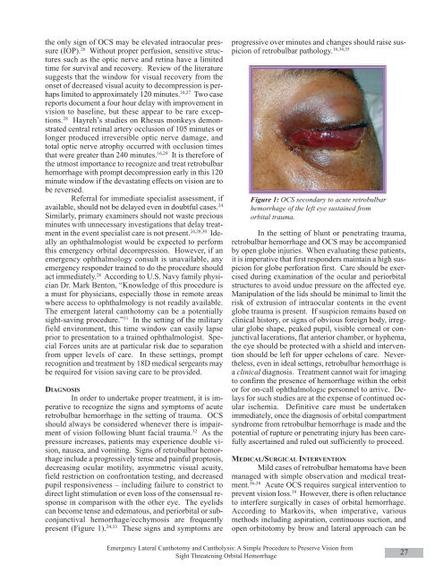
the only sign of OCS may be elevated intraocular pressure(IOP). 26 Without proper perfusion, sensitive structuressuch as the optic nerve and retina have a limitedtime for survival and recovery. Review of the literaturesuggests that the window for visual recovery from theonset of decreased visual acuity to decompression is perhapslimited to approximately 120 minutes. 10,27 Two casereports document a four hour delay with improvement invision to baseline, but these appear to be rare exceptions.28 Hayreh’s studies on Rhesus monkeys demonstratedcentral retinal artery occlusion of 105 minutes orlonger produced irreversible optic nerve damage, andtotal optic nerve atrophy occurred with occlusion timesthat were greater than 240 minutes. 16,29 It is therefore ofthe utmost importance to recognize and treat retrobulbarhemorrhage with prompt decompression early in this 120minute window if the devastating effects on vision are tobe reversed.Referral for immediate specialist assessment, ifavailable, should not be delayed even in doubtful cases. 24Similarly, primary examiners should not waste preciousminutes with unnecessary investigations that delay treatmentin the event specialist care is not present. 24,28,30 Ideallyan ophthalmologist would be expected to performthis emergency orbital decompression. However, if anemergency ophthalmology consult is unavailable, anyemergency responder trained to do the procedure shouldact immediately. 26 According to U.S. Navy family physicianDr. Mark Benton, “Knowledge of this procedure isa must for physicians, especially those in remote areaswhere access to ophthalmology is not readily available.The emergent lateral canthotomy can be a potentiallysight-saving procedure.” 31 In the setting of the militaryfield environment, this time window can easily lapseprior to presentation to a trained ophthalmologist. <strong>Special</strong>Forces units are at particular risk due to separationfrom upper levels of care. In these settings, promptrecognition and treatment by 18D medical sergeants maybe required for vision saving care to be provided.DIAGNOSISIn order to undertake proper treatment, it is imperativeto recognize the signs and symptoms of acuteretrobulbar hemorrhage in the setting of trauma. OCSshould always be considered whenever there is impairmentof vision following blunt facial trauma. 32 As thepressure increases, patients may experience double vision,nausea, and vomiting. Signs of retrobulbar hemorrhageinclude a progressively tense and painful proptosis,decreasing ocular motility, asymmetric visual acuity,field restriction on confrontation testing, and decreasedpupil responsiveness – including failure to constrict todirect light stimulation or even loss of the consensual responsein comparison with the other eye. The eyelidscan become tense and edematous, and periorbital or subconjunctivalhemorrhage/ecchymosis are frequentlypresent (Figure 1). 24,33 These signs and symptoms areprogressive over minutes and changes should raise suspicionof retrobulbar pathology. 16,34,35Figure 1: OCS secondary to acute retrobulbarhemorrhage of the left eye sustained fromorbital trauma.In the setting of blunt or penetrating trauma,retrobulbar hemorrhage and OCS may be accompaniedby open globe injuries. When evaluating these patients,it is imperative that first responders maintain a high suspicionfor globe perforation first. Care should be exercisedduring examination of the ocular and periorbitalstructures to avoid undue pressure on the affected eye.Manipulation of the lids should be minimal to limit therisk of extrusion of intraocular contents in the eventglobe trauma is present. If suspicion remains based onclinical history, or signs of obvious foreign body, irregularglobe shape, peaked pupil, visible corneal or conjunctivallacerations, flat anterior chamber, or hyphema,the eye should be protected with a shield and interventionshould be left for upper echelons of care. Nevertheless,even in ideal settings, retrobulbar hemorrhage isa clinical diagnosis. Treatment cannot wait for imagingto confirm the presence of hemorrhage within the orbitor for on-call ophthalmologic personnel to arrive. Delaysfor such studies are at the expense of continued ocularischemia. Definitive care must be undertakenimmediately, once the diagnosis of orbital compartmentsyndrome from retrobulbar hemorrhage is made and thepotential of rupture or penetrating injury has been carefullyascertained and ruled out sufficiently to proceed.MEDICAL/SURGICAL INTERVENTIONMild cases of retrobulbar hematoma have beenmanaged with simple observation and medical treatment.36-38 Acute OCS requires surgical intervention toprevent vision loss. 39 However, there is often reluctanceto interfere surgically in cases of orbital hemorrhage.According to Markovits, when imperative, variousmethods including aspiration, continuous suction, andopen orbitotomy by brow and lateral approach can beEmergency Lateral Canthotomy and Cantholysis: A Simple Procedure to Preserve Vision fromSight Threatening Orbital Hemorrhage27
- Page 1 and 2: Volume 9, Edition 3 / Summer 09 Jou
- Page 3 and 4: An 18D deworms a camel during a “
- Page 5 and 6: Field Evaluation and Management of
- Page 7 and 8: The circumferential anchoring strip
- Page 9 and 10: In doing so, all the skin is closed
- Page 11 and 12: NATO SOF Transformation and theDeve
- Page 13 and 14: current and future operations, thes
- Page 15 and 16: sion of a physician, and limited pr
- Page 17 and 18: REFERENCES1. James L. Jones, “A b
- Page 19 and 20: This article is the first of two me
- Page 21 and 22: Figure 4 : A Special Forces medic c
- Page 23 and 24: exposure. Conversely, the customary
- Page 25 and 26: 7. Ted Westmoreland. (2006). Attrib
- Page 27 and 28: first three days of injury, althoug
- Page 29: 9. Markgraf CG, Clifton GL, Moody M
- Page 33 and 34: E. The canthotomy allows for additi
- Page 35 and 36: 33. Rosdeutscher, J.D. and Stradelm
- Page 37 and 38: Tinnitus, a Military Epidemic:Is Hy
- Page 39 and 40: The development of chronic NIHL pro
- Page 41 and 42: supplied by diffusion. During expos
- Page 43 and 44: similar to those of other authors,
- Page 45 and 46: promising effect on tinnitus. Howev
- Page 47 and 48: ADDITIONAL REFERENCESHoffmann, G; B
- Page 49 and 50: et al. demonstrated that both right
- Page 51 and 52: TYPICAL CHEST RADIOGRAPH FINDINGS I
- Page 53 and 54: 11. Norsk P, Bonde-Petersen F, Warb
- Page 55 and 56: ABSTRACTS FROM CURRENT LITERATUREMa
- Page 57 and 58: tourniquet times are less than 6 ho
- Page 59 and 60: tal from July 1999 to June 2002. In
- Page 61 and 62: Operation Sadbhavana: Winning Heart
- Page 63 and 64: CENTRAL RETINAL VEIN OCCLUSION IN A
- Page 65 and 66: of the X chromosome. Notable is tha
- Page 67 and 68: AUTHORS*75th Ranger Regiment6420 Da
- Page 69 and 70: Casualties presenting in overt shoc
- Page 71 and 72: PSYCHOLOGICAL RESILIENCE AND POSTDE
- Page 73 and 74: spondents without PTSD (M = 4.6, SD
- Page 75 and 76: patients, whereas the mean score of
- Page 77 and 78: 29. Whealin JM, Ruzek JI, Southwick
- Page 79 and 80: average, time between return from d
- Page 81 and 82:
ing functioning in both PTSD (Zatzi
- Page 83 and 84:
Editorial Comment on “Psychologic
- Page 85 and 86:
Blackburn’s HeadhuntersPhilip Har
- Page 87 and 88:
The Battle of Mogadishu:Firsthand A
- Page 89 and 90:
Task Force Ranger encountered enemy
- Page 91 and 92:
Peter J. Benson, MDCOL, USACommand
- Page 93 and 94:
Numerous military and civilian gove
- Page 95 and 96:
Anthony M. Griffay, MDCAPT, USNComm
- Page 97 and 98:
This is a great read that speaks di
- Page 99 and 100:
and twenty-eight. Rabies immune glo
- Page 101 and 102:
Rhett Wallace MD FAAFPLTC MC SFS DM
- Page 103 and 104:
LTC Craig A. Myatt, Ph.D., HQ USSOC
- Page 105 and 106:
LTC Bill Bosworth, DVM, USSOCOM Vet
- Page 107 and 108:
Europe, Mideast, Africa and SWAU.S.
- Page 109 and 110:
SOF and SOF Medicine Book ListWe ha
- Page 111 and 112:
TITLE AUTHOR ISBNCohesion, the Key
- Page 113 and 114:
TITLE AUTHOR ISBNI Acted from Princ
- Page 115 and 116:
TITLE AUTHOR ISBNRats, Lice, & Hist
- Page 117 and 118:
TITLE AUTHOR ISBNThe Healer’s Roa
- Page 119 and 120:
TITLE AUTHOR ISBNGuerilla warfare N
- Page 121 and 122:
TITLEAUTHORBlack Eagles(Fiction)Bla
- Page 123 and 124:
TITLE(Good section on Merrill’s M
- Page 125 and 126:
GENERAL REFERENCESALERTS & THREATSB
- Page 127 and 128:
Aviation Medicine Resources: http:/
- Page 129 and 130:
LABORATORYClinical Lab Science Reso
- Page 131 and 132:
A 11 year old boy whose tibia conti
- Page 133 and 134:
Meet Your JSOM StaffEXECUTIVE EDITO
- Page 135 and 136:
Special Forces Aidman's PledgeAs a