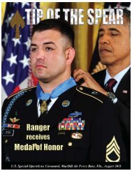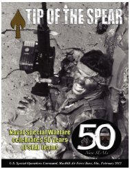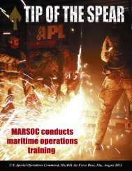
Summer - United States Special Operations Command
Summer - United States Special Operations Command
Summer - United States Special Operations Command

You also want an ePaper? Increase the reach of your titles
YUMPU automatically turns print PDFs into web optimized ePapers that Google loves.
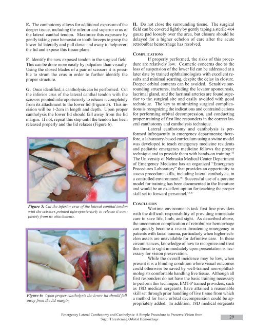
E. The canthotomy allows for additional exposure of thedeeper tissue, including the inferior and superior crus ofthe lateral canthal tendon. Maximize this exposure bygently taking your hemostat or tooth forceps to grasp thelower lid laterally and pull down and away to help evertthe lid and expose this tissue plane.F. Identify the now exposed tendon in the surgical field.This can be done more easily by palpation than visually.Using the closed blades of a pair of scissors it is possibleto strum the crus in order to further identify theproper structure.G. Once identified, a cantholysis can be performed. Cutthe inferior crus of the lateral canthal tendon with thescissors pointed inferoposteriorly to release it completelyfrom its attachment to the lower lid (Figure 5). This incisionwill be 1-2cm in length and depth. Upon propercantholysis the lower lid should fall away from the lidmargin. If not, repeat this step until the tendon has beenreleased properly and the lid relaxes (Figure 6).Figure 5: Cut the inferior crus of the lateral canthal tendonwith the scissors pointed inferoposteriorly to release it completelyfrom its attachments.Figure 6: Upon proper cantholysis the lower lid should fallaway from the lid margin.H. Do not close the surrounding tissue. The surgicalfield can be covered lightly by gently taping a sterile 4x4gauze pad loosely over the area, but closure should bedelayed for a higher echelon of care after the acuteretrobulbar hemorrhage has resolved.COMPLICATIONSIf properly performed, the risks of this procedureare relatively low. Cosmetic concerns due to theloss of suspension of the lower lid can be addressed at alater date by trained ophthalmologists with excellent resultsand minimal scarring, despite the delay in closure.Deeper orbital contents can be avoided. Sensitive surroundingstructures, including the levator aponeurosis,lacrimal gland, and the lacrimal arteries are found superiorto the surgical site and easily avoided with goodtechnique. The key to minimizing surgical complicationsis recognizing the indications and contraindicationsfor performing orbital decompression, and conductingproper training of first line responders in the correct lateralcanthotomy and cantholysis technique.Lateral canthotomy and cantholysis is performedinfrequently in emergency departments; therefore,a laboratory-based curriculum using a swine modelwas developed to teach emergency medicine residentsand pediatric emergency medicine fellows the propertechnique and to provide them with hands-on training. 45The University of Nebraska Medical Center Departmentof Emergency Medicine has an organized “EmergencyProcedures Laboratory” that provides an opportunity toassess procedure skills, including lateral cantholysis, ina controlled environment. 46 Successful use of a porcinemodel for training has been documented in the literatureand would be an excellent option for teaching the properskill set to forward personnel. 45,47CONCLUSIONWartime environments task first line providerswith the difficult responsibility of providing immediatecare to save life, limb, and sight. As described above,the uncommon complication of retrobulbar hemorrhagecan quickly become a vision-threatening emergency inpatients with facial trauma, particularly when higher echelonassets are unavailable for definitive care. In thesecircumstances, knowledge of how to recognize and treatthis threat to sight immediately upon presentation is necessaryfor vision preservation.While the overall incidence may be low, whenpresent it is a blinding condition where visual outcomescould otherwise be saved by well-trained non-ophthalmologistscomfortable handling live tissue. Although allfirst responders do not have the basic training necessaryto perform this technique, EMT-P trained providers, suchas 18D medical sergeants, have attained a reasonableskill set through prior handling of live tissue from whicha method for basic orbital decompression could be appropriatelyadded. In addition, 18D medical sergeantsEmergency Lateral Canthotomy and Cantholysis: A Simple Procedure to Preserve Vision fromSight Threatening Orbital Hemorrhage29