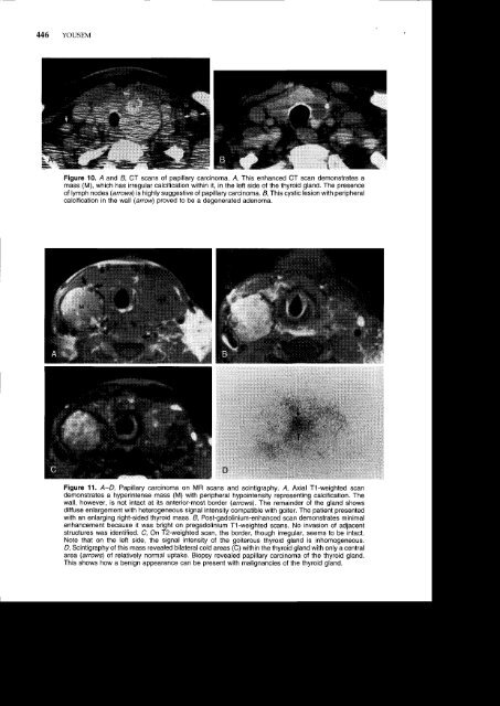
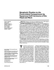
446 YOUSEMFigure 10. A and B, CT scans of papillary carcinoma. A, This enhanced CT scan demonstrates amass (M), which has irregular calcification within it, in the left side of the thyroid gland. The presenceof lymph nodes (arrouvs) is highly suggestive of papillary carcinoma. & This cystic lesion with peripheralcalcification in the wall (arrowy proved to be a degenerated adenoma.Figure 11. A-D, Papillary carcinoma on MR scans and scintigraphy.4, Axial T1-weighted scandemonstrates a hyperintense mass (M) with peripheral hypointensity representing calcification. Thewafl, however, is not intact at its anterior-most border (arrows\. The remainder of the gland showsdiffuse enlargement with heterogeneousignal intensity compatible with goiter. The patient presentedwith an enlarging right-sided thyroid mass. 4 Post-gadolinium-enhanced scan demonstrates minimalenhancement because it was bright on pregadolinium T1-weighted scans. No invasion of adjacentstructures was identified. C On T2-weighted scan, the border, though irregular, seems to be intact.Note that on the left side, the signal intensity of the goiterous thyroid gland is inhomogeneous.D, Scintigraphy of this mass revealed bilateral cold areas (C) within the thyroid gland with only a centralarea (arrows) of relatively normal uptake. Biopsy revealed papillary carcinoma of the thyroid gland.This shows how a benign appearance can be present with malignancies of the thyroid gland.
<strong>PARA<strong>THYROID</strong></strong> <strong>AND</strong> <strong>THYROID</strong> <strong>IMAGING</strong> 447variant" histologic classification, the "mixed cancer"ultimately behaves like a papillary carcinoma(vide infra). Purely follicular carcinoma occursin 5% to 15%. Anaplastic carcinoma represents 3%lo 10% of all malignancies, with m-edullary orHurthle's cell carcinoma accounting for 4Vo lo5Vo.1e'3e Medullary carcinoma may present in associationwith the multiple endocrine neoplasia syndromes(see Table 2) and serologically may expresscalcitonin. The other histologic diagnoses to considerin thyroid malignancies are non-Hodgkin'slymphoma and metastases.Imaging has a well-defined role in the workupof the patient with a solitary nodule in the thyroidgland. Unless a classic appearance of a benign conditionis present, however, pathologic sampling isrequired. This is because the appearance of thyroidmalignancies may simulate many benign processes,and vice versa. The specificity of imaging findingsis low.The detection and characterization of a thyroidneoplasm is not the only role of imaging. Imagingshould also evaluate for infiltration of the adiacentsoft tissue, aerodigestive tract, paraspinal musculature,and carotid arteries. The presence or absenceof adenopathy is also important for prognostic implicationswith thyroid cancer.Papillary CarcinomaThe presence of psammoma bodies (laminatedcalcific spherules in25-40Vo of cases), ground glassnuclei, and a branching pattern with a fibrovascularpapillary stroma are the histologic signatures ofpapillary carcinoma of the thyroid gland.3e As notedearlier, follicular growth patterns may coexist. Cystformation (cystadenocarcinoma), encapsulation,multifocality, and anaplasia may be present withina thyroid gland with papillary carcinoma.Papillary carcinoma is the thyroid malignancythat has the greatest likelihood of spread to lymphnodes; the nodes may be tiny, cystic, hemorrhagic,or calcified (Fig. 12). The incidence of nodal metastasesduring diagnosis is50%, whereas distant metastasesare reported to occur in 4Va to 7Vo, usu.ally tothe lungs, bone, or central nervous system.3e Despitethese features, the 2O-year survival rate is reportedto be as high as 90%. Approximately 10% of papillarycarcinomas are bilateral.eaFollicular CarcinomaPure follicular carcinoma is relatively uncommonwhen the follicular variant of papillary carcinomais excluded. The tumor mav be diffuselv invasiveor well encapsulated. Follicular carcinorna less freqentlyspreads to lymph nodes (2-10%) than doespapillary carcinoma, but disseminates hematogenouslymore readily.3e The prognosis depends onthe presence of hematogenous metastases or localinvasiveness but is not as optimistic as that for papil-lary carcinoma. No distinguishing features on imagingstudies suggest this diagnosis as opposed toother cancers, although, on ultrasonography, follicularcarcinoma is isoechoic in 52% and hypoechoicin44Vo.20'72 Compared with papillary carcinoma, follicularcarcinoma rarely becomes cystic and morefrequently invades vessels.3eAnaplastic CarcinomaThis type of cancer is one of the most aggressivemalignancies of the head and neck with prognosesmarked in months rather than years. Older patientsare usually affected. Anaplastic thyroid carcinomasoccur within a substrate of goiters in 47Vo of cases3eand often coexist with other forms of better differentiatedthyroid cancer. The outcome in mixed tumorcases is dominated by the poor prognosis of anar:lastic^ carcinoma.On ultrasonography, these carcinomas are mostcommonly hypoechoiq20,D,72 whereas on CT, anaplasticcarcinomas show evidence of dense amorphouscalcification in SBVo of cases and necrosis in74Vo.u Metastatic lymph nodes are present in 74Voto 80% of cases and show necrotic areas 50% of thetime.e'82 Invasion into carotid arteries or adjacentaerodigestive structures occurs in 34% to 55% ofpatients, and in 25Vo the primary tumor extendsinto the mediastinum (Fig. 13).q8'?Rapid growth andobliteration of adjacent tissue planes are hallmarksof this deadly tumor with a median survival ofapproximately 7 months.Medullary CarcinomaMedullary carcinoma originates in the parafollicularor "C" cells of the thyroid gland, cells derivedfrom neural crest tissue in the ultimobranchial bodiesof the branchial pouch system. These cells normallvsecrete thvrocalcitonin, which decreases serum'calcium.Eiahty percent to 90% of medullarycarcinomas express calcitonin. Functioning tumorshave a better prognosis.qMedullary carcinomas are usually hypoechoic onultrasonographyzo' zz' 72; however, echogenic focicaused by deposits of calcium may be seen withinthese tumors and metastatic lymph nodes whenpresent.22 These tumors usually do not take up iodinebut may be thallium or gallium avid. Somatostatinreceptor scintigraphy also may detect medullarycarcinoma.ll The tumor is solid on CT and MRimaging and spreads to lymph nodes in more than50Va of cases (Fig. 14).Medullary carcinoma has a familial incidence of"lOVo to 20Vo. Sipple syndrome is the association ofmedullary carcinoma with pheochromocytoma andparathyroid adenoma or hyperplasia. This is alsoknown as the MEN 24. When mucosal neuromasand marfanoid facies coexist, MEN 28 is said to bepresent (see Table 2). Both syndromes have beenlocalized to an abnormal gene on the tenth chromosome.