PARATHYROID AND THYROID IMAGING - Neuroradiology
PARATHYROID AND THYROID IMAGING - Neuroradiology
PARATHYROID AND THYROID IMAGING - Neuroradiology
- No tags were found...

Create successful ePaper yourself
Turn your PDF publications into a flip-book with our unique Google optimized e-Paper software.
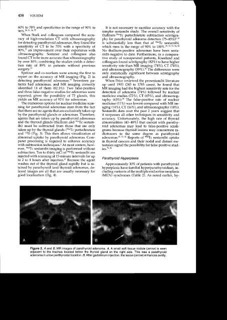
438 YOUSEM60Va to 707o and specificities in the range of 907a to967a.20,21,36, nWhen Stark and colleagues compared the accuracyof high-resolution CT with ultrasonographyfor detecting parathyroid adenomas, they found thesensitivity of CT to be 70% with a specificity o{90Vo,77 an improvernent over their experience withultrasonography. Sommer and colleagues alsofound CT to be more accurate than ultrasonographyby over 10%; combining the studies yields a detectionrate of 89% in patients without previoussurgery.'"Spritzer and co-workers were among the first toreport on the accuracy of MR imaging (Fig. 2) indetecting parathyroid adenomas.T6 Seventeen patientshad adenomas, and MR imaging correctlyidentified 14 of them (82.3Va). Two false-positiveand three false-negative studies for adenomis werereported; given the possibility of 72 glands, thisyields an MR accuracy of 92% for adenomas.The numerous options for nuclear medicine scanningfor parathyroid adenomas stem from the factthat there are no agents that are exclusively taken upby the parathyroid glands or adenomas. Therefore,agents that are taken up by parathyroid adenomasand the thyroid glands (thallium and ee^Tc sestamibi)must be subtracted from those that are onlytaken up by the thyroid glands (ry*Tc pertechnetateand 123I) (Fig. 3). This then allows visualization ofabnormal uptake by parathyroid adenomas. Computerprocessing is required to enhance accuracywith subtraction techniques.2 At most centers, however,ee^Tc sestamibi imiging is performed withoutsubtraction. Ten to thirtv mCi of ee-Tc sestamibi areinjected with scanning a1 1S-minute intervals for uplo 2 to 4 hours after iniection.aT Because the agentwashes out of the thyroid gland rapidly but iJ retainedby parathyroid (and thyroid) adenomas, de-Iayed images are all that are usually necessary forgood localization (Fig. 4).It is not necessary to sacrifice accuracy with thesimpler sestamibi study. The overall sensitivity ofthallium-ee'Tc pertechnetate subtraction scintigraphyfor parathyroid adenoma detection (75-85Vo1z'szis substantially less than that of ee'Tc sestamibi,which runs in the range of 90% to 100%.2e,33,53.54.80No thallium-positive adenomas have been sestamibi-negativeto date. Furthermore, in a comparativestudy of nonoperated patients, Kneeland andcolleagues found scintigraphy (82Vo) to have highersensitivity rate than MR imaging (74%), CT (74%),and ultrasonography (59%).36 The differences wereonly statistically significant between scintigraphyand ultrasonography.When Price reviewed the presestamibi literatureup until 1993 Q43 to 1785-cases). he found thatMR imaging had the highest sensitivity rate for thedetection of adenoma (74%) followed by nuclearmedicine studies (72%), CT (65%), and ultrasonography(63%).58 The false-positive rate of nuclearmedicine (.1.Vo) was lowest compared with MR imaging(14%), CT (16Vo), and ultrasono graphy (187").Sestamibi data over the past 2 years suggest thatit surpasses all other techniques in sensitivity andaccuracy. Unfortunately, the high rate of thyroidabnormalities (40-487") that coexist with parathyroidadenomas may lead to false-positive scintigrams because thyroid lesions may concentrate radiotracersto the same degree as parathyroidadenomas.as,53,sa Reports of ee*Tc sestamibi uptakein thyroid cancers and their nodal and distant metastasesignal the possibility for false-positive studles.'' '"Parathyroid Hype rplasiaApproximately 30% of patients with parathyroidhyperplasia have familial hyperparathyroidism, includingvariants of the multiple endocrine neoplasia(MEN) syndromes (Table 2). As noted earlier, hy-Figure 2. A and B, MR images of parathyroid adenoma. A, A small soft tissue nodule (arror,rr) is seenadjacent to the trachea located below the thyroid gland on the right side. This was a parathyroidadenoma in a low perithyroidal location. A After gadolinium injection, the lesion (arrow'1 enhances avidly.