Exploring the amyloid state of proteins by micro
Exploring the amyloid state of proteins by micro
Exploring the amyloid state of proteins by micro
- No tags were found...

Create successful ePaper yourself
Turn your PDF publications into a flip-book with our unique Google optimized e-Paper software.
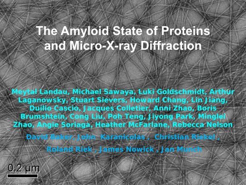
The Amyloid State <strong>of</strong> Proteinsand Micro-X-ray DiffractionQuickTime and adecompressorare needed to see this picture.Meytal Landau, Michael Sawaya, Luki Goldschmidt, ArthurLaganowsky, Stuart Sievers, Howard Chang, Lin Jiang,Duilio Cascio, Jacques Colletier, Anni Zhao, BorisBrumshtein, Cong Liu, Poh Teng, Jiyong Park, MingleiZhao, Angie Soriaga, Hea<strong>the</strong>r McFarlane, Rebecca NelsonDavid Baker, John Karanicolas , Christian Riekel ,Roland Riek , James Nowick , Jan Munch
Micro X-ray Diffractionfor Structural BiologyThe biological problem and ourneed for <strong>micro</strong>-X-ray diffractionWhat <strong>micro</strong>-X-ray diffractionhas allowed us to findFuture opportunities for <strong>micro</strong>-and nano-X-ray diffraction in biology
Amyloid• Unbranched, elongated protein fibrils• Associated with varied diseases (e.g. CJDAlzheimer’s, Dialysis-related <strong>amyloid</strong>osis)• Cross-β diffraction pattern shows β strandsperpendicular to fiber axis: common spineKishimoto, Namba et al (2004)
Amyloid Fibril-Related ConditionsAmyloid (24) Prion (transmissible) Amyloid-likeAlzheimer’s Aβ [Psi+] Sup35Alzheimer’s Tau [Ure2] Ure3 Parkinson’s α-synucleinDiabetes IIAmylinaka IAPPLouGehrig’s(ALS)SuperoxideDismutaseTDP-43InjectionInsulinCJD, GSSPrPHIV SexualSEVI<strong>amyloid</strong>osisKurutransmissionDialysis<strong>amyloid</strong>osisβ2-<strong>micro</strong>globulinBSE, vCJD(mad cow)PrP Cancer p53SenileTrans-<strong>amyloid</strong>osisthyritin
My Scientific Dilemma in 2001Important biological problem—structure <strong>of</strong> <strong>amyloid</strong>5.4 M Alzheimer’s patients in US in 2010~19 M patients expected <strong>by</strong> 2050Economic burden:In 2010 ~$ 183B in US heath care costs~11 M people provide unpaid care <strong>of</strong> AD patientsEssentially no structural informationStructure-based design impossibleAmyloid crystals discovered but 30,000 times smallerthan crystals we had previously worked with
Short segments <strong>of</strong> fiber-forming <strong>proteins</strong>form both <strong>amyloid</strong> fibers and <strong>micro</strong>crystalsGNNQQNYsinglefibril~80ÅBalbirnie et al. PNAS 2001singlefibril~80ÅNNQQNNQQIn both <strong>micro</strong>crystalsand fibrils,β-strands are normalto <strong>the</strong> long axis.NNQQNYGNNQQNY~280Å0.5mmGNNQQNY fibrils exhibit allproperties <strong>of</strong> <strong>amyloid</strong> fibrils:dye binding, cooperativeaggregation kinetics, stability,cross-β diffraction1μm1μmFibers seem to growfrom tips <strong>of</strong> crystals
10-100 mM < 24 hrGNNQQNY <strong>micro</strong>crystalsX-raysCrystal width = ~280 ÅBalbirnie et al. PNAS 2001Research <strong>of</strong> Ruben Diaz & Donald Caspar
Crystal unit cell dimensions and space groupdetermined from X-ray powder diffractionBalbirnie et al. PNAS 2001
Packing <strong>of</strong> GNNQQNY peptides in <strong>micro</strong>crystalsBalbirnie, Gro<strong>the</strong>, & Eisenberg PNAS 2001
Failed Approachesto <strong>the</strong> Structure2001-2005X-ray powder diffractionTextured X-ray powder diffractionElectron diffractionSolid-<strong>state</strong> NMR
Micr<strong>of</strong>ocus is required to reducebackground noiseChristian Riekel100 µm beam diameterStandard at home or synchrotronnly a fraction <strong>of</strong> incoming X-rays impinge crystal.High background obscures reflections1 µm beam diameterESRF ID13All X-rays impinge crystalLow background, good I/σ.
Microcrystals <strong>of</strong>segments <strong>of</strong> 15 <strong>amyloid</strong>forming<strong>proteins</strong> haveyielded >90 steric zippersX-ray beamCrystal50μGlass pin
GNNQQNY, a dry steric zipperFibers and <strong>micro</strong>crystal have 50,000 layersExtended strands, H-bonded 4.8Åapart into in register β-sheetsGln, Asn, Tyr sidechains also H-bondedTwo sheets, interdigitated, with tightlycomplementary sidechains, bonded<strong>by</strong> van der Waals forcesMore tightly complementary thanany previous structure in PDBDry between <strong>the</strong> β-sheetsNelson et al. Nature 2005
caView down <strong>the</strong> fibril axis shows selfcomplementaryinteractions betweenpaired beta sheets <strong>of</strong> <strong>the</strong> steric zipperand weak interactions between pairsOne unit cellNelson et al. Nature, 2005Nine unit cells
As <strong>of</strong> April 2011, ~90 X-raycrystal structures <strong>of</strong> fibrillikedry steric zippers from12 disease-related<strong>proteins</strong>:Sup35 and PrP prionsAβ and Tau - Alzheimer’sα-synuclein - Parkinson’sIAPP - Diabetes type 2SOD - ALSTransthyritin-familial<strong>amyloid</strong>osisLysozyme-lysozyme<strong>amyloid</strong>osisInsulin-injection<strong>amyloid</strong>osisSEVI-HIV transmission
Structure-baseddesign <strong>of</strong> ablocker <strong>of</strong>fibril formationfor TauSievers et al.Nature 2011
D-TLKIVW prevents fibril formation <strong>by</strong> Tau K12K12Fluorescent Units – 510 nmK12 + D-TLKIVWHoursResearch <strong>of</strong> Stuart Sievers & Howard Chang
Defining <strong>the</strong>Amyloid Pharmacophore
GGVVIA +JugloneMeytal LandauGGVVIA + R-(−)-ApomorphineGGVVIA + 1.2,2′-DihydroxybenzophenoneGGVVIA +CurcuminGGVVIA +Chicago SkyBlueGGVVIA +Phenol RedGGVVIA +BenserazideVQIVYK +RhodamineVQIVYK+PhenolRedVQIVYK +NeocuproineVQIVYK +JugloneVQIVYK +PerphenazineVQIVYK +AZETGGVVIA +EGCGKLVFFA +ThTVQIVYK +Phenol RedAbeta17-40+ FDDNPVQIVYK +ThTGVVEVD +Orange GVQIVYK +MethyleneBlueSSTNVG +Phenol redVQIVYK +Azure CVQIVYK +MeclocyclinesulfosalicylateVQIVYK + 1,2-NaphthoquinoneVQIVYK+RhodamineVQIVYK+Hexadecyltrimethylammonium bromideVQIVYK+ApomorphineVQIVYK +RolitetracyclineGGVVIA+ThTGGVVIA +RhodamineGGVVIA +Rifamycin SVGGVVIA + 1,2-NaphthoquinoneSSTNVG+Phenol RedVQIVYK +Chicago skyblue 6BVQIVYK+DobutamineGGVVIA+Chicago SkyBlueGGVVIA +Orange GGGVVIA+Azure CGGVVIA +Trimethyl(tetradecyl)ammoniumbromideGGVVIA + CreosolGGVVIA +Eosin YVQIVYK+FDDNP
KLVFFA (from Amyloid Beta) + Orange G
Structure <strong>of</strong> KLVFFA with Orange GKLVFFA + Orange G 5-10:1mM10-30% w/v Polyethylene glycol 1,500,20-30% v/v GlycerolRmerge = 18.4%; Resolution=1.8A;Completeness=96.4%C2;a, b, c 43.64 26.85 9.55Å;β 91.55 °Rwork/Rfree(%)= 22.6/27.6The asymmetric unit contains:Two peptide segmentsOne Orange GSix water moleculesLandau et al. PLoS BiologyIn press
Towards atomicprotein structures fromnano-crystals
Amyloid-like crystals<strong>of</strong> KLIMY from PAPgrown <strong>by</strong> Anni ZhaoX-ray beam1 µm diameter4
A 3-sheet prionstructure?Only nanocrystalsavailable
Nano-crystals <strong>of</strong>longer (11-residue) segmentPerhaps from <strong>the</strong> toxic segment<strong>of</strong> alpha-synuclein(Parkinson’s disease))
Towards determiningstructures <strong>of</strong> crystals ando<strong>the</strong>r ordered proteinaggregatesfound in human and o<strong>the</strong>ranimal cells
10 <strong>micro</strong>n Mictigen <strong>micro</strong>mesh in cryostream
2Drosophilablood cells1
Drosophila crystal cellon 10 µm mesh10umThe diffraction pattern revealspowder rings at 50, 38, 26, 13,4.2, and 3.7 Å spacingsMichael Sawaya, Mari Gingery, Duilio CascioGroup <strong>of</strong> Utpal Banerjee, UCLAJacques Colletier, Christian Riekel, ESRF-IBS
Eosinophile (type <strong>of</strong> white blood cell) granulesCharacterized <strong>by</strong> George Palade et al. (1965) <strong>by</strong> EMUnit cell dimensions tentatively determined <strong>by</strong>Alice Soragni, Jacques Colletier,Manfred Brunner, Christian Riekel
Cell Types Containing Intracellular Crystalline InclusionsSpecies Cell type Description <strong>of</strong> crystals Protein ReferenceDrosophila Crystal cells Intracellular inclusions [lamellar] Prophenol oxidase (Shrestha & Gateff 1982);T. M. Rizki & R. M. Rizki 1980)HumanRatGuinea pigMouseEosinophil leukocytesMembrane-bound granules [0.3-1.2 um]Granule cores [lamellar]? (Miller et al. 1966)Human B cell lymphomas ER-bound crystal rods [lamellar] Ig (Peters et al. 1984)Human Abnormal mitochondria inmuscle myopathiesCrystal rods in outer mitochondrialmembrane compartmentMitochondrial creatine kinase (Stadhouders et al. 1994)Human Kidney mitochondria Helical crystals in outer mitochondrial ? Jasmin 1978compartment; linear and flexuous crystalsin matrixHumanDogMonkeyLiver mitochondria Intramitochondrial [lamellar] ? Wills 1965Human Ad5-infected KB cells Intranuclear adenovirus-induced inclusions heteromeric capsid protein (Franqueville et al. 2008);formed <strong>of</strong> penton base and (Carstens et al. 1975)fiber subunitsFrog Oocyte mitochondria Intramatrix & intracristae inclusions[lamellar]? Spornitz 1972Armadillo Epididymus Single membrane-bound cytoplasmiccrystalline rods [lamellar]? (Edmonds et al. 1973)Earthworm Spermatazoa Intranuclear inclusions [lamellar] ? (Anderson et al. 1968)Tomato Young leaf mesophyll Intracellular inclusions [Cubic] ? Singh 1976Helicobacter pyloriCausative agent <strong>of</strong> gastricdiseasesCytoplasmic paracrystalline inclusions Pfr, bacterial ferritin (Frazier et al. 1993)Photorhabdus luminescens Entomopathogenic bacteria Intracellular inclusions cipAcipB(Bintrim et al. 1998)Bacillus thuringiensis Insecticidal bacteria parasporal crystals Cry (H<strong>of</strong>te et al. 1989)Paenibacillus popilliae Insecticidal bacteria parasporal crystals ? Weiner 1978Brevibacillus laterosporus Mosquitocidal bacteria parasporal crystals ? (Smirnova et al. 1996); (Orlovaet al. 1998)
Summary•Micro-X-ray diffraction has enabled <strong>the</strong> determination <strong>of</strong><strong>the</strong> atomic structures <strong>of</strong> <strong>the</strong> <strong>amyloid</strong> <strong>state</strong>, includingdesign <strong>of</strong> inhibitors and partial definition <strong>of</strong> <strong>the</strong> <strong>amyloid</strong>pharmacophor•The structures <strong>of</strong> still smaller crystals are needed tounderstand <strong>the</strong> toxic mechanism <strong>of</strong> <strong>amyloid</strong> perhapswith LCLS• LCLS <strong>of</strong>fers <strong>the</strong> possibility <strong>of</strong> learning <strong>the</strong> atomicstructure <strong>of</strong> ordered aggregates within biological cells,including <strong>amyloid</strong> aggregates
The Amyloid State <strong>of</strong> ProteinsUCLA: Rebecca Nelson, Michael Sawaya, Marcin Apostol, Melinda BalbirnieMagdalena Ivanova, Stuart Sievers, Jed Wiltzius, Minglei Zhao, Cong LiuLuki Goldschmidt, Hea<strong>the</strong>r Mcfarlane, Howard Chang, Anni ZhaoLin Jiang, Jiyong Park, Jacques Colletier, Poh Teng, Boris BhrumsteinUniv. <strong>of</strong> Washington: John Karanicolis, David BakerESRF: Christain Riekel ETH: Roland Riek, Alice SoragniStuartSieversMike SawayaMelindaJedBalbirnieWiltziusRebecca NelsonMarcinApostolMarcinApostolJacques ColletierArtLaganowskyDuilio Meytal LandauCascioLukiGoldschmidtPoh Teng
CollaboratorsChristian RiekelESRFDavid BakerUniversity <strong>of</strong>WashingtonJamesNowickUCIRoland Riek (ETH Zurich), John Karanicolas (U. Kansas),Jan Münch (Ulm)