Optical Fiber Sensors for Biomedical Applications
Optical Fiber Sensors for Biomedical Applications
Optical Fiber Sensors for Biomedical Applications

Create successful ePaper yourself
Turn your PDF publications into a flip-book with our unique Google optimized e-Paper software.
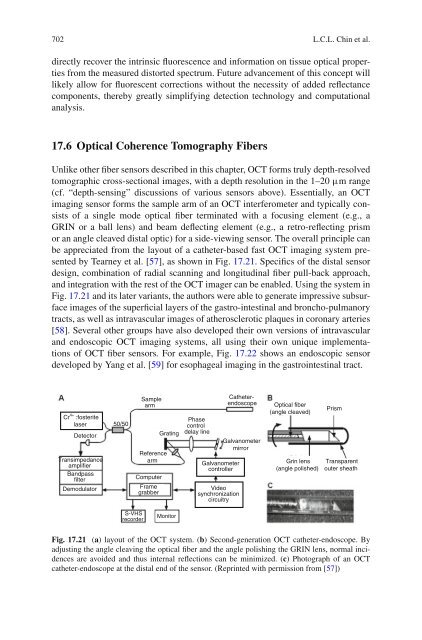
702 L.C.L. Chin et al.directly recover the intrinsic fluorescence and in<strong>for</strong>mation on tissue optical propertiesfrom the measured distorted spectrum. Future advancement of this concept willlikely allow <strong>for</strong> fluorescent corrections without the necessity of added reflectancecomponents, thereby greatly simplifying detection technology and computationalanalysis.17.6 <strong>Optical</strong> Coherence Tomography <strong>Fiber</strong>sUnlike other fiber sensors described in this chapter, OCT <strong>for</strong>ms truly depth-resolvedtomographic cross-sectional images, with a depth resolution in the 1–20 μm range(cf. “depth-sensing” discussions of various sensors above). Essentially, an OCTimaging sensor <strong>for</strong>ms the sample arm of an OCT interferometer and typically consistsof a single mode optical fiber terminated with a focusing element (e.g., aGRIN or a ball lens) and beam deflecting element (e.g., a retro-reflecting prismor an angle cleaved distal optic) <strong>for</strong> a side-viewing sensor. The overall principle canbe appreciated from the layout of a catheter-based fast OCT imaging system presentedby Tearney et al. [57], as shown in Fig. 17.21. Specifics of the distal sensordesign, combination of radial scanning and longitudinal fiber pull-back approach,and integration with the rest of the OCT imager can be enabled. Using the system inFig. 17.21 and its later variants, the authors were able to generate impressive subsurfaceimages of the superficial layers of the gastro-intestinal and broncho-pulmanorytracts, as well as intravascular images of atherosclerotic plaques in coronary arteries[58]. Several other groups have also developed their own versions of intravascularand endoscopic OCT imaging systems, all using their own unique implementationsof OCT fiber sensors. For example, Fig. 17.22 shows an endoscopic sensordeveloped by Yang et al. [59] <strong>for</strong> esophageal imaging in the gastrointestinal tract.Cr 4+ :fosteritelaserDetectorTransimpedanceamplifierBandpassfilterDemodulator50/50SamplearmReferencearmComputerFramegrabberGratingPhasecontroldelay lineGalvanometercontrollerVideosynchronizationcircuitryCatheterendoscopeGalvanometermirror<strong>Optical</strong> fiber(angle cleaved)Grin lens(angle polished)PrismTransparentouter sheathS-VHSrecorderMonitorFig. 17.21 (a) layout of the OCT system. (b) Second-generation OCT catheter-endoscope. Byadjusting the angle cleaving the optical fiber and the angle polishing the GRIN lens, normal incidencesare avoided and thus internal reflections can be minimized. (c) Photograph of an OCTcatheter-endoscope at the distal end of the sensor. (Reprinted with permission from [57])