Optical Fiber Sensors for Biomedical Applications
Optical Fiber Sensors for Biomedical Applications
Optical Fiber Sensors for Biomedical Applications

Create successful ePaper yourself
Turn your PDF publications into a flip-book with our unique Google optimized e-Paper software.
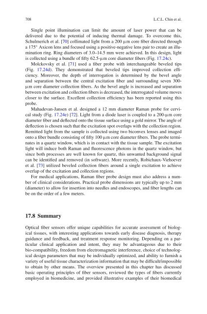
708 L.C.L. Chin et al.Single point illumination can limit the amount of laser power that can bedelivered due to the potential of inducing thermal damage. To overcome this,Schulmerich et al. [70] collimated light from a 200 μm core fiber directed througha 175 ◦ Axicon lens and focused using a positive-negative lens pair to create an illuminationring. Ring diameters of 3.0–14.5 mm were achieved. In this design, lightis collected using a bundle of fifty 62.5-μm core diameter fibers (Fig. 17.24c).Molckovsky et al. [71] used a fiber probe with interchangeable beveled tips(Fig. 17.24d). They demonstrated that beveled tips improved collection efficiency.Moreover, the depth of interrogation is determined by the bevel angleand separation between the central excitation fiber and surrounding seven 300-μm core diameter collection fibers. As the bevel angle is increased and separationbetween excitation and collection fibers is decreased, the interrogated volume movescloser to the surface. Excellent collection efficiency has been reported using thisprobe.Mahadevan-Jansen et al. designed a 12 mm diameter Raman probe <strong>for</strong> cervicalstudy (Fig. 17.24e) [72]. Light from a diode laser is coupled to a 200-μm corediameter fiber and deflected onto the tissue surface using a gold mirror. The angle ofdeflection is chosen such that the excitation spot overlaps with the collection region.Remitted light from the sample is collected using two biconvex lenses and imagedonto a fiber bundle consisting of fifty 100 μm core diameter fibers. The probe terminatesin a quartz window, which is in contact with the tissue sample. The excitationlight will induce both Raman and fluorescence photons in the quartz window, butsince both processes are well known <strong>for</strong> quartz, this unwanted background signalcan be identified and removed (in software). More recently, Robichaux-Viehoeveret al. [73] utilized beveled collection fibers around a single excitation to achieveoverlap of the excitation and collection regions.For medical applications, Raman fiber probe design must also address a numberof clinical considerations. Practical probe dimensions are typically up to 2 mm(diameter) to allow <strong>for</strong> insertion into needles and endoscopes, and fiber lengths canbe on the order of a few meters.17.8 Summary<strong>Optical</strong> fiber sensors offer unique capabilities <strong>for</strong> accurate assessment of biologicaltissues, with interesting applications towards early disease diagnosis, therapyguidance and feedback, and treatment response monitoring. Depending on a particularclinical application and intent, they may be advantageous due to theirbio-compatibility, freedom from electromagnetic interference, choice of technologicaldesign parameters that may be individually optimized, and ability to furnish avariety of useful tissue characterization in<strong>for</strong>mation that may be difficult/impossibleto obtain by other means. The overview presented in this chapter has discussedbasic operating principles of fiber sensors, reviewed the types of fibers currentlyemployed in biomedicine, and provided illustrative examples of their biomedical