
You also want an ePaper? Increase the reach of your titles
YUMPU automatically turns print PDFs into web optimized ePapers that Google loves.
INTRODUCTION<br />
J Int Soc Plastination, Vol 6:38-40, 1992 38<br />
SILICONE TRACHEOBRONCHIAL CASTS<br />
Robert W. Henry<br />
Department of Animal Science, The University of Tennessee,<br />
College of Veterinary Medicine, Knoxville, TN, 37901-1071, USA<br />
Preparation of beautiful and anatomically precise<br />
tracheobronchial casts have an inherent teaching<br />
value. To actually see <strong>the</strong> replica of <strong>the</strong> airway<br />
instead of merely hearing a description of it or only<br />
seeing a figure rendered by an artist is an invaluable<br />
teaching aid. In addition to education, casts have<br />
been used for morphometric studies (Weibel and<br />
Gomez, 1962; Horsefield and Gumming, 1968;<br />
Pump, 1969). Some previously used resins produce<br />
brittle specimens which often shatter and may be<br />
totally destroyed if dropped (Tucker and Krementz,<br />
1957). Frank and Yoder (1966) introduced <strong>the</strong> use<br />
of silicone for forming casts. Kilpper and Stidd<br />
(1973) devised a complicated method for filling <strong>the</strong><br />
airways in a wet state to prevent shrinkage artifact,<br />
while Phalen and coworkers (1973) introduced<br />
making silicone casts of <strong>the</strong> airways in situ. We<br />
have chosen to use air-dried lungs, similar to Wang<br />
and Kraman (1988), to produce <strong>the</strong> casts. There are<br />
several manufacturers of polymer and numerous<br />
polymers to chose from. The properties of <strong>the</strong><br />
polymers are just as diverse. Comparisons of <strong>the</strong><br />
properties of polymers from <strong>the</strong> various<br />
manufacturers are available but not totally accurate.<br />
Five polymers of varying properties were used to<br />
make tracheobronchial casts.<br />
METHODS<br />
Fresh cat, dog, horse, pig and ox lungs were<br />
procured from local abattoirs and from <strong>the</strong><br />
necropsy laboratory. The heart, esophagus, any<br />
o<strong>the</strong>r mediastinal tissue and fat were separated from<br />
<strong>the</strong> lungs and tracheobronchial tree similar to<br />
McKiernan and Kneller (1983). A cannula of <strong>the</strong><br />
appropriate diameter was ligated in <strong>the</strong> trachea. A<br />
water source was attached to <strong>the</strong> cannula and <strong>the</strong><br />
lungs gently inflated to near capacity. After filling,<br />
<strong>the</strong> water source was removed and <strong>the</strong> water,<br />
mucous and blood allowed to flow out of <strong>the</strong><br />
trachea. This flushing procedure was repeated 6 to<br />
10 times, until most of <strong>the</strong> blood and secretions<br />
were cleared from <strong>the</strong> lungs and airways. The<br />
surface of <strong>the</strong> lungs was kept moist during <strong>the</strong><br />
flushing period. After <strong>the</strong> majority of <strong>the</strong> water had<br />
drained from <strong>the</strong> organ, <strong>the</strong> lungs were inflated and<br />
dried using compressed laboratory air directed into<br />
<strong>the</strong> trachea. Smaller lungs were suspended from <strong>the</strong><br />
trachea and larger lungs were placed on <strong>the</strong>ir dorsal<br />
(posterior) surface on a tray to contain <strong>the</strong><br />
extravasated fluid. The air flow was gradually<br />
increased until <strong>the</strong> lungs were inflated and remained<br />
inflated to near capacity. Flow and inflation were<br />
maintained until <strong>the</strong> lungs were thoroughly dried<br />
(Henry and Butler, 1990).<br />
After drying was complete, <strong>the</strong> lung was<br />
suspended in a vertical position. Aided by an<br />
appropriate size funnel, a selected polymer was<br />
poured into <strong>the</strong> trachea and allowed to flow into <strong>the</strong><br />
lung via gravitational forces. Five polymers were<br />
used and two or three casts were made of each<br />
polymer. 1. Biodur - S/10/S3/S2 mixture [Biodur<br />
Products, Dr. Gun<strong>the</strong>r von Hagens, Rathausstrasse<br />
18, Heidelberg, D-6900 GERMANY]. 2. Dow<br />
Corning - Silastic E RTV [Dow Corning Corp.,<br />
Midland, Ml 48640-0994, USA]. 3. General Electric<br />
- RTV 11 [General Electric Co., Silicone Products<br />
Div., Waterford, NY 12188, USA]. 4. & 5. Rhone-<br />
Poulenc - 2 different Rhodorsil Silicones: 4. Rhodorsil<br />
RTV II - # 1556 and 5. Rhodorsil RTV II - # 1547<br />
[Rhone-Poulenc, Inc., Specialty Plastics Division, CN<br />
5266, Princeton, NJ 08543-5266, USA]. All<br />
product components were combined and mixed<br />
using <strong>the</strong> manufacturer's recommendations.<br />
Hardener/polymer ratios were 1:10, except Biodur<br />
which was 1:100 (S3:S10) but <strong>the</strong> S2 was varied<br />
from 0.5:100 to 2:100 in 0.5 increments.<br />
Approximate quantity of polymer used was .2 - .5<br />
kg/ 20 kg dog, 1-2 kg for adult pig, horse or ox. In<br />
larger specimens before <strong>the</strong> silicone hardened, a<br />
heavy wire was placed into <strong>the</strong> lumen of <strong>the</strong> trachea<br />
to give support to <strong>the</strong> silicone cast. The polymer<br />
mixes were allowed to harden over night. The next<br />
morning, <strong>the</strong> lung unit was placed in a vat of<br />
simmering to slowly boiling water for several hours.
39 R. Henry<br />
The specimens were checked in four to five hours to<br />
determine if most of <strong>the</strong> lung and airway tissue had<br />
been macerated away from <strong>the</strong> cast material. Once<br />
most of <strong>the</strong> tissue had been loosened or removed,<br />
<strong>the</strong> cast was removed from <strong>the</strong> hot water and<br />
sprayed with a high pressure hose to remove much<br />
of <strong>the</strong> remaining tissue. If tissue was not removed<br />
with <strong>the</strong> high pressure water, <strong>the</strong> cast was<br />
submerged ei<strong>the</strong>r in hot water again or in a 10%<br />
sodium hydroxide solution until <strong>the</strong> remaining tissue<br />
was released. The specimens were rinsed with<br />
water and allowed to dry before use.<br />
RESULTS<br />
All five polymers produced representative casts of<br />
<strong>the</strong> airways or portions of <strong>the</strong> airways. The more<br />
viscous polymers flowed into <strong>the</strong> terminal bronchi<br />
(Fig. 1) and occasionally into <strong>the</strong> alveoli. The less<br />
viscous polymers flowed freely into <strong>the</strong> alveoli, thus<br />
filling <strong>the</strong> entire airway (Fig. 2). S2 ratios of 1.5 -<br />
2:100 (S10/S3 mix) caused <strong>the</strong> polymer to harden<br />
too quickly, only allowing filling of <strong>the</strong> larger<br />
bronchioles, while casts of S2 0.5:100 ratios<br />
remained tacky. The Biodur mix (0.5-1.0:100)<br />
consistently was <strong>the</strong> least viscous and entered <strong>the</strong><br />
distal airways. The Rhodorsil products usually<br />
entered <strong>the</strong> distal airways. About 30% of <strong>the</strong> time,<br />
Silastic E RTV reached <strong>the</strong> terminal airways. The GE<br />
polymer seldom reached <strong>the</strong> alveoli. Biodur, Silastic<br />
E, and Rhodorsil 1556 produced specimens which<br />
had <strong>the</strong> most elasticity. The GE, Silastic E and<br />
Rhodorsil 1547 casts maintained <strong>the</strong> shape of <strong>the</strong><br />
lung best.<br />
DISCUSSION<br />
All six polymers produced durable, representative<br />
casts of <strong>the</strong> airways. The cranial most portion of <strong>the</strong><br />
cranial (superior) lobes did not fill as uniformly as <strong>the</strong><br />
caudal (inferior) lobes. This was due to <strong>the</strong> lungs<br />
hanging via <strong>the</strong> trachea and <strong>the</strong> least gravitational<br />
force was to this area. The type cast which is to be<br />
produced must be decided before starting <strong>the</strong><br />
project. Polymers low in viscosity flow into <strong>the</strong><br />
alveoli thus filling nearly <strong>the</strong> entire volume of <strong>the</strong><br />
lung, whereas more viscous polymers present a more<br />
clear view of <strong>the</strong> branching pattern of <strong>the</strong> bronchi<br />
because less polymer enters <strong>the</strong> distal airway.<br />
Viscosity of <strong>the</strong> polymer mix may be increased by<br />
increasing <strong>the</strong> time interval between combining <strong>the</strong><br />
polymer and hardener and actually pouring <strong>the</strong> mix<br />
into <strong>the</strong> lungs. Therefore, distal airway filling can be<br />
decreased or eliminated by waiting 15 minutes to one<br />
hour before filling <strong>the</strong> airway. Thus a cast is<br />
produced which allows better visualization of <strong>the</strong><br />
branching pattern of <strong>the</strong> airways. All polymer mixes<br />
benefited by having a stiff wire inside <strong>the</strong> tracheal<br />
cast for support. The tracheas with <strong>the</strong> stiff wire<br />
insert were better suited to support <strong>the</strong> airways in a<br />
horizontal position, however, steel wire rusted during<br />
<strong>the</strong> maceration process. To prevent rust, a<br />
galvanized wire should be used ra<strong>the</strong>r than a steel<br />
wire. Cost of various polymers is similar, $35.00/kg.<br />
Recently, we have used "general household sealant"<br />
a 100% silicone rubber product in a caulking tube by<br />
General Electric Corporation. The results look<br />
promising, especially considering <strong>the</strong> lower cost,<br />
$10.00/kg.<br />
ACKNOWLEDGMENTS<br />
A special thanks to Kreis Weigel and Dr. M.<br />
McCracken for help with <strong>the</strong> photography.<br />
REFERENCES<br />
FRANK NR, RE Yoder: A method of making a flexible<br />
cast of <strong>the</strong> lung. J Appl Physiol 21:1925-1926,<br />
1966.<br />
HENRY RW, J Butler: Room-temperature "forced air"<br />
impregnation of dried lungs with S10/S3-xylene<br />
mix. J Int Soc Plastination 4:14-15,23, 1990.<br />
HORSEFIELD K, G Gumming: Morphology of <strong>the</strong><br />
bronchial tree in man. J Appl Physiol 24:373-383,<br />
1968.<br />
KILPPER RW, PJ Stidd: A wet-lung technique for<br />
obtaining silastic rubber casts of <strong>the</strong> respiratory<br />
airways. Anat Rec 176:279-287, 1973.<br />
MCKIERNAN BC, SK Kneller: A simple method for<br />
<strong>the</strong> preparation of inflated air-dried specimens. Vet<br />
Rad 24(2):58-62, 1983.<br />
TUCKER Jr. JL, ET Krementz: Anatomical corrosion<br />
specimens. Anat Rec 127:655-676, 1957.<br />
PHALEN RF, HC Yeh, OG Raabe, DJ Velasquez:<br />
Casting <strong>the</strong> lungs in situ. Anat Rec 177:255-263,<br />
1973.<br />
PUMP KK: Morphology of <strong>the</strong> acinus of <strong>the</strong> human<br />
lung. Dis Chest 56: 126-134, 1969.<br />
WANG PM, SS Kraman: Construction of a flexible<br />
airway model for teaching. Anat Rec 221:780-<br />
781, 1988.<br />
WEIBEL ER, DM Gomez: Architecture of <strong>the</strong> human<br />
lung. Science 137:577-585, 1962.
J Int Soc Plastination, Vol 6:38-40, 1992 40<br />
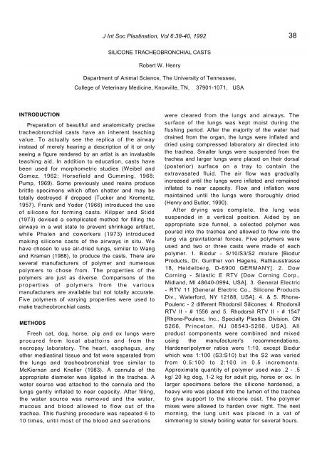
Figure 1. Silicone tracheobronchial cast from a dog. The airways are of # 1547 Rhone-Poulenc polymer<br />
mixture (10:1). Note, <strong>the</strong> airways are filled distally into <strong>the</strong> alveoli.<br />
Figure 2. Silicone tracheobronchial from a dog. The airway cast is made of Silastic E RTV polymer mixture<br />
(10:1). Note, only <strong>the</strong> larger airways are filled and <strong>the</strong> branching pattern is easily observed.