- Page 3: Laboratory handling of renal Bx:•
- Page 7 and 8: Our Medical Laboratory Scientist re
- Page 9 and 10: Tissue for light microscopy:• des
- Page 11 and 12: A paraffin embedded renal biopsy co
- Page 13 and 14: Immunohistochemical stains:Native
- Page 15 and 16: Examination of H&E stains:• on lo
- Page 17 and 18: Medium power examination of H&E sta
- Page 19 and 20: High power examination of H&E stain
- Page 21 and 22: H&E x2This is an H&E stain of a ren
- Page 23 and 24: H&E x10This is a higher power view.
- Page 25 and 26: H&E x10This is renal medulla. Most
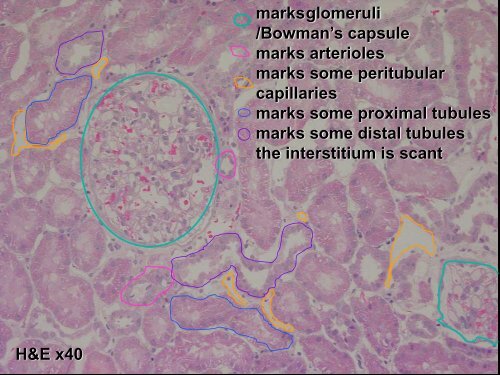
- Page 27: marksglomerulimarks arteriolesmarks
- Page 31 and 32: H&E x40marks arteriolesoutlines par
- Page 33 and 34: tubular polevascular pole
- Page 35 and 36: peritubular capllariesarteriolessma
- Page 37 and 38: H&E x2H&E x10This is a biopsy from
- Page 39 and 40: atrophic tubules in anarea of inter
- Page 41 and 42: segmental sclerosisfocal mild incre
- Page 43 and 44: the mesangium is expandedby amorpho
- Page 45 and 46: PAS x10
- Page 47 and 48: Bowman’s capsulemesangiumtubular
- Page 49 and 50: PAS x40Glomerulus sectioned through
- Page 51 and 52: segmental sclerosisfocal hyalinosis
- Page 53 and 54: OVG x40 - interlobulararteryOVG x40
- Page 55 and 56: Masson x40 -normal glomerulusMasson
- Page 57 and 58: Masson x40Masson x40variations in s
- Page 59 and 60: Masson x40: glomerulus with mesangi
- Page 61 and 62: AgMT x40 - small arteryAgMT x40 - g
- Page 63 and 64: AgMT x100Can you identify mesangial
- Page 65 and 66: This biopsy is from someonewith the
- Page 67 and 68: AgMT x100 -variations in staining f
- Page 69 and 70: mesangial cell nucleismallsubepithe
- Page 71 and 72: AgMT x100:there is fuzzy materialor
- Page 73 and 74: Congo red stain showing theapple gr
- Page 75 and 76: Examination of immunohistochemical
- Page 77 and 78: IF IgG x40Normal kidney -immunofluo
- Page 79 and 80:
IF fibrin x40Normal kidney -immunof
- Page 81 and 82:
IF C1q x40Normal kidney -immunofluo
- Page 83 and 84:
IP IgA x40Normal kidney - immunoper
- Page 85 and 86:
IP IgG x40Normal kidney - immunoper
- Page 87 and 88:
IP IgM x40Normal kidney - immunoper
- Page 89 and 90:
IP C3 x10
- Page 91 and 92:
IP C1q x40IP C1q x10Normal kidney -
- Page 93 and 94:
IP kappa x40IP lambda x40Immunopero
- Page 95 and 96:
IP C4d x40Immunoperoxidase for C4d
- Page 97 and 98:
IP SV40 x40 biopsy BImmunoperoxidas
- Page 99 and 100:
Electron microscopy:• a piece of
- Page 101 and 102:
Toluidine blue x40Toluidine blue x2
- Page 103 and 104:
Assessment of the glomerulus in ele
- Page 105 and 106:
This is a low power electron microg
- Page 107 and 108:
This glomerulus is normocellular, w
- Page 109 and 110:
mesangial matrixepithelial cellfoot
- Page 111 and 112:
The Banff Classification of Renal A
- Page 113 and 114:
Specimen adequacy(a necessary prere
- Page 115 and 116:
Quantitative criteria for mononucle
- Page 117 and 118:
Quantitative criteria for arteriola
- Page 119 and 120:
Quantitative criteria for peritubul
- Page 121 and 122:
Quantitative criteria for interstit
- Page 123 and 124:
Quantitative criteria for fibrous i
- Page 125 and 126:
C4d scoring :% biopsy area(cortex a
- Page 127 and 128:
2. Antibody-mediated changes:may co
- Page 129 and 130:
4. T-cell-mediated rejection (TCMR)
- Page 131 and 132:
6. Other:• changes not considered
- Page 133 and 134:
References:• Banff 07 Classificat