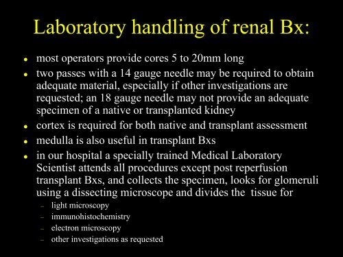
- Page 6 and 7: Petri dish, filter paper moistened
- Page 8 and 9: The divided tissue is place in appr
- Page 10 and 11: Immunohistochemistry:• we general
- Page 12 and 13: Sections & stains for light microsc
- Page 14 and 15: A tray of renal biopsy slides withH
- Page 16 and 17: Low power examination of H&E stains
- Page 18 and 19: High power examination of H&E stain
- Page 20 and 21: High power examination of H&E stain
- Page 22 and 23: H&E x2The biopsy is all cortex butc
- Page 24 and 25: marksglomerulimarks an arterymarks
- Page 26 and 27: H&E x20Can you find glomeruli, arte
- Page 28 and 29: H&E x40This is renal cortex. Can yo
- Page 30 and 31: H&E x40Find afferent/efferent arter
- Page 32 and 33: H&E x40H&E x40These glomeruli have
- Page 34 and 35: This section is too thick and it is
- Page 36 and 37: proximal tubules withcytoplasmic eo
- Page 38 and 39: some groups of tubulesare atrophics
- Page 40 and 41: What abnormalities can you seen in
- Page 42 and 43: What abnormalities can you seen in
- Page 44 and 45: Examination of PAS stains:• PAS s
- Page 46 and 47: PAS x40 - small arteryPAS x40 - glo
- Page 48 and 49: PAS x40PAS x40slight variations in
- Page 50 and 51: What abnormalities can you seen in
- Page 52 and 53:
Examination of OVG stains:• OVG s
- Page 54 and 55:
Examination of Masson stains:• Ma
- Page 56 and 57:
Masson x40 -normal glomerulus
- Page 58 and 59:
Masson x40 -variations in staining
- Page 60 and 61:
subepithelial depositsubendothelial
- Page 62 and 63:
AgMT x100AgMT x100This section is t
- Page 64 and 65:
AgMT x100marks mesangialcell nuclei
- Page 66 and 67:
tubular basement membraneproximal t
- Page 68 and 69:
AgMT x40: glomerulus with deposits,
- Page 70 and 71:
AgMT x100:What abnormalities can yo
- Page 72 and 73:
Additional stains which may be help
- Page 74 and 75:
IP SV40 x40 biopsy AIP SV40 x10 bio
- Page 76 and 77:
IF IgA x40Normal kidney -immunofluo
- Page 78 and 79:
IF IgM x40Normal kidney -immunofluo
- Page 80 and 81:
IF C3 x40Normal kidney -immunofluor
- Page 82 and 83:
IP IgA x10
- Page 84 and 85:
IP IgG x10
- Page 86 and 87:
IP IgM x10
- Page 88 and 89:
IP fibrin x40IP fibrin x10Normal ki
- Page 90 and 91:
IP C3 x40Normal kidney - immunopero
- Page 92 and 93:
IP IgM x40IP C1q x40Immunoperoxidas
- Page 94 and 95:
IP C4d x40Normal kidney - immunoper
- Page 96 and 97:
IP SV40 x40 biopsy BIP SV40 x40 bio
- Page 98 and 99:
IP SV40 x40 biopsy CPAS x40 biopsy
- Page 100 and 101:
Slide with semithin sections of the
- Page 102 and 103:
Assessment of electron micrographs:
- Page 104 and 105:
Assessment of the glomerulus in ele
- Page 106 and 107:
mesangial matrixred cellepithelial
- Page 108 and 109:
This tissue has been reprocessedfro
- Page 110 and 111:
This glomerulus has normalnumbers o
- Page 112 and 113:
Specimen adequacy and lesion scorin
- Page 114 and 115:
Quantitative criteria for tubulitis
- Page 116 and 117:
Quantitative criteria for the early
- Page 118 and 119:
Quantitative criteria for intimal a
- Page 120 and 121:
Quantitative criteria for allograft
- Page 122 and 123:
Quantitative criteria for tubular a
- Page 124 and 125:
Quantitative criteria for mesangial
- Page 126 and 127:
Diagnostic categories for renal tra
- Page 128 and 129:
3. Borderline changes:• suspiciou
- Page 130 and 131:
5. Interstitial fibrosis and tubula
- Page 132 and 133:
The transplant biopsy report:• th
- Page 134:
Acknowledgements:• Staff of the A