Answers to HW Questions
Answers to HW Questions
Answers to HW Questions

You also want an ePaper? Increase the reach of your titles
YUMPU automatically turns print PDFs into web optimized ePapers that Google loves.
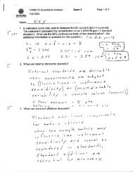
<strong>Answers</strong>Chem 250 Unit 1 – Proteomics by Mass SpectrometryArticle #1 Quantitative MS for proteomics: teaching a new dog old tricks.MacCoss MJ, Matthews DE., Anal Chem. 2005 Aug 1;77(15):294A-302A.1. <strong>Questions</strong>1.1. Define resolution for MS and for chroma<strong>to</strong>graphy. Resolving power orresolution may be defined for one peak relative <strong>to</strong> another or simply based on asingle peak.MSCHROMATOGRAPHYRESOLUTIONΔ m defined arbitrarily byα ≡ tR2 − tR 2distinguishability of 2 peaksRESOLVINGPOWERmR ≡ where Δm isΔmdefined arbitrarily asdistinguishableN16t≡wR1.2. What is an internal standard and what does it accomplish analytically.Internal standards are standards added intentionally <strong>to</strong> all samples, standards andblanks. The ideal internal standard is completely resolved from the unknown analyteby the measuring instrument but is chemically ‘identical’ <strong>to</strong> the analyte. Identicalmeans that it experiences the same losses during sample preparation, the same matrixeffects and responds <strong>to</strong> changes in instrument sensitivity in the same way as theanalyte.1.3. Does an internal standard account for losses during sample preparation? If so,how?The internal standard ideally is lost at the same rate during sample preparation byaccidental dilutions, chemical degradation, evaporation, pipeting losses, adsorption <strong>to</strong>surface etc. This is not so hard <strong>to</strong> imagine as the IS is mixed with the sample beforethe sample preparation is begun.1.4. Discuss iso<strong>to</strong>pe dilution from the perspective of a ‘perfect internal standard’.Mass spec is unique in its ability <strong>to</strong> distinguish identical chemicals of different mass(iso<strong>to</strong>pomers). Most iso<strong>to</strong>pomers have chemical and physical properties that areindistinguishable. For example, CH 3 37 Cl and CH 3 35 Cl have indistinguishable boilingpoints, electronic spectra, dielectric constants, reactivities, solubilities etc., but arecompletely resolved in even inexpensive mass spectrometers.
1.5. Distinuish between a stable iso<strong>to</strong>pic label and a radioiso<strong>to</strong>pic label. Why arestable iso<strong>to</strong>pic labels used in MS?Radioiso<strong>to</strong>pes are those that decay by alpha, beta, gamma, electron capture, positronemission etc. All but pure gamma emitters change chemical identities and massesduring this transition. The transition can make the radioiso<strong>to</strong>pe detectable by avariety of means (scintillation counting, x-ray film etc.) But these mass changesdefeat the resolution desired in MS iso<strong>to</strong>pe dilution. Radioiso<strong>to</strong>pes are dangerous,expensive and require special handling1.6. Define proteomics.Proteomics refers <strong>to</strong> the measurement of the broad inven<strong>to</strong>ry of proteins expressed ina given organism or cell line. For example, proteomics may refer <strong>to</strong> theidentification, detection and quantitation of all of proteins and post-translationalmodifications observed in a given cell line at a given time.1.7. Define polynucleotide, polypeptide and protein.DNA nucleotides comprise adenine (A), guanine (G), thymine (T) and cy<strong>to</strong>sine (C).Polynucleotides comprise polymers with variable A,G,T and C subunits linked byphosphate esters of deoxyribose (RNA uses uracil (U) and ribose). Oligonucleotidesare short (less than about 20 or so units) of AGTC.Proteins are formed from amino acids linked by peptide (amide) bonds. Proteins aremade by living cells – polypeptides are short (< about 20?) chains of amino acids thatmay or may not be biogenic.1.8. Would you describe the contents of a cell as:1.8.1. A solution of one or two different proteins.1.8.2. A solution of a few polynucleotides and polypeptides.1.8.3. A solution with a few mono and polysaccharides, lipids, glycolipids,proteins, glycoproteins, peptides, RNA, DNA, small molecule electrolytesand metabolites and basically a very complex thing.1.9. How does chroma<strong>to</strong>graphy help <strong>to</strong> simplify the analysis of a cell’s contents?Analysis of a cellular contents is a very challenging problem – usually requiring avariety of fractionations such as centrifugation <strong>to</strong> separate organelles, salting out ofproteins, dialaysis <strong>to</strong> remove salts and small molecules etc. This may reduce thedimension of the problem <strong>to</strong> just 10’s <strong>to</strong> hundreds of abundant chemical species.Chroma<strong>to</strong>graphy (HPLC in one of its many incarnations) can then reduce this by afac<strong>to</strong>r of ten or better.1.10. How does mass spectrometry help?MS gives sensitively and possibly quantitatively reports the masses of thebiomolecule constituents of the mixture. If two biomolecules have different massesthen they are resolved even if they co-elute. Mass may not seem like the most amplydescriptive property of a biomolecule, but it’s not bad! Fragmentation patterns fromprotein databases can be identified by computer analysis rapidly and faithfully.
<strong>Questions</strong> that are more specific <strong>to</strong> MS:1.11. How is product ion scanning superior <strong>to</strong> single quadrupole analysis of aprotein mixture for the purpose of identifying or quantifying a given componen<strong>to</strong>f a complex biological sample?In the product ion scan, one can screen the mixture for the mass of the component ofinterest in Q1, fragment it in Q2 and then scan for the fingerprint of the fragmentationproducts in Q3. This is powerful if you know what you are looking for! Even if Q1admits some interfering species, the fingerprint in Q3 is likely <strong>to</strong> be interpretable.1.12. Given the exact structure of a small molecule, say, CO, and the mass spectrumcomprising just the monocation, CO+, how many peaks do you expect <strong>to</strong> find inthe MS of this molecule? Assume you can measure within about 100:1 signal <strong>to</strong>noise ratio. If more than one, then with what relative intensities?It’s kind of a weak question, I admit – but the idea is that CO + should show up in fourplaces corresponding <strong>to</strong> 12 C 16 O + , 13 C 16 O + , 12 C 18 O + and 13 C 18 O + . The abundance of13 C is about 1%, so this one should be seen at SNR=100. But 18 O is rarer and theiso<strong>to</strong>pomers with 18 O would not normally be observed unless the sample wereenriched in 18 O. One <strong>to</strong> two peaks should be observed for CO + .1.13. Apply the same reasoning <strong>to</strong> the analysis of a 5 kDa protein (not in detail, justdiscuss the general idea).The basic idea then is this – the 5 kDa peak will have many sidebands comprisingsmall and variable amounts of 13 C and 2 H. So, for example, if the resolving power ofan instrument is 10,000 at 5kDa, then while it is conceivable that one might resolvethe centers of the distributions formed by two different proteins of nominal mass5000 Da and a 4,998 Da proteins, their individual iso<strong>to</strong>per masses will not beresolved. Take home message: Proteins will produce iso<strong>to</strong>pic distributions aroundtheir nominal masses that are normally unresolved. Even if an instrument has theresolving power <strong>to</strong> distinguish two proteins, they actual resolution may be difficult <strong>to</strong>these distribution functions.
1.14. Contrast quadrupole MS detection with fluorescence and absorbancespectroscopy in terms of:Figure of Merit q-Mass Spec Fluorescence AbsorbanceSensitivity+++++++++++milli -3 micro -6nano -9 pico -12fem<strong>to</strong> -15 at<strong>to</strong> -18yoc<strong>to</strong> -21SelectivityGeneralityng (10 -9 g)++++++++Especially withfragmentationpattern recognition.+++++++++++++Pretty muchanything can give aion fragment!yg (10 -21 g)+++++Fluorescence is notthat common.+?+?+?+?+Only selective if afluorphore can beattached selectively <strong>to</strong>your target.Destructive or non? destructive semi-destructivethrough labelnM conc. DL+Many interferantsnormally possible.++Lots of moleculesexhibit a UV abs.,all have IR abs.non-destructiveSample Size RequirednL (MALDI) <strong>to</strong>100 uL (ESI)pL + microscope0.1 mL – 1.0 mLSpeedMedium <strong>to</strong> fast(instrumentdependent, sampleprep dependent)Slow(assumebiolabeling <strong>to</strong> makeselectivity)Fast(simplemeasurement)CostHigh(instrument)Medium(instrument +enzymes + labor)lowInstrument size big med Small
<strong>Questions</strong> we have answered:Chem 250 Fall 2008<strong>Answers</strong> <strong>to</strong> questions given in class 8.27.081.15. A strong cation exchange resin contains sulfonic acid (a strong acid) residues.Assume the solution pH is 7.5.1.15.1. Will all proteins bind <strong>to</strong> this resin?No, a cation exchange resin has anionic (strong acid) groups on the surface such as R-SO3 - . Therefore it will bind proteins that are either net cationic or that have at least somepatches of positive charge on the surface. It will not bind purely anionic proteins. It willnot bind proteins with a pI above the buffer pH = 7.5.1.15.2. How will proteins bind <strong>to</strong> this resin?Electrostatic attraction between cationic (lysine and arginine) groups on the protein’speriphery and sulfonates on the resin.1.15.3. Will this be a mono- or multi-valent binding interaction? (Note thatmultivalent interactions such as DNA duplexation have characteristicallysharp association isotherms.)The likelihood is that this will be multivalent. The more lysine groups on the proteinsurface, the more points of attraction and the stronger the binding.1.15.4. Why would increasing ionic strength of, e.g. NaCl decrease thestrength of this binding interaction?Increasing ionic strength will tend <strong>to</strong> elute the protein. One can think of this as acompetitive binding effect (like the Na + in the solution competes with lysine + for thesulfonate) or as a charge screening effect wherein at high ionic strength ions in solutionsimply tend <strong>to</strong> cancel electric fields and prevent the normal electrostatic interactions fromextending beyond a few angstroms.1.15.5. On what basis will this separation work?The separation will work on the basis of the strength of the cation-anion attractionbalanced against the solvating (enthalpic) and liberating (entropic) energies of releasingthe protein.1.16. On what basis does reverse-phase chroma<strong>to</strong>graphy separate analytes?Hydrophobicity – or, the tendency of non-polar components of the solution <strong>to</strong> stick <strong>to</strong> orpartition in<strong>to</strong> the oily coating on the chroma<strong>to</strong>graphic stationary phase.
1.17. Will ionic strength have a dominant influence on reverse-phasechroma<strong>to</strong>graphic separation carried out in 100% aqueous buffer?Ionic strength will only slightly modify the retention in the reverse phase systemcompared <strong>to</strong> the hydrophobic/hydrophilic quality of the protein (solute / analyte). Even ifit does change the retention properties, the idea is that the basis of the separation on thereverse phase column will be different <strong>to</strong> that of the ion exchange column.1.18. How does the combination of strong cation exchange followed by reversephaseLC equate <strong>to</strong> a 2-D separation? Just sketch the outcome.absorbancet R / minμ = 0.01μ = 0.05Ionicstrength(μ) / Mμ = 0.1μ = 0.5