Magnetic anisotropy of textured CrO2 thin films investigated ... - KOPS
Magnetic anisotropy of textured CrO2 thin films investigated ... - KOPS
Magnetic anisotropy of textured CrO2 thin films investigated ... - KOPS

Create successful ePaper yourself
Turn your PDF publications into a flip-book with our unique Google optimized e-Paper software.
e. goering 1,✉<br />
m. justen 1<br />
j. geissler 1<br />
u. rüdiger 2<br />
m. rabe 2<br />
g. güntherodt 2<br />
g. schütz 1<br />
Received: 8 January 2002/Accepted: 8 January 2002<br />
Published online: 26 March 2002<br />
ABSTRACT Highly a-axis-<strong>textured</strong> <strong>CrO2</strong> <strong>films</strong> have been deposited<br />
on Al2O3 (0001) substrates by chemical vapor deposition.<br />
<strong>CrO2</strong> has been found to have highly a-axis (010)-oriented<br />
columnar growth on a Cr2O3 (0001) initial layer. The sixfold<br />
surface symmetry <strong>of</strong> the Cr2O3 initial layer leads to three<br />
equivalent in-plane orientations <strong>of</strong> the a-axis-oriented <strong>CrO2</strong> unit<br />
cell. We report Cr L2,3 X-ray magnetic circular dichroism data<br />
along the surface normal and at 60 ◦ <strong>of</strong>f-normal sample orientation.<br />
For a 60 ◦ sample alignment, a strong increase <strong>of</strong> the<br />
projected orbital moment could be observed for unoccupied majority<br />
t2g states using moment analysis. Therefore, the c axis is<br />
identified as the intrinsic magnetic easy axis <strong>of</strong> <strong>CrO2</strong>. In addition,<br />
a small spin moment and a very strong magnetic dipole<br />
term Tz have been found.<br />
PACS 75.30.Cr; 75.30.Gw; 78.20.Lj; 78.70.Dm; 78.20.Bk<br />
1 Introduction<br />
The theoretically predicted 100% spin polarizationattheFermienergy,εF,<br />
<strong>of</strong><strong>CrO2</strong> makes it a promising<br />
material for magnetoelectronic devices [1–3]. According<br />
to Jullière’s model, the magnetoresistance (MR) <strong>of</strong><br />
a ferromagnet/insulator/ferromagnet tunnel junction depends<br />
on the spin polarization <strong>of</strong> the electrode material<br />
used [4]. The MR increases with increasing spin polarization<br />
<strong>of</strong> the ferromagnetic electrode material. Also, injection<br />
<strong>of</strong> spins from ferromagnetic metals into semiconductors<br />
may be allowed only for highly polarized metallic electrodes<br />
[5]. Additionally, transition-metal oxides like <strong>CrO2</strong>,<br />
high-temperature superconductors and colossal magnetoresistance<br />
lanthanum-based manganates have attracted a lot <strong>of</strong><br />
theoretical interest due to the complex interplay between orbital,<br />
structural and electronic degrees <strong>of</strong> freedom. This has<br />
revived research on half-metallic ferromagnetic transitionmetal<br />
oxides, especially <strong>CrO2</strong>, in the past few years. Unfortunately,<br />
<strong>CrO2</strong> is metastable at room temperature and<br />
atmospheric conditions and typically a <strong>thin</strong> Cr2O3 layer covers<br />
the <strong>CrO2</strong> surface. Methods like photoemission (PES),<br />
✉ Fax: +49-711/689-1912, E-mail: goering@mf.mpg.de<br />
<strong>Magnetic</strong> <strong>anisotropy</strong> <strong>of</strong> <strong>textured</strong> <strong>CrO2</strong> <strong>thin</strong><br />
<strong>films</strong> <strong>investigated</strong> by X-ray magnetic circular<br />
dichroism<br />
1 Max-Plank-Institut für Metallforschung, Heisenbergstraße 1, 70569 Stuttgart, Germany<br />
2 Physikalisches Institut, Rheinisch-Westfälische Technische Hochschule Aachen, 52056 Aachen, Germany<br />
X-ray absorption (XAS) or X-ray magnetic circular dichroism<br />
(XMCD) spectroscopy could not easily be used for examination<br />
<strong>of</strong> basic properties <strong>of</strong> <strong>CrO2</strong>, because <strong>of</strong> their intrinsic<br />
surface sensitivity. Therefore, only a few experimental results<br />
on single crystals or epitaxial-grown <strong>thin</strong> <strong>films</strong> have been published.<br />
Nevertheless, spectroscopic methods are able to give<br />
quantitative information about intrinsic magnetic properties<br />
<strong>of</strong> <strong>CrO2</strong>.<br />
2 Experiment and results<br />
<strong>CrO2</strong> crystallizes in a tetragonal rutile-type structure,<br />
where chromium atoms form a body-centered tetragonal<br />
unit cell. Cr sites are octahedrally surrounded by oxygen<br />
atoms. The apical axis <strong>of</strong> the oxygen octahedra is oriented<br />
along the [110]([110]) direction for the edge (bodycentered)<br />
Cr ions. The lattice parameters are a = b = 4.421 Å<br />
and c = 2.916 Å [6, 7]. <strong>CrO2</strong> is a metastable Cr-oxide phase<br />
at room temperature, orders ferromagnetically at TC = 393 K<br />
and degrades above 400 K at ambient atmospheric pressures<br />
into the thermodynamically stable Cr2O3 phase. The <strong>CrO2</strong><br />
film has been prepared by a chemical vapor deposition (CVD)<br />
technique proposed by Ishibashi et al. [8]. Al2O3 (0001)<br />
substrates have been used, which were annealed at 1000 ◦ C<br />
for several hours before deposition. During the deposition,<br />
the substrate was oriented at an angle <strong>of</strong> 30 ◦ relative to<br />
the horizontal axis <strong>of</strong> the tube furnace. CrO3 decomposes<br />
at a temperature <strong>of</strong> 260 ◦ C wi<strong>thin</strong> a two-zone tube furnace.<br />
A well-controlled flow <strong>of</strong> oxygen and a stabilized substrate<br />
temperature <strong>of</strong> 390 ◦ C lead to approximately 200-nm-thick<br />
<strong>CrO2</strong> <strong>films</strong>. Characterization by X-ray diffractometry showed<br />
only <strong>CrO2</strong> (020) and (040) reflexes, indicating a preferred<br />
a-axis growth. Electron-diffraction analysis exhibits an artificial<br />
six-fold in-plane symmetry and a formation <strong>of</strong> twin<br />
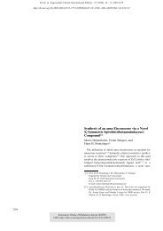
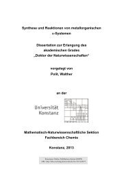
boundaries [9]. A scanning electron microscopy (SEM) image<br />
<strong>of</strong> (010)-oriented <strong>CrO2</strong> crystallites on the Cr2O3 (0001) initial<br />
layer is shown in Fig. 1. The six-fold in-plane symmetry<br />
<strong>of</strong> the (0001)-oriented Cr2O3 initial layer (which is<br />
isostructural to Al2O3) leads to three equivalent in-plane<br />
orientations for the (011) plane <strong>of</strong> the <strong>CrO2</strong> unit cell. Transmission<br />
electron microscopy (TEM) analysis shows a small<br />
amount <strong>of</strong> Cr2O3 columns wi<strong>thin</strong> the <strong>CrO2</strong> layer. More details<br />
<strong>of</strong> sample growth, characterization and magnetotransport<br />
properties have been published elsewhere [10]. The sam-
748<br />
FIGURE 1 Scanning electron microscopy image <strong>of</strong> (010)-oriented <strong>CrO2</strong><br />
crystallites on a Cr2O3 (0001) initial layer [10]<br />
ple has been exposed to air. No further treatment has been<br />
performed.<br />
X-ray absorption spectra were recorded at the bending<br />
magnet beamline SX 700 III at BESSY I. The monochromator<br />
energy resolution was set to E/∆E ≈ 1200 and the<br />
degree <strong>of</strong> circular polarization was 0.69 ± 0.05. Two Keithley<br />
6517A electrometers were used for simultaneous measurement<br />
<strong>of</strong> the total drain sample current and the I0 current from<br />
an Au-coated Cu grid. The I0 grid was positioned in a µ-metal<br />
cylinder in order to prevent deviations produced by magnetic<br />
fields. A split-coil superconducting magnet system was used<br />
with a center bore <strong>of</strong> 5cmand a maximum magnetic field <strong>of</strong><br />
30 kOe. All spectra have been recorded at magnetic fields <strong>of</strong><br />
±5kOe. The XMCD signal was carefully checked for selfabsorption<br />
effects. Atomic absorption values <strong>of</strong> Cr and O have<br />
been extracted from tabulated values [11] and scaled to atomic<br />
densities in <strong>CrO2</strong>. We shifted and scaled our measured XAS<br />
spectra for both magnetization directions to fit the pre-edge region<br />
<strong>of</strong> oxygen and the post-edge region <strong>of</strong> Cr L2,3 edges <strong>of</strong><br />
the calculated tabulated values. Due to this procedure, the resonant<br />
behavior <strong>of</strong> the absorption in the near-edge region could<br />
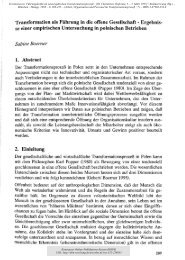
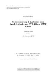
be estimated quantitatively. The resulting absorption coefficient<br />
is shown in Fig. 2. We extracted an absolute absorption<br />
length for the maximal absorption <strong>of</strong> 35 nm, which is significantly<br />
larger than the expected effective mean free path length<br />
<strong>of</strong> about 1.5nm[12, 13]. After that, we modified our magnetic<br />
XAS spectrum (parallel and antiparallel alignment <strong>of</strong> helicity<br />
and magnetization) with the above-derived absolute absorption,<br />
using equation 5 and 7 from [13]. We compared observed<br />
small changes in the XAS spectra from normal to 60 ◦ incidence<br />
with simulated self-absorption-induced changes and<br />
could obtain the best agreement with our XAS data for an effective<br />
electron mean free path <strong>of</strong> only 0.7nm. We applied<br />
sum rules to the original and self-absorption-modified spectra<br />
and compared orbital and spin contributions. Absolute<br />
changes between normal and 60 ◦ incidence are for the spin<br />
moment <strong>of</strong> −0.005µB (−0.009µB) and for the orbital moment<br />
<strong>of</strong> −0.0004µB (−0.0008µB) using an effective electron mean<br />
free path [12, 13] <strong>of</strong> 0.7nm(1.5nm). In comparison to the results<br />
<strong>of</strong> Nakajima et al. [13], self-absorption effects are not as<br />
Absorbtion (1/µm)<br />
30<br />
20<br />
10<br />
0<br />
CrO 2<br />
Scaled XAS<br />
Henke Tables<br />
500 600 700 800<br />
Photon Energy (eV)<br />
FIGURE 2 XAS spectra scaled to absolute <strong>CrO2</strong> absorption values averaged<br />
for parallel and antiparallel alignments <strong>of</strong> the photon k-vector and the<br />
sample magnetization for normal incidence (full line) compared to <strong>CrO2</strong><br />
adapted Henke table [11] values (dotted line)<br />
strong as expected for a 3d 2 system. Two different reasons are<br />
responsible for this reduction. <strong>CrO2</strong> has hybridization-related<br />
broadened L2 and L3 white lines, and therefore a strongly reduced<br />
maximal absorption compared to a pure metal spectrum<br />
with the same number <strong>of</strong> 3d holes. On the other hand, the chromium<br />
density and the Cr-related absorption coefficient are<br />
strongly reduced by dilution. Due to the nonlinear behavior,<br />
self-absorption is magnetically more effective for large differences<br />
between parallel and antiparallel orientations <strong>of</strong> sample<br />
magnetization and photon helicity, which is present in Fe or<br />
Co spectra but not in our <strong>CrO2</strong> spectra.<br />
To prevent XMCD <strong>of</strong>fset signals, we applied a small<br />
asymmetry <strong>of</strong> about 0.2kOe in the external magnetic field,<br />
which does not influence the total sample magnetization [14].<br />
To get a derivative-free XMCD signal the external magnetic<br />
field was flipped at each energy position. All shown XAS<br />
and XMCD spectra have been divided by the I0 current,<br />
background-subtracted and edge-normalized. For parallel and<br />
antiparallel field alignments we used exactly the same background<br />
and the same normalization factor; therefore the shape<br />
<strong>of</strong> the dichroism spectrum is not affected. The photon beam<br />
was parallel aligned to the magnetic field. We show X-rayabsorption<br />
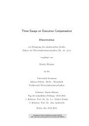
data for normal incidence Φ = 0 ◦ and more grazing<br />
incidence Φ = 60 ◦ (the geometry <strong>of</strong> the experimental<br />
setup is shown in Fig. 3).<br />
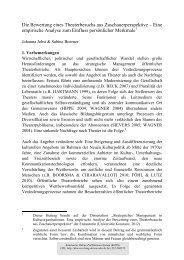
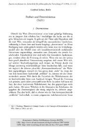
The upper part <strong>of</strong> Fig. 4 shows the Cr L2,3 normalincidence<br />
absorption spectra <strong>of</strong> <strong>CrO2</strong> for parallel and antiparallel<br />
alignments <strong>of</strong> the photon k-vector and the applied<br />
magnetic field. In the lower part <strong>of</strong> Fig. 4 the corresponding<br />
XMCD signal for normal and grazing (60 ◦ ) incidence<br />
is shown. An unusually clear and pronounced change in the<br />
spectral shape <strong>of</strong> the XMCD signal is clearly observable.<br />
For a quantitative analysis, we calculated the projected orbital<br />
moments for the two given angles <strong>of</strong> incidence using<br />
sum rules [15, 16]. With a number <strong>of</strong> core holes nh = 8 (3d 2<br />
configuration) we extract raw values <strong>of</strong> projected orbital moments<br />
for normal incidence Lz(0 ◦ ) =−0.041µB and for more<br />
grazing incidence Lz (60 ◦ ) =−0.016µB. Unfortunately, we
FIGURE 3 Schematic image <strong>of</strong> the experimental geometry<br />
XAS (edge normalized)<br />
XMCD (edge normalized)<br />
18<br />
16<br />
14<br />
12<br />
10<br />
8<br />
6<br />
4<br />
2<br />
0<br />
0.5<br />
0.0<br />
-0.5<br />
-1.0<br />
Cr<br />
L 3 L 2<br />
↑↑<br />
↑↓<br />
0° 5kOe<br />
60° 5kOe<br />
570 575 580 585 590 595 600<br />
Photon Energy (eV)<br />
FIGURE 4 Normalized Cr L2,3 absorption signal for parallel and antiparallel<br />
alignments <strong>of</strong> the photon k-vector and the sample magnetization for<br />
normal incidence (upper part). Lower part shows the XMCD signal for normal<br />
(0 ◦ : solid line) and grazing (60 ◦ : dotted line) incidence<br />
could not achieve technical saturation in 5-kOe external fields<br />
for the case <strong>of</strong> normal incidence due to the shape <strong>anisotropy</strong><br />
<strong>of</strong> the sample. From detailed SQUID M(T, H) measurements<br />
(not shown), we correct for T = 0K saturation magnetization<br />
and obtain for 0 ◦ (60 ◦ ) a correction factor <strong>of</strong> 1.78 (1.41).<br />
The 0 ◦ value is slightly enhanced, due to the described shape<strong>anisotropy</strong><br />
correction. The 60 ◦ correction factor <strong>of</strong> 1.41 is<br />
related only to the temperature dependence, which is also<br />
the dominating part in the perpendicular-to-plane measurement.<br />
For thick samples, closure domains could be present<br />
at the sample surface and differences between surface-related<br />
XMCD TEY (Total Electron Yield)-projected magnetizations<br />
and SQUID-derived values could appear. This is not the case<br />
in our measurement. In Fig. 5 we show in-plane and out<strong>of</strong>-plane<br />
SQUID M(H) curves and XMCD L3-edge-related<br />
out-<strong>of</strong>-plane hysteresis loops. The SQUID and XMCD curves<br />
are in perfect agreement and therefore our above-described<br />
SQUID-derived normal-incidence absolute correction factor<br />
M/M s<br />
1.5<br />
1.0<br />
0.5<br />
0.0<br />
-0.5<br />
-1.0<br />
CrO 2 at RT<br />
XMCD at Cr L 3<br />
SQUID out <strong>of</strong> plane<br />
SQUID in plane<br />
749<br />
-1.5<br />
-15 -10 -5 0 5 10 15<br />
H [kOe]<br />
FIGURE 5 XMCD L3-edge-related out-<strong>of</strong>-plane hysteresis loop (straight<br />
line) compared to SQUID-derived out-<strong>of</strong>-plane (hollow rectangles) andinplane<br />
(filled circles) curves<br />
<strong>of</strong> 1.71 is not disturbed by this possible TEY-related artefact.<br />
The technique used to obtain high-quality TEY-related<br />
XMCD hysteresis loops has been described in detail elsewhere<br />
[17]. By further correcting for the degree <strong>of</strong> circular polarization,<br />
we can obtain Lz(0 ◦ ) =−0.105µB and Lz(60 ◦ ) =<br />
−0.034µB.<br />
3 Momentanalysis<br />
3.1 Introduction to moment analysis for 3d transition<br />
metals<br />
We analyze the L2,3 dichroism spectra in terms <strong>of</strong><br />
ground-state moments [18, 19]. The advantage <strong>of</strong> this method<br />
is the possibility to get quantitative values even for the light<br />
transition-metal (TM) ions with a small p-shell spin–orbit<br />
coupling (SOC). In principle, moment analysis tries to gain<br />
information from the shape <strong>of</strong> the XMCD spectrum. This<br />
is an advantage compared to sum-rule analysis, which only<br />
uses integral values for the L2 and L3 parts <strong>of</strong> the XMCD<br />
spectrum separately. Therefore sum rules could not give reasonable<br />
values for the projected spin moments if L2 and L3<br />
XMCD features overlap in energy. Another advantage <strong>of</strong> this<br />
moment analysis is the possibility to gain quantitative information<br />
about spin and other ground-state moments, which<br />
have contributions to the observed XMCD signal like the<br />
number <strong>of</strong> core holes and the spin–orbit term. While this<br />
method is related to an atomic picture, which takes into account<br />
the coupling <strong>of</strong> the core hole to the final-state d shell,<br />
band-structure effects could be considered by Lorentzian or<br />
Gaussian broadening and by the use <strong>of</strong> different energy levels.<br />
This could yield, for example, to magnetization values for<br />
spectrally resolved and well-separated sublevels, like t2g or<br />
eg contributions in many TM oxides. This has been demonstrated<br />
for the ferrimagnetic iron garnets, where octahedrally<br />
and tetrahedrally coordinated Fe 3+ ions could be identified<br />
separately by XMCD [20, 21]. We believe that the Cr L2 − L3<br />
edge separation is strong enough to apply moment analysis.<br />
Experimental results for Cr metal from O’Brien et al. [22]<br />
clearly show that XMCD features from the L3 and L2 edges<br />
are separated. The results <strong>of</strong> van der Laan [18, 19] and the results<br />
<strong>of</strong> O’Brien et al. show exactly the same behavior. So,
750<br />
the present broadening in the <strong>CrO2</strong> L2,3 spectra compared<br />
to the pure metal spectra is related to hybridization effects.<br />
It is clear that, in a covalent type <strong>of</strong> bonding, different electron<br />
configurations could be present (for example d 1 , d 2 or<br />
d 3 ), which could yield to correlation effects. These results in<br />
multiplet-like features, which are considered by a configuration<br />
interaction (CI) ligand-field multiplet calculation. Early<br />
calculations <strong>of</strong> de Groot et al. [23] have shown that the cubic<br />
crystal field is very strong. Recent results <strong>of</strong> van Elp and<br />
Tanaka [24] clearly mention the dominating part <strong>of</strong> the crystal<br />
symmetry and the strength <strong>of</strong> oxygen hybridization to O<br />
K-edge spectral features <strong>of</strong> some TM oxides. From this point<br />
<strong>of</strong> view, crystal-field effects are strong and the resulting energies<br />
<strong>of</strong> the CI calculations nearly represent band-structure<br />
center-<strong>of</strong>-gravity positions. The below-described quite good<br />
agreement between experiment and fit, the direct correspondence<br />
to the band-structure-related projected density <strong>of</strong> states<br />
(DOS) and the reduced number <strong>of</strong> free fitting parameters give<br />
strong support that the moment analysis is a good representation<br />
<strong>of</strong> the XMCD spectra that leads to well-separated L2 and<br />
L3 spectral features.<br />
3.2 Application to <strong>CrO2</strong><br />
It has been verified that oxygen K-edge spectra<br />
represent the 2p hybridized part <strong>of</strong> the band structure and give<br />
an image <strong>of</strong> the unoccupied projected O2p DOS [25]. Influences<br />
<strong>of</strong> the core hole could be considered by a configurationinteraction<br />
scheme [24]. Due to this projection behavior, the<br />
main features could be understood by quantitative comparison<br />
to recent band-structure calculations [1–3, 26]. O K spectra<br />
and band-structure calculations could give similar information.<br />
We have separated O K-edge spectra <strong>of</strong> <strong>CrO2</strong> into<br />
two different regions. First, 1eV above threshold, which is<br />
unambiguously identified as unoccupied t2g majority states<br />
and, second, a broader feature around 3eVabove, which corresponds<br />
to a mixture <strong>of</strong> t2g eg minority and the eg majority<br />
states [27]. The latter represents band-structure-related<br />
values [1–3, 26]. This energy separation is also present in<br />
<strong>CrO2</strong> L2,3 spectra, which have a more complicated shape<br />
compared to simple pure and metallic TM spectra. The total<br />
width <strong>of</strong> the broader structure is about 7eV[1], but the dominating<br />
part has a FWHM <strong>of</strong> only 3eV. Considering the given<br />
arguments, we try to interpret the L2,3 XAS and XMCD spectra<br />
by the use <strong>of</strong> only two different energies for the moment<br />
Energy 1 FWHM 1 Energy 2 FWHM 2 2p SOC Eff. 2p3d exch.<br />
577.6eV 1.3 eV 580.5eV 3.3eV 9.3eV 0.45 eV<br />
TABLE 1 Fixed-fit parameters for moment analysis<br />
E0 = 577.6eV E1 = 580.5eV<br />
Angle w000 w011 w101 w000 w011 w101 w110 ∝ nh ∝ Sz ∝ Lz ∝ nh ∝ Sz ∝ Lz ∝ L<br />
Sz0 + Sz1 Lz0 + Lz1<br />
∗S (arb. units) (µB) (µB) (arb. units) (µB) (µB) (arb. units) (µB) (µB)<br />
0◦ 1.03 0.25 0.05 49.8 0.73 –0.15 3.3 0.98 –0.1<br />
60◦ 1.29 0.12 0.11 44.8 0.34 –0.14 21.9 0.46 –0.03<br />
analysis, which are separated by 2.9eV, and only Lorentzian<br />
line broadening has been applied. For a reduction <strong>of</strong> free<br />
fitting parameters, the energy position, line width, effective<br />
2p3d exchange and the 2p SOC energy (see Table 1)<br />
are previously estimated and have been held fixed. Also, the<br />
sum <strong>of</strong> the projected orbital moments was constrained to the<br />
above-discussed sum-rule values. All fixed parameters are<br />
shown in Table 1. The FWHM values have been chosen to fit<br />
the XMCD data and to match roughly band-structure-related<br />
values. The value for the 2p SOC energy has been taken<br />
from [28, 29].<br />
For the moment-fitting procedure the relative energydependent<br />
ratios for the atomic-like different sublevels have<br />
been taken from [18, 19]. For data fitting we used the w 000 ,<br />
w 011 , w 101 and w 110 moments, which are proportional to the<br />
number <strong>of</strong> holes in the d shell (nh), the spin moment Sz, the<br />
orbital moment Lz and to L · S, respectively. The areas corresponding<br />
to the w 011 and w 101 moments have been used<br />
for sum-rule analysis. In Table 2 we show the fitted moment<br />
XMCD (edge normalized)<br />
XMCD (edge normalized)<br />
0.5<br />
0.0<br />
-0.5<br />
-1.0<br />
1.0<br />
0.5<br />
0.0<br />
-0.5<br />
-1.0<br />
0°<br />
60°<br />
570 575 580 585 590 595 600<br />
Photon Energy (eV)<br />
XMCD<br />
Energy 1<br />
Energy 2<br />
Fit<br />
FIGURE 6 Moment-analysis fit results for normal (above) and grazing (below)<br />
incidence (circle: measurement; dotted: fit at energy 577.6eV;dashed:<br />
fit at energy 580.5eV;solid line: fitted sum)<br />
TABLE 2 Moment-analysis fit results
data, while spin and orbital moments have been corrected for<br />
T = 0Ksaturation.<br />
In Fig. 6 we show fit results <strong>of</strong> our moment analysis for<br />
each energy and the sum <strong>of</strong> both for 0 ◦ and 60 ◦ angles <strong>of</strong> incidence,<br />
which represent our XMCD spectra nearly perfectly.<br />
All structures and tendencies could be reproduced by this simple<br />
two-energy fit.<br />
Four fit parameters change significantly with rotation: the<br />
spin and the orbital contribution <strong>of</strong> the lower energy, and the<br />
spin and the SOC (Spin-Orbit-Interaction) contribution <strong>of</strong> the<br />
higher-energy part in the XMCD spectrum.<br />
4 Discussion<br />
4.1 Spin <strong>anisotropy</strong> and magnetic dipole term Tz<br />
First, we want to discuss the projected spin moments.<br />
For both energies the observed projected spin moments<br />
change by a factor <strong>of</strong> two and the sum decreases from approximately<br />
1.0µB to 0.5µB (see Table 2). This is quite unusual<br />
compared to XMCD-related spin values <strong>of</strong> other transition<br />
metals, where only very small changes have been observed for<br />
saturated samples. To obtain the moment-analysis spin values,<br />
we simply used conventional sum rules by artificially separating<br />
the L3 from the L2 edge at a splitting energy <strong>of</strong> 584.9eV.<br />
The unknown <strong>of</strong>fshoot from the L3 edge is present at the L2<br />
XMCD area and therefore is missing at the L3 edge. Compared<br />
to moment-analysis-derived spin values, this results in<br />
an increase <strong>of</strong> 10% for the spin moment, but the trends and the<br />
general reduction by a factor <strong>of</strong> two are similar to momentanalysis-derived<br />
values. As discussed above, we could not<br />
perfectly saturate the sample along the normal direction. This<br />
simply should result in a reduced spin moment but not in<br />
a strong increase by a factor <strong>of</strong> two.<br />
This angle-dependent change suggests a very strong magnetic<br />
dipole term Tz, which is directly related to a quadrupolar<br />
spin distribution, originated by a reduced symmetry that is<br />
less than cubic. For the case <strong>of</strong> strong symmetry breaking,<br />
given at an interface, where the octahedral surface symmetry<br />
is broken by the adjacent constituent, Tz could be significantly<br />
increased. Calculations have shown that for a 3d 2<br />
configuration this surface-induced symmetry breaking gives<br />
a Tz <strong>of</strong> about 20% <strong>of</strong> the spin moment [30]. In the case <strong>of</strong><br />
<strong>CrO2</strong> the local octahedral environment is distorted in the rutile<br />
structure; therefore the Tz (index z is related to a local<br />
distortion) value could be significantly enhanced. Estimating<br />
now the spin moment, the 3d symmetry relation 〈Tz〉x +<br />
〈Tz〉y +〈Tz〉z = 0 has been applied, which has been published<br />
by Stöhr and König [31] and verified on a Au/Co/Au wedge<br />
by Weller et al. [32]. Transferring this equation to the rutiletype<br />
unit cell yields 2〈Tz〉a +〈Tz〉c = 0. Measuring along the<br />
aaxis(0 ◦ ), only 〈Tz〉a-axis contributions are present due to<br />
the film’s texture, while more grazing incidence gives rise<br />
to a more complicated mixture <strong>of</strong> a- and c-axis magnetic<br />
dipole term contributions. For 60 ◦ grazing incidence and<br />
a random in-plane c- (a-) axis distribution, there is an inplane<br />
mixture <strong>of</strong> a and c axes and an effective 〈Tz〉 projection<br />
<strong>of</strong> sin(60 ◦ )(〈Tz〉a +〈Tz〉c)2/π, while the <strong>of</strong>f-plane contribution<br />
is simply cos(60 ◦ )〈Tz〉a. To estimate the zero-〈Tz〉 condition<br />
we have to calculate Sz(60 ◦ ) + Sz(0 ◦ )(sin(60 ◦ )2/π −<br />
cos(60 ◦ )), which cancels out 〈Tz〉 contributions and normal-<br />
751<br />
izes by (1 + sin(60 ◦ )2/π − cos(60 ◦ )). This results in a corrected<br />
spin moment without any magnetic dipole term contribution<br />
<strong>of</strong> Szeff = 0.49µB. For a fully spin-polarized system<br />
with an empty minority band a d 2 configuration yields a spin<br />
moment <strong>of</strong> 2µB, which has been experimentally and theoretically<br />
verified. SQUID measurements performed on the sample<br />
used exhibit the same value. A correct treatment <strong>of</strong> the 120 ◦<br />
c-axis in-plane distribution instead <strong>of</strong> the random model used<br />
above leads to a spin moment from 0.46 to 0.50µB, depending<br />
on the unknown in-plane orientation <strong>of</strong> the sample relative to<br />
the photon beam.<br />
The extracted effective XMCD spin moment <strong>of</strong> 0.49µB<br />
is about a factor <strong>of</strong> 4.1 too small compared to SQUID data,<br />
which is a rather large deviation.<br />
In this section some possible explanations for this strong<br />
reduction will be discussed.<br />
Due to the small spin–orbit splitting for light 3d transition<br />
metals, there are two dominating problems. Firstly, spectral<br />
features <strong>of</strong> the L2 and the L3 edges are overlapping and therefore<br />
sum rules could give wrong values. Due to the moment<br />
analysis, we could avoid this problem. Secondly, the final<br />
states could not be considered as pure L2 or L3 states because<br />
<strong>of</strong> quantum-mechanical mixing. Therefore, the spectral intensity<br />
in the L3 region has L2 contributions and vice versa. This<br />
has been calculated for the case <strong>of</strong> Cr 2+ (3d 4 ), where a reduction<br />
<strong>of</strong> about 50% has been proposed [30], but this strong<br />
reduction has been obtained by sum-rule application to theoretical<br />
XMCD spectra. Following the spectral simulation in<br />
this reference, it is not clear whether the dominant part <strong>of</strong> the<br />
reduction is related to overlapping features, discussed above,<br />
or to real L2–L3 final-state mixing. Assuming such a mixing<br />
present in <strong>CrO2</strong>,wehaveaCr d 2 configuration with the same<br />
L2,3-edge spin–orbit splitting, and the theoretically proposed<br />
effect could be a suitable explanation <strong>of</strong> this strong reduction<br />
in the observed spin moment.<br />
A third point is related to the spin–orbit coupling in the<br />
3d shell. If a spin and an orbital moment are present, the<br />
octahedral symmetry could be reduced by SOC itself and<br />
a nonvanishing Tz is present. This has also been calculated by<br />
Crocombette et al. [30]. If the SOC is isotropic, the Tz moment<br />
is always oriented along the spin moment and varies with the<br />
sample magnetization. The sample orientation will not change<br />
this spin-induced Tz; therefore the above-used symmetry relation<br />
〈Tz〉x +〈Tz〉y +〈Tz〉z = 0 is no longer valid and Tz is<br />
always present. Reported calculations for a 3d 2 configuration<br />
have shown that SOC itself could give a Tz contribution up<br />
to 13% <strong>of</strong> the spin moment, but only at zero temperature. For<br />
room-temperature measurements this SOC-induced value is<br />
strongly decreased to about 1% relative to the spin part. Our<br />
experiments have been performed at room temperature; therefore<br />
we conclude that this SOC-induced Tz is not the origin <strong>of</strong><br />
the observed reduction <strong>of</strong> the spin moment, despite the possibility<br />
that for <strong>CrO2</strong> the SOC could be strongly enhanced.<br />
A fourth and probably most interesting point for the effective<br />
XMCD spin-moment reduction is related to hybridization.<br />
Due to hybridization, the wave functions <strong>of</strong> the 3d electrons<br />
are partly delocalized and form a mixture <strong>of</strong> Cr 3dand<br />
O2p-like states. Therefore, parts <strong>of</strong> the electronic wave<br />
function will not overlap any more with 2p initial-state wave<br />
functions. This would induce a decrease in the XMCD ef-
752<br />
fect for high-bandwidth metals like Fe or Co, corresponding<br />
to the strongly delocalized behavior <strong>of</strong> the 3d electrons,<br />
which has not been observed. In fact, for pure Fe and Co,<br />
nearly perfect agreement could be achieved between sumrule-related<br />
magnetic moments and other experimental and<br />
theoretical values [33]. But, the mixing or hybridization <strong>of</strong> the<br />
3d electrons in a pure metal happens between Fe–Fe nearest<br />
neighbors and, therefore, the electrons are bisected between<br />
different Fe ions. On average, all 3d electrons are located only<br />
on Fe sites. The 3d band-like electron wave functions are <strong>of</strong><br />
dominating Fe 3d character and no reduction <strong>of</strong> the magnetic<br />
moment has been observed [33]. This argument is supported<br />
by XMCD measurements performed on rare-earth iron garnets<br />
(Re3Fe5O12 or ReIG; Re: rare earth). For HoIG [20],<br />
DyIG [21] and GdIG [34, 35], the Fe L2,3 XMCD has been<br />
studied for different temperatures and sum rules have been<br />
applied at the Fe L2,3 edges. In contrast to the Fe metal<br />
XMCD data <strong>of</strong> Chen et al. [44], a reduction <strong>of</strong> the spin moment<br />
by a factor <strong>of</strong> two for the Fe L2,3 XMCD has been<br />
observed [20, 21]. For the pure Fe system nearly no reduction<br />
<strong>of</strong> the magnetic moment due to overlapping L2,3 features or<br />
L2,3 final-state mixing is present [15, 30]; therefore, the reduction<br />
<strong>of</strong> the magnetic moment present in the garnets could<br />
not be related to this issue. We have also performed XMCD<br />
measurements and moment analysis, as described above, at<br />
the Mn L2,3 edges <strong>of</strong> La1−xCaxMnO3, and again a reduction <strong>of</strong><br />
the spin moment by a factor <strong>of</strong> two has been observed [36, 37].<br />
XMCD <strong>of</strong> ‘ferromagnetic’ Cr could be used in principle for<br />
comparison, but the Cr ground state is antiferromagnetic. To<br />
achieve ferromagnetically ordered Cr, it has to be deposited<br />
on or diluted in a ferromagnet [22, 38–40]. For Co and Fe substrates,<br />
Cr is ordered antiferromagnetically with respect to the<br />
ferromagnetic substrate. The absolute moment used for normalization<br />
<strong>of</strong> the XMCD spectra is still questionable, because<br />
the magnetic moment <strong>of</strong> the reference system, a FeCr alloy<br />
with low Cr concentration, is not well defined [41]. In this reference<br />
system Cr is dominantly hybridized with Fe ions and<br />
the influence <strong>of</strong> hybridization on the Cr XMCD is not known.<br />
To summarize the discussion <strong>of</strong> the spin moment, we believe<br />
that hybridization with oxygen plays a dominant role<br />
and the observed reduction in the magnetic spin moment<br />
in combination with bulk magnetization data and theoretical<br />
calculations could be used to investigate hybridization and<br />
oxygen coupling. Residual Cr2O3 spectral weight, further discussed<br />
below, reduces the XMCD spin moment as well as<br />
L2,3 mixing with an unknown reduction strength. Further investigations<br />
<strong>of</strong> epitaxially grown <strong>CrO2</strong> <strong>thin</strong> <strong>films</strong> on TiO2<br />
substrates with a probably lower Cr2O3 contribution could<br />
give more insight for quantitative estimation <strong>of</strong> the magnetic<br />
dipole term Tz.<br />
A further argument for XMCD spin reduction is related to<br />
Cr2O3 spectral contributions, which will be discussed below.<br />
4.2 Orbital <strong>anisotropy</strong><br />
In this section the observed changes <strong>of</strong> the orbital<br />
moment will be discussed.<br />
Using conventional sum-rule analysis, we find negative orbital<br />
moments for pure a-axis projections and for increased<br />
c-axis contributions (see above or Table 2), which is con-<br />
sistent with Hund’s-rule coupling for a less than half-filled<br />
3d band. From a simple sum-rule analysis, we get a decreased<br />
absolute projected orbital moment along the magnetic<br />
easy c-axis and a higher one along the hard a-axis, which<br />
is quite unexpected, because the easy axis for a nearly unstrained<br />
<strong>thin</strong> film is the c-axis [27, 42]. This is in contradiction<br />
to Bruno’s model for the intrinsic origin <strong>of</strong> magnetocrystalline<br />
easy-axis behavior with increased and nondecreased<br />
absolute orbital projections along the easy axis [43, 44]. Focusing<br />
on the lower part <strong>of</strong> Fig. 4, we can visually identify<br />
spectral changes present at 577.6eV and 586.9eV,which<br />
are separated by 9.3eV (related to 2p SOC). This behavior<br />
suggests a dominant change in the projected orbital moment<br />
and is reflected by the moment-analysis fit in Table 2,<br />
where only the narrow-band unoccupied majority t2g feature<br />
at 577.6eVshows significant changes in the orbital contribution<br />
<strong>of</strong> 0.06µB. In contrast, the broad-band feature at 580.5eV<br />
has a nearly constant orbital moment. This result is quite<br />
important, because it confirms Bruno’s model. A positive<br />
orbital moment is strongly increased along the easy c-axis<br />
while a constant negative orbital background, corresponding<br />
to a broad majority eg and minority t2g eg mixture, is<br />
present. Bruno pointed out that quenching <strong>of</strong> the orbital moment<br />
is related to the ratio <strong>of</strong> the 3d effective SOC constant<br />
ξ to the effective bandwidth W. For the unoccupied majority<br />
t2g feature at 577.6eV, the fitted effective bandwidth is only<br />
W = 1.3eV and a corresponding large residual orbital moment<br />
is present, while the broad-band feature at 580.5eVhas<br />
a higher bandwidth <strong>of</strong> W = 3.3eVand, therefore, a smaller<br />
orbital contribution.<br />
The orbital moment is not influenced by L2,3 mixing and<br />
overlapping problems, because both edges have the same sign<br />
and a mixing will not reduce the XMCD-related projected orbital<br />
moment.<br />
Due to the strong shape <strong>anisotropy</strong>, we were unable to<br />
identify unambiguously the magnetic easy axis <strong>of</strong> the sample.<br />
We fitted our hysteresis SQUID data with a Stoner–Wohlfart<br />
model, which suggests an intrinsic easy axis along the c-axis.<br />
Summarizing the discussion <strong>of</strong> the orbital moment, large<br />
changes in the orbital contribution by increased c-axis projections<br />
have been identified. Using the technique <strong>of</strong> moment<br />
analysis, we identified those changes with an unoccupied<br />
majority t2g feature at 577.6eV, while the higher-energy<br />
mixed states provide a nearly constant orbital background<br />
with a negative sign.<br />
4.3 Cr2O3 contributions<br />
For a quantitative estimation <strong>of</strong> the Cr2O3 spectral<br />
weight in our measurements we measured O K-edge spectra<br />
and fitted the normalized XAS data to digitized reference<br />
spectra for <strong>CrO2</strong> and Cr2O3 [27]. This reference data has been<br />
slightly broadened to fit our experimental conditions. Afterwards,<br />
a simulated <strong>CrO2</strong> normal-incidence and 60 ◦ spectrum<br />
was synthesized by a weighted-average sum <strong>of</strong> pure a- and<br />
c-axes spectra. Figure 7 shows O K-edge XAS for normal incidence<br />
and the fit result. <strong>CrO2</strong> and Cr2O3 contributions are<br />
also shown and an effective O K-edge spectral weight ratio <strong>of</strong><br />
<strong>CrO2</strong>/Cr2O3 = 1.3 ± 0.2 has been extracted. Due to this contamination,<br />
a Cr2O3-related nonmagnetic resonant part in the
1.5<br />
1.0<br />
0.5<br />
0.0<br />
OK-edge<br />
XAS 0°<br />
CrO 2 + Cr 2 O 3<br />
CrO 2 Simulated<br />
Cr 2 O 3<br />
528 530 532 534 536<br />
Photon Energy (eV)<br />
FIGURE 7 Normal-incidence oxygen K-edge XAS spectrum (open circles);<br />
<strong>CrO2</strong> simulated for circularly polarized light [27] with equal a- and<br />
c-axis contributions (dotted), Cr2O3 (dashed) and the weighted fit results<br />
(solid)<br />
Cr L2,3 XAS spectrum (Fig. 4, upper part) is present, which<br />
is not related to <strong>CrO2</strong>. Therefore, the resonant XAS intensity<br />
used by the sum-rule analysis is overestimated and the Cr<br />
3d magnetic moments are underestimated. Assuming that this<br />
ratio stays constant for O K and Cr L2,3 edges, we could estimate<br />
the nonmagnetic Cr2O3-related spectral weight in the<br />
nonmagnetic XAS resonance lines and calculate a correction<br />
factor <strong>of</strong> 1 + (1/1.3) = 1.8(−0.2/ + 0.3) for all magnetic moments,<br />
which have been extracted by sum-rule or moment<br />
analysis. The error bar has been calculated by quantitative estimation<br />
<strong>of</strong> the maximal Cr2O3 contamination spectral weight<br />
in the <strong>CrO2</strong> reference spectra [27], which leads to an underestimation<br />
<strong>of</strong> Cr2O3. Using intensity relations from Nakajima<br />
et al. [13] and an effective mean free path <strong>of</strong> 1.5nm,we<br />
could obtain a maximal Cr2O3 thickness <strong>of</strong> 0.4nm,whichis<br />
0.6-nm smaller than the original tunneling-derived thickness<br />
value.<br />
With this correction the effective absolute averaged projected<br />
Cr d spin moment will be 0.9 ± 0.2µB and the total<br />
change in the orbital moment is 0.12µB.<br />
5 Conclusions<br />
We identified the intrinsic magnetic easy axis <strong>of</strong><br />
<strong>CrO2</strong>, which is oriented along the rutile c-axis, by an increase<br />
in orbital contributions for unoccupied majority t2g<br />
electrons. This result could only be obtained by the use <strong>of</strong><br />
an XMCD analysis in terms <strong>of</strong> ground-state moments and<br />
demonstrates the possibility to derive much more quantitative<br />
information from XMCD measurements, even without<br />
further theoretical support. Sum-rule-related spin values<br />
could be obtained, which take into account spectral overlap<br />
in light TM oxides. An unusually large Tz term has been<br />
found.<br />
ACKNOWLEDGEMENTS We would like to thank T. Kachel<br />
and H. Gundlach for kind user support at BESSY, and S. Gold, A. Bayer,<br />
F. Weigand and A. Peter for helpful discussions. This work was performed<br />
at and supported by BESSY. Additional financial support was by BMBF<br />
(05SC8WW1) and DFG (Schu 964/2-3).<br />
REFERENCES<br />
753<br />
1 M.A. Korotin, V.I. Anisimov, D.I. Khomskii, G.A. Sawatzky: Phys. Rev.<br />
Lett. 80, 4305 (1998)<br />
2 S.P. Lewis, P.B. Allen, T. Sasaki: Phys. Rev. B 55, 10 253 (1997)<br />
3 K.Schwarz:J.Phys.F:Met.Phys.16, L211 (1986)<br />
4 M. Jullière: Phys. Lett. 54A, 225 (1975)<br />
5 G. Schmidt, D. Ferrand, L.W. Molenkamp, A.T. Filip, B.J. van Wees:<br />
Phys. Rev. B 62, R4790 (2000)<br />
6 P. Porta, M. Marezio, J.P. Reimeika, P.D. Dernier: Mater. Res. Bull. 7,<br />
157 (1972)<br />
7 B.J. Thamer, R.M. Douglass, E. Staritzky: J. Am. Chem. Soc. 79, 547<br />
(1957)<br />
8 S. Ishibashi, T. Namikawa, M. Satou: Mater. Res. Bull. 14, 51 (1979)<br />
9 M. Rabe, J. Pommer, K. Samm, B. Özyilmas, C. König, M. Fraune,<br />
U. Rüdiger, G. Güntherodt, S. Senz, D. Hesse: J. Phys.: Condens. Matter<br />
14, 7 (2002)<br />
10 U. Rüdiger, M. Rabe, K. Samm, B. Özyilmas, J. Pommer, M. Fraune,<br />
G. Güntherodt, S. Senz, D. Hesse: J. Appl. Phys. 89, 7699 (2001)<br />
11 B.L. Henke, E.M. Gullikson, J.C. Davis: At. Data Nucl. Data Tab. 54,<br />
181 (1993)<br />
12 V. Chakarian, Y.U. Idzerda: J. Appl. Phys. 81, 4709 (1997)<br />
13 R. Nakajima, J. Stöhr, Y.U. Idzerda: Phys. Rev. B 59, 6421 (1999)<br />
14 E. Goering, S. Gold, A. Bayer, G. Schuetz: J. Synchrotron Radiat. 8, 434<br />
(2001)<br />
15 P. Carra, B.T. Thole, M. Altarelli, X. Wang: Phys. Rev. Lett. 70, 694<br />
(1993)<br />
16 B.T. Thole, P. Carra, F. Sette, G. van der Laan: Phys. Rev. Lett. 68, 1943<br />
(1992)<br />
17 E. Goering, A. Fuss, W. Weber, J. Will, G. Schuetz: J. Appl. Phys. 88,<br />
5920 (2000)<br />
18 G. van der Laan: Phys. Rev. B 55, 8086 (1997)<br />
19 G. van der Laan: J. Phys.: Condens. Matter 9, L259 (1997)<br />
20 E. Goering, S. Gold, G. Schuetz: J. Synchrotron Radiat. 8, 422 (2001)<br />
21 S. Gold, E. Goering, A. Bayer, G. Schuetz: (2001) in preparation<br />
22 W.L. O’Brien, B.P. Tonner, G.R. Harp, S.S.P. Parkin: J. Appl. Phys. 76,<br />
6462 (1994)<br />
23 F.M.F. de Groot, J.C. Fuggle, B.T. Thole, G.A. Sawatzky: Phys. Rev. B<br />
42, 5459 (1990)<br />
24 J. van Elp, A. Tanaka: Phys. Rev. B 60, 5331 (1999)<br />
25 E. Goering, O. Müller, M. Klemm, M.L. den Boer, S. Horn: Philos. Mag.<br />
B 75, 229 (1997)<br />
26 D.I. Khomskii, G.A. Sawatzky: Solid State Commun. 102, 87 (1997)<br />
27 C.B. Stagarescu, X. Su, D.E. Eastman, K.N. Altmann, F.J. Himpsel,<br />
A. Gupta: Phys. Rev. B 61, R9233 (2000)<br />
28 J.A. Bearden, A.F. Burr: Rev. Mod. Phys. 39, 125 (1967)<br />
29 J.C. Fuggle, N. Martensson: J. Electron. Spectrosc. Relat. Phenom. 21,<br />
275 (1980)<br />
30 J.P. Crocombette, B.T. Thole, F. Jollet: J. Phys.: Condens. Matter 8, 4095<br />
(1996)<br />
31 J. Stöhr, H. König: Phys. Rev. Lett. 75, 3748 (1995)<br />
32 D. Weller, J. Stöhr, R. Nakajima, A. Carl, M.G. Samant, C. Chappert,<br />
R. Megy, P. Beauvillain, P. Veillet, G.A. Held: Phys. Rev. Lett. 75, 3752<br />
(1995)<br />
33 C.T. Chen, Y.U. Idzerda, H.J. Lin, N.V. Smith, G. Meigs, E. Chaban,<br />
G.H. Ho, E. Pellegrin, F. Sette: Phys. Rev. Lett. 75, 152 (1995)<br />
34 P. Rudolf, F. Sette, L.H. Tjeng, G. Meigs, C.T. Chen: J. Appl. Phys. 70,<br />
6338 (1991)<br />
35 P. Rudolf, F. Sette, L.H. Tjeng, G. Meigs, C.T. Chen: J. Magn. Magn.<br />
Mater. 109, 109 (1992)<br />
36 E. Goering, J. Will, G. Schuetz: (2001) in preparation<br />
37 J. Will: Diploma Thesis, Würzburg (1999)<br />
38 Y.U. Idzerda, L.H. Tjeng, H.J. Lin, C.J. Gutierrez, G. Meigs, C.T. Chen:<br />
Phys. Rev. B 48, 4144 (1993)<br />
39 K.M. Kemner, Y.U. Idzerda, V.G. Harris, V. Chakarian, W.T. Elam,<br />
C.-C. Kao, E. Johnson, Y.C. Feng, D.E. Laughlin, C.T. Chen, K.-B. Lee,<br />
J.C. Lodder: J. Appl. Phys. 81, 1002 (1997)<br />
40 M.A. Tomaz, W.J. Antel, Jr., W.L. O’Brien, G.R. Harp: Phys. Rev. B 55,<br />
3716 (1997)<br />
41 M.B. Stearns, L.A. Feldkamp: Phys. Rev. B 13, 1198 (1976)<br />
42 X.W. Li, A. Gupta, G. Xiao: Appl. Phys. Lett. 75, 713 (1999)<br />
43 P. Bruno, J. Seiden: J. Phys. Colloq. 49, 1645 (1989)<br />
44 P. Bruno: Phys. Rev. B 39, 865 (1989)