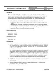
8<strong>Wingspan</strong>campus news<strong>December</strong> 6, 2010wingspan.lccc.wy.eduEconomic study plannedResults could draw employers,give real numbers to LegislatureBy Seneca FlowersCo-EditorLaramie CountyCommunity College hasgreat financial impact uponCheyenne, Laramie Countyand the state; a new studywill soon reveal just howgreat the impact is.Nov. 15 was the kick-offof an economic impactstudy for LCCC. Due inmid-March, the finalimpact study is part of agreater statewide collegeeffort to show how theeconomy is affected by havinga college in an area. Theeconomic impact is madeby not only direct employmentof college faculty andstaff but also from studentsand revenue made fromgraduates who remain inthe region.The presidents of thecolleges in Wyomingdecided to request studiesto show all their economiccontributions.In 2008, an EconomicModeling Specialists Inc.study showed the impactof Casper Collegeupon its community was$304 million annually. Italso showed for every $1invested in Casper College,the community receives$2.20 back in the form ofcumulative tax receipts andavoided social costs.This study may havehelped influence NatronaCounty residents subsequentlyto pass a multimilliondollar bond issue forCasper College.LCCC’s president Dr.Darrel Hammon, is hopingthe current LCCC study willbring similar results.Hammon said the resultsfor the study will give statelawmakers real numbersto see the impact of LCCC.“It’s easier to show legislatorswhy we ought to fund aparticular project,” he said.“It validates communitycolleges.”Hammon said the collegeis an incentive for somecompanies to come toCheyenne as it can providetraining and qualifiedemployees. He added theflexibility of being a comprehensivecommunitycollege allows for classesto be specifically tailoredto a business’s needs. Hesaid some business suchas the National Center forAtmospheric Research(NCAR) estimated to beconstructed by August 2011in Cheyenne could potentiallycreate work/educationaloutles. Hammon saidalthough LCCC does notdirectly produce scientistswith master’s degrees, LCCCdoes offer other partnershipsthat may be of interest suchas internships and computertechnology degrees.Hammon said he feltLCCC, as a comprehensivecommunity college,creates a greater economicimpact than just aregular junior college thatfocuses on transfer only.Comprehensive communitycolleges offer technicalcertificates, GED, ESL, partnershipsetc., in addition totransfer.Hammon said he feltLCCC will favor competitivelywith other regionalcolleges because of thenew businesses moving toCheyenne such as oil companies.Hammon added theoil companies will greatlycontribute to LCCC and thecourses LCCC can providefor them.Associated Student GovernmentTwo senators electedBy SenecaFlowersCo-EditorAt a Nov. 30, meeting,the AssociatedStudent Governmentannounced recentelections filled two ofthe four open senatorpositions at LaramieCounty CommunityCollege.The newly electedsenators are CameronGreen and Emma LeePetty.Green said he waslooking forward to“just being able tohelp voice the studentopinion to the rest ofthe LCCC community.”ASG failed to fillall four open studentsenator positions forthe spring semester.The positions wereopen after threeresignations and oneimpeachment.ASG promotedelections throughEaglesEye, posters inall buildings on campus,an applicationdrive in the studentlounge and distributionof applicationsto those who seemedinterested.Online votingthrough EaglesEyespanned Nov. 22–23.ASG adviser DaveGaer said the holidaysand finals may havehindered the electionprocess.ASG PresidentAlex Barker addedshe was pleased withthe two new senatorselected who bothhave Campus ActivityBoard experience.Petty is currently thepresident of CAB.At the Nov. 16 ASGmeeting, the groupbegan preparation fora campus forum toreceive informationfrom various bodies atthe college.Seneca FlowersReporting for duty:Newly elected senator CameronGreen listens at his first ASG meeting.ASG will host “Aska senator day” 11a.m.–1 p.m. Tuesday,Dec. 7 in the studentlounge, The forum willbe held to raise studentawareness of thestudent governmentas well as to connectwith issues oncampus. Barker saidthe open discussion ispart of a greater goalto enhance campuscommunications withstudents.Earlier in thatmeeting, the senateapproved thepurchasing of eightadditional suggestionboxes to be placed invarious departmentson campus.Big city food, small town service.30 minutes east of Cheyenne.711 Parsons Pine Bluffs, WY 82082307-245-3111638-8591620 Central Ave.For a Little “Spice of Life”LosAmigosCheyenne.comTuesday–SaturdayLunchDinnerClosed11 a.m. to 2 p.m.4:30 p.m. to 8:30 p.m.Sunday & Monday
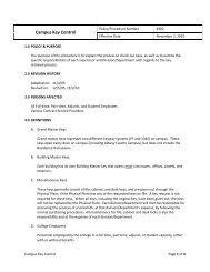
3220 SECTION XVII ● Pediatric UrologyA B CFigure 120–11. A, Vertical flap technique may be used when a dependent ureteropelvic junction is situated at the medial margin of a large,box-shaped extrarenal pelvis. In contrast to the spiral flap, the base of the vertical flap is situated more horizontally on the dependentaspect of the renal pelvis, between the ureteropelvic junction and the renal parenchyma. The flap itself is formed by two straight incisionsconverging from the base vertically up to the apex on either the anterior or posterior aspects of the renal pelvis. As for the spiral flap, theposition of the apex determines the length of the flap, which should be a function of the length of proximal ureter to be bridged. Themedial incision of the flap is carried down the proximal ureter completely through the strictured area into normal-caliber ureter. B, Theapex of the flap is rotated down to the most inferior aspect of the ureterotomy. C, The flap is closed by approximating the edges withinterrupted or running fine absorbable sutures.A B CFigure 120–12. Anderson-Hynes dismembered pyeloplasty. A, Traction sutures are placed on the medial and lateral aspects of thedependent portion of the renal pelvis in preparation for dismembered pyeloplasty. A traction suture is also placed on the lateral aspect ofthe proximal ureter below the level of the obstruction. This suture will help maintain proper orientation for the subsequent repair. B, Theureteropelvic junction is excised. The proximal ureter is spatulated on its lateral aspect. The apex of this lateral, spatulated aspect of theureter is then brought to the inferior border of the pelvis while the medial side of the ureter is brought to the superior edge of the pelvis.C, The anastomosis is performed with fine interrupted or running absorbable sutures placed full thickness through the ureteral and renalpelvic walls in a watertight fashion. In general, the authors prefer to leave an indwelling internal stent in adult patients. The stent isremoved 4 to 6 weeks later.aspect of the anastomosis at the vertex of the spatulationis critical to ensuring a watertight closure.11. Before the repair is completed, the renal pelvis is irrigatedto remove any blood clots or debris that could obstruct theUPJ. If an indwelling double-J ureteral stent is employed,it should be placed now, with care taken to place the stentinto the bladder and renal pelvis without kinking it.12. A Penrose drain is placed adjacent to the repair and broughtout through a separate stab wound.13. The kidney is returned to its native position, and perinephricfat, if available, is placed over the anastomosis.14. Closure of the three fascial layers is readily accomplished,followed by closure of the Scarpa fascia and subcuticularskin.
- Page 2 and 3: 2Wingspancampus newsDecember 6, 201
- Page 4 and 5: 4Wingspancampus newsDecember 6, 201
- Page 6 and 7: 6Wingspancampus newsDecember 6, 201
- Page 11 and 12: December 06, 2010wingspan.lccc.wy.e
- Page 13 and 14: December 6, 2010wingspan.lccc.wy.ed
- Page 15 and 16: December 6, 2010wingspan.lccc.wy.ed
- Page 17 and 18: 18 WingspanDecember 6, 2010wingspan
- Page 19 and 20: 20 WingspanDecember 6, 2010wingspan
- Page 21 and 22: 22 Wingspancampus newsDecember 6, 2
- Page 23 and 24: 24Wingspana&eDecember 6, 2010wingsp
- Page 25 and 26: 26 WingspansportsDecember 6, 2010wi
- Page 27 and 28: 28WingspansportsDecember 6, 2010win
- Page 29 and 30: 30WingspansportsDecember 6, 2010win
- Page 31 and 32: 32WingspansportsDecember 6, 2010win
- Page 33 and 34: 2BWingspanA ChangeWestNorthSouthEas
- Page 35 and 36: 4BWingspanA ChangeDecember 6, 2010w
- Page 37 and 38: December 6, 2010wingspan.lccc.wy.ed