New Horizons - myESR.org
New Horizons - myESR.org
New Horizons - myESR.org
- No tags were found...

You also want an ePaper? Increase the reach of your titles
YUMPU automatically turns print PDFs into web optimized ePapers that Google loves.
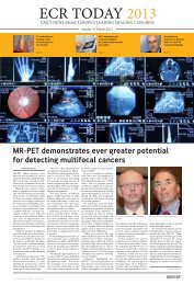
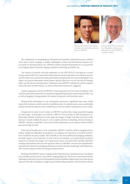
www.<strong>myESR</strong>.<strong>org</strong> | EUROPEAN CONGRESS OF RADIOLOGY 2013Professor Bernd Hamm from Berlin,Germany, will chair the <strong>New</strong> <strong>Horizons</strong>Session on MR/PET.Professor Heinz-Peter Schlemmerfrom Heidelberg, Germany, willtalk about the role of MR/PET inoncologic imaging.The combination of morphological, functional and metabolic information proves useful atevery step of cancer imaging. It enables radiologists to detect and characterise tumours veryaccurately, by showing tumour size, infiltrative pattern and growth potential. It is very good forlocal staging, distant metastatic staging, treatment monitoring, and follow-up.“For instance in patients with pole melanoma, we use FDG PET/CT with glucose to assesstumour spread. MR is very important to detect tumour spread in the brain, liver and bone marrowand PET allows us to see tumours early, particularly in lymph nodes. If you put it all together, youobtain very precise information about tumour spread, which you can use not only for therapyfollow-up, but also for early detection,” Schlemmer said. MR/PET could also be used for patientswith other types of solid tumour, e.g. breast and prostate tumours, he suggested.Another argument in favour of MR/PET is that it generates very low levels of radiation. Thiscould be particularly important for repeated imaging during therapy monitoring and follow-upas well as imaging of young patients, for instance young men with testicular cancer.Having all the information in one examination represents a significant time-saver, whichmeans that treatment could be started or modified earlier. It would also prove more comfortableand less stressful for patients, who wouldn’t have to wait for another examination or new results.It might also be easier to put in place an MR/PET machine than merge sequential imagingat a later stage. “In principle, you could do a PET/CT first and then an MR examination; sophisticatedsoftware would process and merge the images. It might work that way, but in dailypractice it sounds unlikely because it is too complex and time consuming, whereas having anMR/PET machine would allow you to have all the information in 45 minutes without furtherpost-processing,” he said.If the clinical benefits prove to be worthwhile, MR/PET could be useful in imaging tumourentities, despite the difficulties, he predicted. “According to my experience, it would be worth it,but it would also be quite complex. PET and MR are the most sophisticated imaging modalities,and both require a lot of expertise. To have it all in one machine, you would need even greaterexpertise to interpret the images. You would need to know a lot about PET and MR, and aboutoncology and treatment. But once the expertise is there, it could offer a very fast and comprehensiveassessment for detection and staging of tumours as well as therapy monitoring,” said Schlemmer,who trained in physics before becoming a medical doctor.Working with MR/PET means working closely with physicists, radiopharmacists, oncologistsand, naturally, nuclear doctors, especially for making diagnoses. The level of cooperation betweenradiologists and nuclear physicists demanded by hybrid modalities might require more than just aninterest in the other’s discipline; it might require further subspecialisation. Only the future will tell.Preliminary Programme EdiciÓn Invierno | ECR 2013 31